檢測人類子宮內膜異位症的典型鈣粘蛋白並與子宮腔內之子宮內膜作一比較; IDENTIFICATION OF THE CLASSICAL CADHERIN SUBTYPES PRESENT IN THE HUMAN ENDOMETRIOSIS AND IN COMPARISON WITH THE EUTOPIC ENDOMETRIUM
84
0
0
全文
(2) 誌謝 自從中國醫藥學院醫學系畢業,即從事於婦產科臨床醫學工作至 今。有感於臨床治療,往往是發病之後的補救措施。因此,一直希望有機 會能夠再由基礎醫學的研究來探討疾病的形成,以期由細胞或分子層面來 治療或預防疾病。終於,再前年得償夙願,能夠進入母校醫學研究所從事 研究工作。 匆匆兩年過去了,要感謝的人實在太多。首先感謝蔡長海董事長 能夠提供及鼓勵臨床醫師進修的機會。也感謝指導教授蔡鴻德主任不管在 臨床工作或是基礎研究的提攜及督促,還有共同指導教授陳澤昭博士和張 文正教務長的殷切指導。也感謝科內葉聯舜主任、林武周主任及楊東川主 任等學長提供研究的經驗,還有吳慧音同學及黃鈺媚小姐在實驗過程中的 鼎力幫助使得整個實驗得以完成。 雖然初步的研究告一段落,課程也劃上休止符,但卻是我個人從 事更深入研究的開始,藉由兩年師長所傳授的經驗,建立起自己基礎醫學 研究的磐石,相信在未來能夠使自己往前邁進,不懼艱難。 最後感謝我摯愛的妻子以及兩位嗷嗷待哺的小公主,有他們的陪 伴此論文才得以順利完成。. 2.
(3) 中文摘要 背 景 子宮內膜異位症(endometriosis)是指子宮內膜的腺體及其基質出現 在子宮腔外,在生育年齡的婦女其發生率將近 3~10%,然而在不孕症的婦 女,其發生率則高到 25~35%。子宮內膜異位症造成的骨盆腔疼痛及不孕 等症狀,是病人及醫療的極大負擔。至目前為止,其致病的機轉尚未明瞭, 最被廣為接受的理論為經血逆流所造成,然而逆流的子宮內膜組織如何附 著並侵入腹膜並得以增殖和成長,仍然是一個謎。鈣粘蛋白(cadherin)是一 種細胞沾粘分子[cell adhesion molecules (CAM)],其功能為細胞沾粘作 用,同時在胚胎發育中主導不同組織及器官的形成,同時它們與癌細胞的 侵襲性也有關。因此我們認為鈣粘蛋白可能與子宮內膜異位症的形成、侵 入、及成長有關。本論文乃探討子宮內膜異位症中鈣粘蛋白的表現,並分 析它們與子宮腔內之子宮內膜(eutopic endometrium)的不同表現,以瞭解 子宮內膜異位形成的沾粘機轉(adhesive mechanism)。. 方法 收集九位子宮內膜異位症的病人,依其月經週期分為增殖期(n=5)及 分 泌 期 (n=4)。 首先利用 degenerate reverse transcription-polymerase chain reaction (RT-PCR) 來 檢 測 子 宮 內 膜 異 位 組 織 中 典 型 鈣 粘 蛋 白 (classical cadherin)的種類。之後,再以 semiquantitative RT-PCR 的方法, 比較同一病人其子宮內膜異位組織與子宮腔內之子宮內膜組織中,這些鈣 粘蛋白 mRNA 的表現差異。. 3.
(4) 結果 我們確認子宮內膜異位組織中有六種典型鈣粘蛋白存在,它們分別為 Epithelial-cadherin(E-cad). 、. 、. Placental-cadherin(P-cad). Neural-cadherin(N-cad)、cadherin-6、cadherin-9、和 cadherin-11。比較 這六種鈣粘蛋白在子宮腔內之子宮內膜與子宮內膜異位的表現差異時,我 們發現 P-cad mRNA 表現在子宮內膜異位組織中較子宮腔內之子宮內膜組 織有明顯增多。這種表現差異不論是增殖期或是分泌期都相同。. 結論 我們確認六種典型鈣粘蛋白在子宮內膜異位組織。其中只有 P-cad 在子宮內膜異位組織中比子宮腔內之子宮內膜高出許多。因此,我們認為 P-cad 可能在子宮內膜異位症的形成之沾粘機轉中扮演著重要的角色。. 4.
(5) 目錄 頁數 誌謝-----------------------------------------------------------------------------------------1 中文摘要-----------------------------------------------------------------------------------2 目錄-----------------------------------------------------------------------------------------4 圖目錄--------------------------------------------------------------------------------------5 表目錄--------------------------------------------------------------------------------------6 符號與縮寫--------------------------------------------------------------------------------7 第一章 前言------------------------------------------------------------------------------9 第二章 檢測子宮內膜異位組織中的典型鈣粘蛋白種類-----------------------21 第三章 子宮內膜異位組織與子宮腔內之子宮內膜鈣粘蛋白之比較--------40 第四章 討論、總結與未來研究方向-----------------------------------------------52 第五章 參考文獻-----------------------------------------------------------------------56 英文摘要---------------------------------------------------------------------------------81 作者簡歷---------------------------------------------------------------------------------82. 5.
(6) 圖目錄 Figure 1.. Schematic cadherin structure model------------------------------14. Figure 2.. Schematic representation of classical cadherin structure----15. Figure 3.. Schematic representation of PCR-II vector----------------------31. Figure 4.. Agar plate for transformed bacteria selection-------------------33. Figure 5.. Photomicrographs. plasmid. DNA. and. cloned. PCR. products-38 Figure 6.. Validation of semiquantitative RT-PCR for GAPDH and E-cad------------------------------------------------------------------------43. Figure 7.. Validation of semiquantitative RT-PCR for P-cad and N-cad--------------------------------------------------------------------------------44. Figure 8.. Validation of semiquantitative RT-PCR for cad-6 and cad-9---------------------------------------------------------------------------------45. Figure 9.. Validation of semiquantitative RT-PCR for cad-11-------------46. Figure 10. Comparison of the cadherin mRNA levels present in eutopic endometrium. or. endometriosis. 6. obtained. during. the.
(7) proliferative phase------------------------------------------------------48 Figure 11. Comparison of the cadherin mRNA levels present in eutopic endometrium or endometriosis obtained during the secretary phase----------------------------------------------------------------------50. 7.
(8) 表目錄 Table 1.. The primers used in degenerate or semiquantitative RT-PCR------------------------------------------------------------------------------27. Table 2.. The PCR conditions---------------------------------------------------28. Table 3.. Analysis of cadherin cDNA generated from endometriotic lesions---------------------------------------------------------------------39. 8.
(9) 符號與縮寫 Ala ANOVA Bp BSA °C CaCl2 Cad CAM CAR CDNA DDT DNA Dsc Dsg EB EC E-cad ECM ER IL G GAPDH His Kb KCl MgCl2 Min Ml MM MMLV MMPs MRNA µl µM N-cad PBS P-cad Pcdh. Alanine Analysis of variance Base pair Bovine serum albumin Degrees centigrade Calcium chloride Cadherin Cell adhesion molecule Cell adhesion recognition Complementary DNA Dithiothreatol Deoxyribonucleic acid Desmocollins Desmogleins Ethidium bromide Extracellular Epithelial cadherin Extracellular matrix Estrogen receptor Interlukin Grams Glyceraldehyde-3-phosphate dehydrogenase Histidine Kiobases Potassium chloride Maginum chloride Minutes Milliter Millimolar Moloney Murine Leukemia Virus Matrix metalloproteinases Messenger RNA Microliter Micromolar Neural cadherin Phosphate-buffered saline Placental cadherin Protocadherin 9.
(10) PCR PR RNA RNAsin RT Tris-HCl Val VEGF. Polymerase chain reaction Progesterone receptor Ribonucleic acid Ribonuclease inhibitor Reverse transcriptase Tris(hydroxymethyl)-aminomethane-hydrochloric acid Valine Vascular endothelial growth factor. 10.
(11) 第一章 前言. 1-1: 導論(Introduction): 子宮內膜異位症(endometriosis)在生育年齡的婦女是十分常見的 疾 病 , 其發生率將近 3~10%,然而在不孕症的婦女其發生率將高到 25~35%[1,2]。由於子宮內膜異位症會引起疼痛症狀及不孕症問題,因此常 造成婦女很大的困擾。但由於子宮內膜異位症的發病原理及致病機轉至今 尚並不完全明瞭,因此也導致治療上的困難。遠在 1860 年,Rokitansky 即提及子宮內膜異位症[3],但直到 1921 年 Sampson 才描述,具有病理構 造及功能的子宮黏膜出現在子宮外,稱為『子宮內膜異位症』[4]。子宮內 膜異位症最常見的症狀是經痛(dysmenorrhea)其次為不孕症(infertility)、性 交疼痛(dyspareunia)、下腹痛、下背痛。子宮內膜異位症可發生在身體任 何器官[5],疾病若發生在肺部則月經來潮時會有咳血或是氣胸[6-8],若發 生在泌尿道則會有血尿或是小便疼痛甚至導致輸尿管阻塞[9-11],若發生在 胃腸道則會有血便、大便疼痛,甚至會引起腸阻塞[12-16]。. 11.
(12) 1-2: 子宮內膜異位症之病理機轉: 自 1927 年 Sampson 對於子宮內膜異位症的病理機轉提出假說至 今,共有幾項,(一)、植入學說(Implantation theory):子宮內膜組織經由輸 卵管逆流至腹腔,附著、增殖、成長於腹膜中,進而形成子宮內膜異位症 [17-22],(二)、胚胎化生學說(Coelomic metaplasia theory):腹腔的胚腔上 皮細胞(coelomic epithelium)經由內分泌或發炎反應的刺激,而化生成為子 宮內膜細胞[23-26]。(三)、直接擴散學說(Direct extension theory):子宮內 膜細胞直接侵入子宮肌肉層,形成子宮腺肌症,甚至侵入膀胱、輸尿管、 尿道或腸子[27]。(四)、淋巴管和血管轉移學說(Lymphatic and vascular metastasis theory):子宮內膜組織經由淋巴液和血液,轉移至遠方的器官 [5-16,28-33]。(五)、機械式植入學說(Mechanical transplantation theory): 例如開刀或生產而產生子宮內膜異位症[34]。(六)、複合式學說 (Composite theory):認為是由以上機轉所共同形成,非單一學說可解釋所有疾病[35]。 以上以植入學說較被接受且有動物實驗証明。 然而報告指出婦女經血逆流幾乎都會發生,但是為何只有 3~10% 的婦女產生子宮內膜異位症,因此推測可能有某些因素促使子宮內膜異位 症的產生。例如,(一)、遺傳基因(Genetic factors):有些學者認為子宮內 膜異位症可能是遺傳疾病,因而導致免疫系統改變使得異位的子宮內膜細 胞得以成長[36,37]。(二)、免疫系統因素(Immune system factors):子宮內. 12.
(13) 膜 異 位 症 的 病 人 , 腹 腔 體 液 (peritoneal fluid) 中 發 現 巨 噬 細 胞 (macrophage)、輔助性 T 細胞(helper T-cell )及自然殺手細胞(natural killer cell, [NK cell])增加情形,而這些免疫細胞會產生細胞激素(cytokines),包 括 interleukin-1(IL-1)、tumor necrosis factor-á(TNF-á)、IL-6、IL-8。而這 些細胞激素與免疫補體細胞(immunocompetent cells)的成長及分化,以及 嗜中性白血球(neutrophils)和血管生成(angiogenic)有關[38-45]。(三)、血管 生成因素(Angiogenic factors):血管內皮細胞成長因素 vascular endothelial growth factor(VEGF)是一種血管生成因素,可刺激血管內皮細胞成長及分 化。有些學者發現血管內皮細胞成長因素可能是由被活化的巨噬細胞所分 泌,它可以使腹膜上植入的子宮內膜細胞形成新生血管 (neovascularization),而且腹腔體液中血管內皮細胞成長因素的濃度與子 宮 內 膜 異 位 症 的 嚴 重 程 度 有 關 [46-49]。 (四 )、 類 固 醇 接 受 器 (Steroid receptors):子宮內膜異位症的病人其植入腹膜的子宮內膜細胞上發現有 estrogen. receptor(ER),. progesterone. receptor. (PR),. androgen. receptor(AR),且發現 P-450 aromatase mRMA 的轉錄(transcripts),這可 能代表子宮內膜細胞可局部製造雌激素促進子宮內膜細胞的成長及增殖 [50-53]。(五)、附著沾黏及侵入(Adhesion and invasion):腹腔的子宮內膜 細胞藉 CAM 如 intergins、鈣粘蛋白及 maxtrix metalloproteinases(MMPs) 等,與腹膜的細胞外基質(extracellular matrix)相結合促進子宮內膜細胞侵. 13.
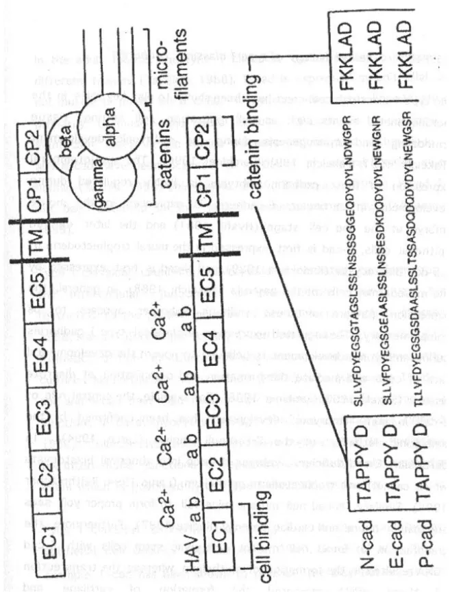
(14) 入[54-73]。 基於子宮內膜異位症的起源,逆流的子宮內膜細胞必須先和腹膜 細胞沾粘,進而互動(interaction) 才能形成子宮內膜異位症病灶。因此我們 著手研究細胞沾粘物質在子宮內膜異位症的發生和生長所扮演的角色。尤 其是細胞與細胞間的沾粘物質-鈣粘蛋白。. 1-3: 鈣粘蛋白: 鈣粘蛋白是一群位於細胞膜上的穿膜醣蛋白,至今至少有四十多 種鈣粘蛋白被發現,它們主導同類細胞之間的粘附作用,在鈣離子的環境 下(calcium-dependent)藉由同種親和的方式(homophilic manner)相結合 [74-77]。(見 Figure 1) 一 般 來 說 , 不同的鈣粘蛋白仍然有類似的初級結構(primary structure) 。 其 構 造 可 分 為 三 部 份 : N- 端 的 細 胞 外 部 分 (N-terminal extracellular domain)、穿細胞膜部分(transmembrane domain)及 C-端細胞 質部分(C-terminal cytoplasmic domain)[74-77]。而鈣粘蛋白其胺基酸的保 留性(conserved)大約為 43~58%,而最高保存性的區域(conserved region) 為 C-端細胞質部分,次高保留性的區域為靠近 N-端的細胞外部分。在細胞 外部分,是由幾組大約 110 個胺基酸重覆序列(cadherin-repeat sequence) 所構成,而鈣離子則附著於胺基酸重覆序列間。不同的鈣粘蛋白其胺基酸. 14.
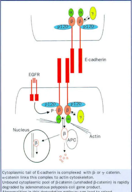
(15) 重覆序列亦不同,同時也表現不同的特性及生物功能。 鈣 粘 蛋 白 可 規 納 為 四 大 類: ( 一 )、 典 型 鈣 粘 蛋 白 (classical cadherins): 其中包括 type 1 及 type 2[74-77], (二 )、 Desmosomal cadherins (三)、Protocadherins[78],(四)、Cadherin-related proteins: 其中包括 truncated cadherins 及 unclassified family members。. 1-3-1: Type 1 典型鈣粘蛋白(見 Figure 2): Type 1 典型鈣粘蛋白:包括 E-cadherin (E-cad)、N-cadherin (N-cad)、 P-cadherin (P-cad) 及 R-cadherin (R-cad)。 主 要 由 五 組 細 胞 外 部 分 (domain)、一組穿細胞膜部分及二組細胞質部分所組成[79],五組細胞外部 分中間有四個鈣離子連接區域(calcium binding regions)。此外,在靠近 N 端的細胞外部分含有 cell adhesion recognition (CAR) sequence,它是由 三個胺基酸 His、Ala、Val (HAV)所構成[80]。藉由 CAR,相同的鈣粘蛋白 彼此接合形成拉鍊狀(zipper-like)的構造[81]。細胞質部分能夠與至少四種蛋 白 相 互 作 用 , 此 四 種 蛋 白 為 α-catenin 、 β-catenin 、 γ-catenin 及 p120cas [82-84]。靠近 C-端細胞質部分與β-catenin 或γ-catenin 接合。而 α-catenin 則一端與β-catenin 或γ-catenin 接合,而另一端與細胞內骨架 (actin cytoskeleton)接合,像這種 cadherin-catenin complexes 才能在細胞 接合、分化及發育中發揮功能。. 15.
(16) Figure 1. From:. Schematic cadherin structure model Wijnhoven: Lancet, Volume 354(9176). July 31, 1999.356-357. 16.
(17) Figure 2.. Schematic representation of classical cadherin structure. 17.
(18) Type1 典 型 鈣 粘 蛋 白 其 生 物 功 能 主 要 是 促 進 細 胞 接 合 (homophilic cell adhesion),在胚胎發育(embryonic development) 中主導 組織器官的形成,並和細胞的移動(cell migration)、組織的分化(tissue remodeling) 有關 [74,75,85]。在胚胎發育時,不同的鈣粘蛋白在適當時間 及地點的表現(spatiotemporal expression) 受到高度調控,引導不同組織的 成型。例如在老鼠胚胎的一個細胞期間(one-cell stage)有 E-cad 之表現 [86],而 N-cad 則表現在腸胚(gastrula)的中胚層細胞[87],P-cad 則表現在 blastocyst 的滋養外胚層(trophectoderm)[88]。Type 1 典型鈣粘蛋白在表 皮細胞的極性(polarity)之維持,佔有重要的地位[89,90]。而且 E-cad 的表 現可維持表皮細胞的分化(differentiated)及非侵襲性 (non-invasive)的 型 態,例如在成熟的表皮細胞若喪失 E-cad 的功能則細胞會發育成像纖維母 細 胞 (fibroblast-like)的型態,且具有侵襲性 [91],甚至導致贅瘤的形成 (neoplastic transformation)[92,93],現今研究指出缺少 E-cad 可能與直腸 癌[94,95]、胃癌[96,97]、胰臟癌[98]、食道癌[99,100]、肝癌[101]、肺癌 [102,103]、膀胱癌[104,105]、前列腺癌[106-108]、乳癌[109-115]、子宮癌 [116]、卵巢癌[117]、甲狀腺癌[118]、皮膚癌及口腔癌等有關[119-123]。而 N-cad 則 與 中 胚 層 及 神 經 外 胚 層 的 腫 瘤 有 關 , 如 pleural mesothelioma[124]. 、. astrocytoma. 、. oligoblastoma[125]. 、. rhadomyosacroma[126]。而 P-cad 則與膀胱癌[127]、乳癌[128]等有關。. 18.
(19) 1-3-2: Type 2 典型鈣粘蛋白: Type 2 典型鈣粘蛋白:包括人類 cadherin-5、-6、-8、-9、-11、 -12 、 -14[129] 及 其 他 動 物 鈣 粘 蛋 白 如 rodent[130] 、 chicken[131] 及 Xenopus[132]。Type 2 典型鈣粘蛋白 與 type 1 典型鈣粘蛋白的結構類似 只是在靠近 N 端的細胞外部分不含有 HAV 的 CAR sequence。 Type 2 典型鈣粘蛋白其生物功能與 type-1 典型鈣粘蛋白類似, 和細胞接合及組織形成(morphogenesis)及腫瘤的形成(tumorigenesis)有 關。例如以往的報告顯示,cadherin-6 與人類及齧鼠類(rodent)腎臟的發育 有關[133,134],且 cadherin-6 與腎臟癌的形成有關。Cadherin-11 則與齧 鼠 類 (rodent)骨 頭 的 形 成 [135]及 中 樞 神 經 的 發 育 有 關[136,137], 而 且 cadherin-11 可能與 trophoblast cell 及胎盤的形成有關[138,139]。另外 cadherin-11 也與子宮內膜基質細胞的分化有關[138-140]。. 1-3-3: Desmosomal 鈣粘蛋白: Desmosomal 鈣粘蛋白:包括 desmocollins (Dscs 1,2,3) 及 desmogleins (Dsgs 1,2,3),其結構與典型鈣粘蛋白類似,但細胞質部分較 長 且 胺 基 酸 較 少 雷 同 。 Desmosomal 鈣 粘 蛋 白 主 要 功 能 則 是 形 成 desmosomes,以接受外力的機械性壓力(mechanical stress)保持細胞的完 整性 ,如位於皮膚、牙齦及子宮頸 ,若受到損傷會導致皮膚的病變如. 19.
(20) pemphigus vulgaris、severe skin blistering[141]。. 1-3-4: Protocadherins: Protocadherins:包括 Pcdh-1、 –2、-3、-8、-9,其結構與典型 鈣粘蛋白類似,但細胞外部分其 cadherin-repeats sequence 超過五組,且 protocadherins 不與 catenins 相接合,此可能反映出 protocadherins 與其 他的鈣粘蛋白有不同的生物特性[78]。Protocadherins 可存在於非脊椎動物 (invertebrates)中,與胚胎腦部及中樞神經的發育有關[142]。. 1-3-5: Cadherin-related proteins: Truncated 鈣 粘 蛋 白 : 包括 T-cad (cadherin-13)、 H-cad 及 L-cad,此群鈣粘蛋白缺少細胞外部分,此外 T-cad 尚缺少穿細胞膜部分而 以 phosphoglycolipid 與細胞膜接合。至於 truncated 鈣粘蛋白的功能,至 目前為止並不十分確定。. 1-4:鈣粘蛋白與子宮內膜異位症的關係: 過去只有少數文獻提及 E-cad、N-cad、P-cad 和子宮內膜異位症 [58-70]。 Gaetje 提及 E-cad-negative 的 子 宮 內 膜 與 轉 移 的 癌 症 細 胞 (metastatic carcinoma cell)具有侵襲的特性(invasive phenotype)[68],而. 20.
(21) Starzinski 也有相似的報告[63]。Gaetje 同時指出在子宮內膜異位症的 E-cad-negative 細胞較正常子宮內膜增加[68],這與 Scotti 的發現相似 [61]。但 Beliard 則發現,E-cad 在子宮內膜細胞與子宮內膜異位細胞中, 未有明顯的改變[69]。Ota 則指出 E-cad 在而分泌期較增殖期增加,且子宮 內膜異位細胞皆較子宮內膜細胞稍多[66]。Peralta 則指出在子宮內膜異位 症其 N-cad 的表現(expression)與 cystoadenoma、borderline ovarian tumor 相類似,因此認為子宮內膜異位症具有贅瘤特性(neoplastic potentially), 且卵巢的子宮內膜異位瘤可能與其他腫瘤一般,是由 mesodermal cells 化 生(metaplasia)來[67]。Van der Linden 認為 P-cad 存在於增殖期子宮內膜 及子宮內膜異位並推論它與子宮內膜異位症可能有關[73]。. 1-5: 假說與基本原理(Hypothesis and Rationale): 子宮內膜異位症雖然是一種良性的疾病,但其特性確是十分具有侵襲 性,即使經過外科手術治療,仍然有很高的復發的機率。由於其發生的病 理機轉至今尚未十分清楚,而異位的子宮內膜細胞,其表現的特性有些類 似癌細胞的增生和侵犯性。為了瞭解異位的子宮內膜細胞如何附著宿主細 胞及增生,研究其沾粘的機轉是有必要的。本研究選擇的鈣粘蛋白為研究 方向,希望找出異位子宮內膜細胞中典型鈣粘蛋白的種類,及子宮內膜異 位組織與子宮腔內之子宮內膜細胞間的典型鈣粘蛋白的表現差異,以便瞭. 21.
(22) 解 子 宮 內 膜 異 位 症 的 形 成 。 本 研 究 分 為 兩 個 階 段 : 第一階段 : 利 用 degenerate reverse transcription-polymerase chain reaction (RT-PCR) 的方法找出子宮內膜異位組織的典型鈣粘蛋白的種類。第二階段: 以 semi-quantitative RT-PCR 的方法分析子宮內膜異位組織與子宮腔內之子 宮內膜中各種典型鈣粘蛋白的表現差異。. 22.
(23) 第二章 鑑定人類子宮內膜異位症的典型鈣粘蛋白 (Identification of Classical Cadherin subtypes expressed in endometriosis). 2-1: 材料與方法 2-1-1: 檢體與取樣(tissues) 本實驗檢體取自在中國醫藥學院附設醫院婦產科接受子宮內膜異 位囊腫切除手術所之檢體,選擇三位患者,年齡分別為 23、28、32 歲,開 刀前三個月未曾使用過賀爾蒙藥物。取出之檢體剝取內膜囊腫的裏層子宮 內膜異位組織,而子宮腔內之子宮內膜的組織則以刮匙刮取少許子宮腔內 之子宮內膜,部份以液態氮急速冷凍隨即保存於-80°C 冰箱中,另一部份以 4% formaldehyde/2% glutaraldehyde 固定,以 hematoxylin & eosin 染色, 確定子宮內膜的組織是處於增殖期或分泌期,其中增殖期為 2 位,分泌期 1 位。. 23.
(24) 2-1-2. RNA 之萃取(Trizol RNA extraction): 2-1-2-A: 儀器 1.. Source capture system (Germfree Laboraties). 2.. Centrifuge 5810R (Eppendorf, Germany). 3.. Microcentrifuge (Uover Laboratories, USA). 4.. Spectrafuge 16M (National Labnet, USA). 5.. TM firestek B206 (Firstek Scientific). 6.. Minigel Migration Trough Mupid-2 (Cosmo Bio Co LTD, USA). 7.. Rocker platform (Bellco Biotechnology, USA). 8.. UV box TFX 20 M (vukber Lourmat, France). 9.. DS34 Polaroid Electrophoresis hood (UK). 2-1-2-B: 試劑 1.. Trizol Reagent (Life Technologies, Inc., Gaithersburg, MD). 2.. 1X TBE Buffer. 3.. 2 % Agarose. 4.. Chloroform. 5.. Isopropanal. 6.. Ethyl glycol. 7.. Diethyl pyrocarbonate. 24.
(25) 8.. Ethidium bromide (10mg/ml) u. Ethidium bromide. u. Dissolved in 100 ml distilled H2O , store at room. 1 g. temperature,避光。 2-1-2-C: 步驟 â. 100 mg 的組織剪碎在加入 1 ml Trizol reagent,再進一步磨碎。. â. 於 4°C 、10000 rpm 離心 1 分鐘後,取上清萃取液。. â. 加入 0.2 ml chloroform 混合均勻,室溫靜置 5 分鐘後。. â. 於 4°C 、12000 rpm 離心 15 分鐘後,取最上面透明之 RNA 萃取液。. â. 再加入 0.5 ml cold isopropanol 混合均勻,室溫靜置 10 分鐘後。. â. 於 4°C 、13000 rpm 離心 8 分鐘後,移除上清液,留下沉澱物。. â. 加入 1 ml 100% alcohol,混合均勻。. â. 於 4°C 、10000 rpm 離心 5 分鐘,去除上清液,再靜置 5 分鐘(需 避免沉澱物過度乾燥)。. â. 加入 40 µl RNAse and DNAse free 水。. â. 於 58°C 加熱 10 分鐘使沉澱物溶解。. â. 將所得的 RNA 產物以電泳分析法來評估含量。. â. 置於-80°C 冰箱中保存。. 2-1-2: 反轉錄(Reverse transcription, Promega):. 25.
(26) 2-1-2-A:儀器: 1.. TM firestek HB-100 (Firstek Scientific). 2.. TM firestek B206 (Firstek Scientific). 2-1-2-B: 試劑: 1.. Random primer (0.5µg/µl of Promega). 2.. Oligo-dT primer (0.5µg/µl of Promega). 3.. MMLV RT buffer (Promega). 4.. 0.1 M DDT(dithiothreitol). 5.. dNTP(100 mM, 25mM each, Protech). 6.. RNAsin (Invitrogen, 10 units/µl). 7.. MMLV RTase (Promega, 200 units/µl). 2-1-2-C: 步驟: â. 將 1-5 µg 之 RNA (2-23µl) 加 RNAse and DNAse free 水至 23µl。. â. 加入 2µl random primer 及 0.5µl Oligo-dT primer 總共 25.5µl。. â. 加熱 70°C 5 分鐘之後,置於冰上 2 分鐘,然後短暫離心。. â. 再加入 8µ MMLV RT buffer、2µl 0.1 M DDT、2µl dNTP、0.5µl RNAsin、2µl MMLV RTase,總共 40µl。. â. 於 37°C 置放 3 小時。. â. 於 94°C 加熱 5 分鐘。. 26.
(27) â. 置於-80°C 冰箱中保存。. 2-1-3:Polymerase chain reaction using degenerate primers 2-1-3-A:儀器: 1.. PCR system 2400 (Perkin Elmer, USA). 2.. Minigel Migration Trough Mupid-2 (Cosmo Bio Co LTD, USA). 3.. Rocker platform (Bellco Biotechnology, USA). 4.. UV box TFX 20 M (vukber Lourmat, France). 5.. DS34 Polaroid Electrophoresis hood (UK). 2-1-3-B:試劑 1.. 10 X PCR buffer. 2.. 2.5 mM dNTP. 3.. MgCl2. 4.. 20 µM cadherin degenerate forward primer (sequence 見 Table 1). 5.. 20 µM cadherin degenerate forward primer (sequence 見 Table 1). 6.. Taq polymerase. 27.
(28) 7.. DNA- Gel-loading buffer (6X) u. Bromophenol blue. 0.25 ml. u. Xylene cyanol FF. 0.25 ml. u. Ficoll. 15 ml. u. Add H2O to 100 ml. 2-1-3-C:步驟: â. 將 2-4 µl cDNA 加入 ddH2O 至 35 µl. â. 加入 5µl of 10X PCR buffer、5µl of 2.5 mM dNTP、2µl of MgCl2、 1 µl of 20 µM forward primer、1µl of 20 µM reverse primper、1µl of Taq polymerase,總共 50µl。. â. 以聚合連鎖反應(PCR)之儀器進行反應。(反應條件見 Table 2). â. 將所得的 DNA 產物將用來植入 PCR-II vector。(見 Figure 3). 28.
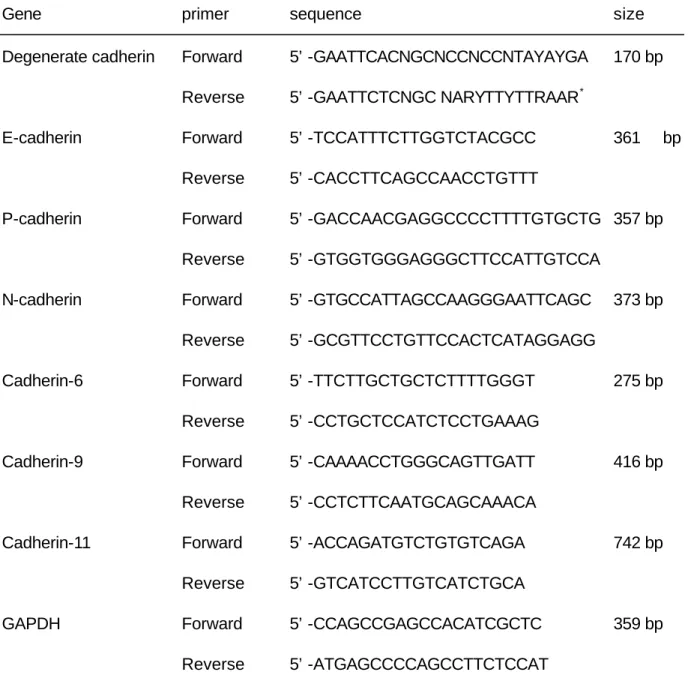
(29) Table 1. Primers used in degenerate and semiquantitative RT-PCR assays. Gene. primer. sequence. size. Degenerate cadherin. Forward. 5’-GAATTCACNGCNCCNCCNTAYAYGA. 170 bp. Reverse. 5’-GAATTCTCNGC NARYTTYTTRAAR *. Forward. 5’-TCCATTTCTTGGTCTACGCC. Reverse. 5’-CACCTTCAGCCAACCTGTTT. Forward. 5’-GACCAACGAGGCCCCTTTTGTGCTG 357 bp. Reverse. 5’-GTGGTGGGAGGGCTTCCATTGTCCA. Forward. 5’-GTGCCATTAGCCAAGGGAATTCAGC. Reverse. 5’-GCGTTCCTGTTCCACTCATAGGAGG. Forward. 5’-TTCTTGCTGCTCTTTTGGGT. Reverse. 5’-CCTGCTCCATCTCCTGAAAG. Forward. 5’-CAAAACCTGGGCAGTTGATT. Reverse. 5’-CCTCTTCAATGCAGCAAACA. Forward. 5’-ACCAGATGTCTGTGTCAGA. Reverse. 5’-GTCATCCTTGTCATCTGCA. Forward. 5’-CCAGCCGAGCCACATCGCTC. Reverse. 5’-ATGAGCCCCAGCCTTCTCCAT. E-cadherin. P-cadherin. N-cadherin. Cadherin-6. Cadherin-9. Cadherin-11. GAPDH. *R = either A or G, Y = either C or T, N = either A, C, G, or T. 29. 361. bp. 373 bp. 275 bp. 416 bp. 742 bp. 359 bp.
(30) Table 2, PCR conditions Cad-11. Cad-6、Cad-9、E-、P-、N-cad. 94°C. hold 5 min. 94°C. hold 5 min. 94°C. 30 sec. 94°C. 30 sec. 55°C. 30 sec. 60°C. 30 sec. 72°C. 2 min. 72°C. 1 min. 30-40 cycles 72°C. 30-40 cycles 8 min. 72°C. 8 min. Cad Degenerate PCR. GAPDH. 95°C. hold 5 min. 94°C. hold 5 min. 95°C. 1.5 min. 94°C. 30 sec. 45°C. 2 min. 60°C. 30 sec. 72°C. 3 min. 72°C. 1 min. 35 cycles 72°C. 20-30 cycles 8 min. 72°C. 30. 10 min.
(31) 2-1-5:Ligation、transformation and bacterial growth 2-1-5-A:儀器 1.. TA cloning Kit Dual Promoter. 2.. Microcentrifuge. 3.. Thermocycler. 4.. 14°C water bath. 5.. 42°C water bath. 6.. 37°C water bath. 7.. Bucket with ice. 2-1-5-B:藥劑 1.. Taq DNA polymerase. 2.. TE buffer. 3.. Mineral oil. 4.. Luria-Bertani (LB) medium and agar. 5.. Dimethylformamide (DMF). 6.. 40 mg/ml 5-bromo-4-chloro-3indolyl-β-galactoside (X-Gal) in DMF. 7.. 50 mg/ml ampicillin stock. 8.. 50 mg/ml kanamycin. 9.. 100 mM isopropyl-β-D-thiogalactoside (IPTG). 31.
(32) 10. One Shot cells ( INVáF’ One Shot Competent Cells & TOP10F’ One Shot Competent Cells ) 2-1-5-C: 步驟 [. Ligation (Clone into PCR II vector ) â. 離心一瓶(vial)PCR II (見 Figure 3. ),然後收集上清液。. â. 計算 PCR product 所需的量(公式 X ng PCR product = Y bp PCR product 乘以 x 50 ng PCR vector 除以 size in bp of the PCR II vector:~3900). â. 轉換成為體積 X µl. â. PCR product X µl 加入 10 X ligation buffer 1µl 再加入 PCR II vector(25ng/µl) 2µl. â. 加入無菌的純水(sterile water)至 9µl. â. 加入 T4 DNA ligase (4.0 Weiss units) 1µ總共 10µl. â. 放置於 14°C 水槽(water bath)至少 4 小時,最好 overnight. â. store -20°C 冰箱中。. 32.
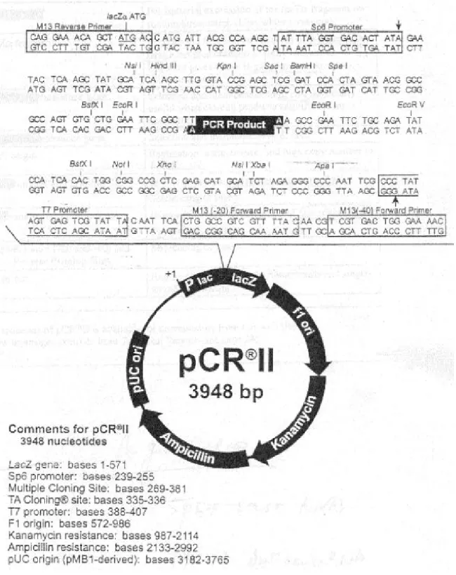
(33) Figure 3. Schematic representation of PCR-II vector. 33.
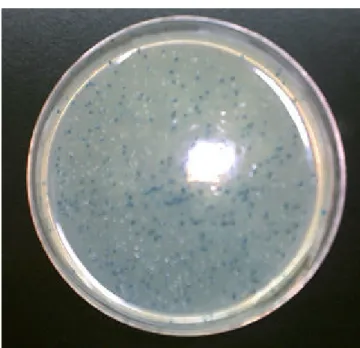
(34) [. Transformation â. 解凍先前已被 PCR product clone 的 PCR II vector(ligation reaction). â. 離心,置於冰上. â. 解凍 50µl One Shot competent cells ( TOP110F’ or INVαF’) 及 0.5M β-mercaptoethanol (β-ME). â. INVαF’ cells only:加入 2µlβ-mercaptoethanol (β-ME),輕輕混 合。. â. 加入 2µl ligation reaction 至 competent cells,輕輕混合。. â. 置於冰上 30 分鐘。. â. 置於 42°C 水槽 30 秒,之後置於冰上。. â. 加入 250µl SOC medium(室溫). â. 置於 37°C shaking incubator 以 225rpm 平行搖晃 1 小時。. â. 取 50 及 200µl 分別塗於 LB agar plates 上(含有 X-Gal 及 50µg/ml kanamycin or ampicillin). â. 每種至少放入兩種不同量,至少有一種 well-spaced colonies. â. 確定凝固後,將 plates 顛倒置於 37°C 的 incubator 至少 18 小 時. â. 置於 4°C 冰箱 2~3 小時以利顏色之形成。(見 Figure 4). 34.
(35) Figure 4.. Agar plate for transformed bacterial selection. 當 被 含 有. cadherin product 所 clone 的 PCR II vector 轉植成功的 TOP10F’細胞則形 成白色菌落,若不含有 cadherin product 所 clone 的 PCR II vector 轉植成 功的 TOP10F’細胞則形成藍色菌落,若轉植不成功的 TOP10F’細胞則無法 成長形成菌落。. 35.
(36) [. Bacterial growth â. 取出至少 10 個白色菌落(white colonies)(本篇為 30 個). â. 置於 2~5 ml LB broth ( 加入 50µg/ml ampicillin or 50µg/ml kanamycin) overnight 日後做 plasmid 分析。. 2-1-6:Plasmid DNA extraction and DNA sequence 2-1-6-A:儀器 1.. Microcentrifuge. 2.. Gene-spin Miniprep Purification Kit( Beckman CEQ2000, ABI377, LICOR IR2, Pharmacia A.L.F.). 2-1-6-B:藥劑 1.. Gene-Spin Kit reagents u. Solution I. u. Solution II. u. Solution III. u. Wash Solution. 36.
(37) 2.. 3.. Restriction Enzyme 10 X buffer H u. 900mM Tris-HCl (pH 7.5). u. 500mM NaCl. u. 100mM MgCl2. Enzymes storage buffer u. 10mM Tris-HCl (pH 7.4). u. 400mM NaCl. u. 0.1mM EDTA. u. 1mM DTT. u. 0.15 % Triton X-100. u. 0.5mg/ml BSA. u. 50 % glycerol. 2-1-6-C:步驟 [. Plasmid DNA extraction â. 取 1~2 ml 放置 overnight 的 culture. â. 置於 12000~14000rpm 離心 30~60 秒. â. 加入 200 µl Solution I,Vortex 直至細胞完全溶解. â. 加入 200 µl Solution II,輕輕上下搖晃 5~6 次. â. 加入 200 µl Solution III,輕輕上下搖晃 5~6 次. 37.
(38) â. 置於 12000~14000rpm 離心之 5 分鐘. â. 將 spin column 放入收集管(collection tube),小心將上清液移 入 spin column. â. 離心 30 秒. â. 將 spin column 移出收集管. â. 拋棄過濾液,再加入 700µl washing solution,離心 60 秒. â. 拋棄過濾液,離心 3 分鐘,已去除殘餘的 ethanol. â. 將 spin column 移出,. â. 再將新的 microcentrifuge tube 放入 column. â. 加 50~100 µl H2O or TE 至 column ( 若 plasmid > 7kb 則使用 60~70°C 的 H2O or TE. [. â. 離心 1 分鐘,以移出 DNA,置於-20°C 冰箱保存。. â. 重覆上述兩個步驟可多得 10~15% DNA. DNA sequencing â. 將所得到的 plasmid DNA 取出 1µg. â. 加入 EcoR I restriction Enzyme 1µl. â. 加入 assay buffer 50 µl. â. 加入 Acetylated BSA 直至 0.1 mg/ml. â. 置於 37°C 1 小時. 38.
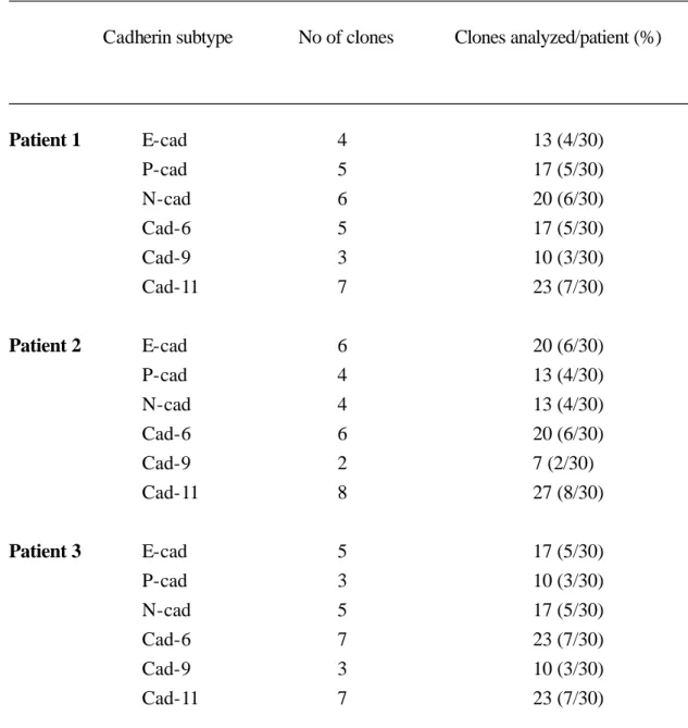
(39) â. 將所得的產物以電泳分析是否含有約 170 bp 的典型鈣粘蛋白 (見 Fig 5). â. 使用 automated DNA sequncer 分析 DNA sequence. â. 得到的 sequence 利用網路上 BLAST computer program 與 Genbank/EMBL 的資料比對以確認找到的鈣粘蛋白亞型. 2-2: 結果 第一階段實驗我們發現在子宮內膜異位組織共有六種典型鈣粘蛋 白亞型:分別為 E-cad、N-cad、P-cad、Cad-6、Cad-9、Cad-11 其出現 比例如 Table 3。. 39.
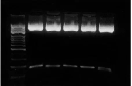
(40) Figure. 5.. A. representative. photomicrographs. of. ethidium. bromide-stained gels containing plasmid DNA (3.9 kb of PCR II vector) and PCR products (~170 bp) generated by using degenerate primers, which extracted from transformed cell and digested by using the restriction enzyme EcoRI.. 40. . ~3.9 kb. . ~170 bp.
(41) Table 3. Analysis of cadherin cDNA clones generated from endometriotic lesions ________________________________________________________________________ Cadherin subtype. No of clones. Clones analyzed/patient (%). ________________________________________________________________________ Patient 1. E-cad P-cad N-cad Cad-6 Cad-9 Cad-11. 4 5 6 5 3 7. 13 (4/30) 17 (5/30) 20 (6/30) 17 (5/30) 10 (3/30) 23 (7/30). Patient 2. E-cad P-cad N-cad Cad-6 Cad-9 Cad-11. 6 4 4 6 2 8. 20 (6/30) 13 (4/30) 13 (4/30) 20 (6/30) 7 (2/30) 27 (8/30). Patient 3. E-cad 5 17 (5/30) P-cad 3 10 (3/30) N-cad 5 17 (5/30) Cad-6 7 23 (7/30) Cad-9 3 10 (3/30) Cad-11 7 23 (7/30) ________________________________________________________________________. 41.
(42) 第三章:六種鈣粘蛋白在子宮內膜異位組織與正常子 宮內膜的不同表現 (Comparison of Six Cadherin Subtypes in Endometriosis and Eutopic Endometrium). 3-1: 材料與方法 2-1-1: 檢體與取樣(tissues) 本實驗檢體取自在中國醫藥學院附設醫院婦產科接受子宮內膜異 位囊腫切除手術所之檢體,患者年齡為 20~42 歲的生殖年齡(reproductive age),開刀前三個月未曾使用過賀爾蒙藥物。取出之檢體剝取內膜囊腫的 裡層子宮內膜異位組織,而正常子宮內膜的組織則以刮匙刮取少許子宮腔 內之子宮內膜,部份以液態氮急速冷凍隨即保存於-80°C 冰箱中,另一部份 以 4% formaldehyde/2% glutaraldehyde 固定,以 hematoxylin & eosin 染 色,確定子宮內膜的組織是處於增殖期或分泌期,其中增殖期 5 位,分泌 期 4 位。. 42.
(43) 3-1-2:RNA 之萃取(Trizol RNA extraction): 其所需儀器、藥劑及步驟如前所述(見第 22 頁) RNA 產物之定量 â. 2µl RNA +3µl ddH2O + 5µl RNA denature buffer. â. 加熱 58°C15 分鐘. â. 置於冰上 2 分鐘. â. 短暫離心. â. 加入 2µl RNA loading buffer. â. 跑膠. â. 置於 EBTBE 內染色 30 分鐘. â. 置於 UV box 照相. â. 評估 RNA 相對含量。. 3-1-3: RT(Reverse transcription, Promega) 其所需儀器、藥劑及步驟如前所述(見第 24 頁). 43.
(44) 3-1-4:PCR 其所需儀器、藥劑及步驟如前所述(見第 25 頁) â. 首先以 PCR 的方式得到的 GAPDH(a house keeping gene),再以 不同的 PCR cycle 求得最適當的 cycles 為 25 cycles. â. 再以相同的 PCR 的方式求得 E-cad、P-cad、N-cad、cad-6、cad-9、 cad-11,最適當的 cycles。. â. 結果以不同的 PCR cycle 求得最適當的 cycles 分別為 GAPDH 25 cycles,E-cad、P-cad、N-cad、cad-6、cad-9、cad-11 為 35 cycles (見 Figure 6-9). â. 算出子宮內膜異位細胞與子宮腔內之子宮內膜細胞之間所需的 mRNA 量. â. 以六種不同鈣粘蛋白亞型的specific cadherin primers 製造鈣粘蛋白 產物(cadherin products). â. 將所得的鈣粘蛋白產物以電泳分析法來評估 DNA 之含量。 u. DNA 產物之定量 â. 加入 2µl DNA loading buffer. â. 跑膠,然後置於 EBTBE 內染色 30 分鐘. â. 置於 UV box 照相. â. 評估 PCR product (DNA)相對含量(見 3-1-5). 44.
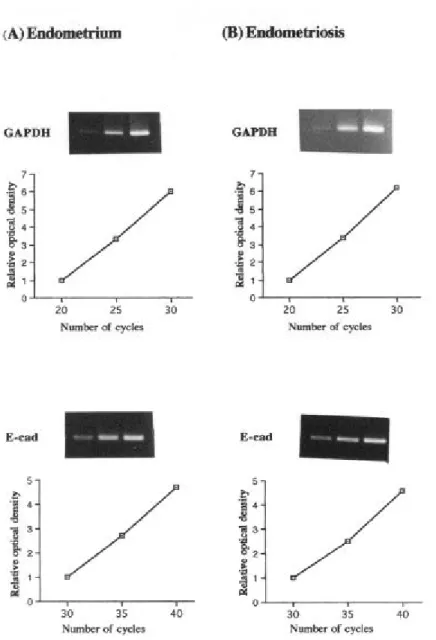
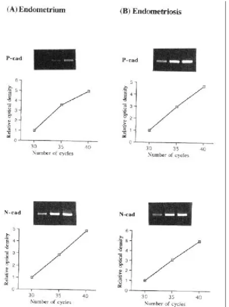
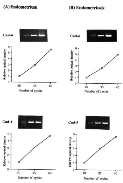
(45) Figure 6. Validation of semiquantitative RT-PCR for GAPDH or E-cad. Total RNA isolated from eutopic endometrium (A) and endometriotic lesion (B) were reverse transcribed. An aliquot of first strand cDNA was amplified for GAPDH or E-cad using different numbers of PCR cycles. A linear relationship was observed between PCR products and amplification cycle when plotted with other two independent experiments (data not shown). 45.
(46) Figure 7. Validation of semiquantitative RT-PCR for P-cad or N-cad. Total RNA isolated from eutopic endometrium (A) and endometriotic lesion (B) were reverse transcribed. An aliquot of first strand cDNA was amplified for P-cad or N-cad using different numbers of PCR cycles. A linear relationship was observed between PCR products and amplification cycle when plotted with other two independent experiments (data not shown). 46.
(47) Figure 8. Validation of semiquantitative RT-PCR for cad-6 or cad-9. Total RNA isolated from eutopic endometrium (A) and endometriotic lesion (B) were reverse transcribed. An aliquot of first strand cDNA was amplified for cad-11 using different numbers of PCR cycles. A linear relationship was observed between PCR products and amplification cycle when plotted with other two independent experiments (data not shown). 47.
(48) Figure 9. Validation of semiquantitative RT-PCR for cad-11. Total RNA isolated from eutopic endometrium (A) and endometriotic lesion (B) were reverse transcribed. An aliquot of first strand cDNA was amplified for cad-11 using different numbers of PCR cycles. A linear relationship was observed between PCR products and amplification cycle when plotted with other two independent experiments (data not shown). 48.
(49) 3-1-5:以半定量 RT-PCR 分析內膜異位組織和子宮內膜組織中六種鈣黏蛋 白亞型的不同表現 â. 將 所 得 到 的 鈣 粘 蛋 白 產 物 (cadherin products) 相 片 以 Hewlett Packer Digital scanner 掃瞄至電腦分析之. â. 使用 UN-SCAN-IT Gel Version 5.1 system (Silk Scientific, Orm, Utah)分析求得 Cadherin/GAPDH mRNA 之比值 。 ( Figure 10 & 11). 3-1-6:統計方法 以 Wilcoxon signed ranks test 來作檢定,以 p<0.05 為有顯著意義。. 3-2: 結果 第二階段實驗我們發現這六種鈣粘蛋白同樣也存在子宮腔內之 子宮內膜組織中。其中 N-cad 和 Cad-9 文獻上尚未有人報告過存在於子宮 內膜,屬於新的發現。還有,E-cad、N-cad、Cad-6、Cad-9 及 Cad-11 的 表現,在子宮內膜異位組織與子宮腔內之子宮內膜之間並無統計學上的差 異。唯有 P-cad 在子宮內膜異位組織的表現較較子宮腔內之子宮內膜高, 呈統計學上有意義的差別,這樣的差異無論是在增生期或分泌期都是存在 的(見 Figure 10 & 11)。. 49.
(50) Figure 10. Comparison of the cadherin mRNA levels present in eutopic endometrium or endometriosis obtained during the proliferative phase of the menstrual cycle.. Photomicrographs of ethidium bromide-stained. gels containing PCR products generated using primers for E-cad P-cad or N-cad at 361, 357 and 373 base pairs respectively (Panel A) or cad-6, cad-9, or cad-11 at 275, 416 and 742 base pairs respectively (panel B). For each cadherin subtype, PCR products were generated using template cDNAs generated from eutopic endometrium (lane a) or endometriotic lesions (lane b). A PCR reaction in which the first strand cDNA was omitted was performed as a negative control (lane c). Products amplified using GAPDH specific primers at 359 base pairs are also shown for each sample. DNA markers are presented on the left hand-side of each gel. The absorbance values obtained for each cadherin subtype were normalized to the values obtained from for the corresponding GAPDH. The results derived from this analysis as well as from two other independent. experiments. (photomicrographs. not. shown). were. standardized to the control. The values obtained from this study, as well as from 4 other studies are presented (mean ±SEM; n=5) in the bar graphs. (*p<0.05).. 50.
(51) Figure 10. Comparison of the cadherin mRNA levels present in eutopic endometrium or endometriosis obtained during the proliferative phase of the menstrual cycle.. 51.
(52) Figure 11. Comparison of the cadherin mRNA levels present in eutopic endometrium or endometriosis obtained during the secretary phase of the menstrual cycle.. Photomicrographs of ethidium bromide-stained. gels containing PCR products generated using primers for E-cad P-cad or N-cad at 361, 357 and 373 base pairs respectively (Panel A) or cad-6, cad-9, or cad-11 at 275, 416 and 742 base pairs respectively (panel B). For each cadherin subtype, PCR products were generated using template cDNAs generated from eutopic endometrium (lane a) or endometriotic lesions (lane b). A PCR reaction in which the first strand cDNA was omitted was performed as a negative control (lane c). Products amplified using GAPDH specific primers at 359 base pairs are also shown for each sample. DNA markers are presented on the left hand-side of each gel. The absorbence values obtained for each cadherin subtype were normalized to the values obtained from for the corresponding GAPDH. The results derived from this analysis as well as from two other independent. experiments. (photomicrographs. not. shown). were. standardized to the control. The values obtained from this study, as well as from 4 other studies are presented (mean ±SEM; n=4) in the bar graphs. (*p<0.05).. 52.
(53) Figure 11. Comparison of the cadherin mRNA levels present in eutopic endometrium or endometriosis obtained during the secretary phase of the menstrual cycle.. 53.
(54) 第四章 討論、總結與未來研究方向. 4-1:討論 為了瞭解子宮內膜異位症的形成機轉,探討它們的細胞接合物質 有助於解開為什麼有 3~10%的婦女,她們的月經逆流會產生子宮內膜異位 症這個謎。本研究檢測出六種 classical cadherins 存在於子宮內膜異位症的 組織中,除了 E-cad、N-cad、P-cad 曾被報告存在子宮內膜異位細胞外, 我們還首先指出有 Cad-6、Cad-9、和 Cad-11。隨後在比較子宮腔內之子 宮內膜與異位子宮內膜時,這六種鈣黏蛋白也都存在子宮腔內之子宮內膜 中。過去的文獻未曾報告 N-cad 或 cad-9 在子宮腔內之子宮內膜當中。本 研究也首次分析出這兩種鈣黏蛋白存在於子宮內膜,雖然子宮內膜上皮細 胞或基質細胞並未曾被測出含有 N-cad,但是血管的內皮細胞則具有 N-cad [143],因此我們認為本研究的 N-cad 可能來自組織中的血管上皮細胞。同 樣地,cad-9 在文獻中未曾有人報告存在子宮內膜上皮或基質細胞[139], 因此,cad-9 在子宮內膜的分佈與功能有待往後繼續研究。 本研究的結果,我們認為這六種鈣粘蛋白負責子宮內膜異位組織 的成長和維持。雖然我們僅分析 mRNA,但是過去我們以及別人的研究報 告均顯示 mRNA 和蛋白質的表現是具有良好的對應性的[127, 144-146]。 過去的文獻中Gaetje 曾指出在子宮內膜異位細胞 E-cad-negative. 54.
(55) 較子宮內膜細胞增加[68],而 Scotti [61] 報告 E-cad 在子宮內膜異位症的 表現略為減少。然而 van der Linden 和 Beliard 則發現,E-cad 在子宮腔內 之子宮內膜細胞與子宮內膜異位細胞中,未有明顯的改變[69, 72]。Ota 則 指出 E-cad 在而分泌期較增殖期多,且子宮內膜異位細胞皆較子宮腔內之 子宮內膜細胞稍多[66]。本研究發現 E-cad 不管在增殖期或是分泌期,於子 宮內膜異位組織與子宮腔內之子宮內膜之間的表現,並無明顯的差別。因 此我們認為 E-cad 在子宮內膜的形成可能不是關鍵性的角色。這些研究結 果的差異有可能是來自於使用不同的研究方法學或是取樣時的差異。 另外 cad-6、cad-9 和 cad-11,不管在增殖期或是分泌期,於子 宮內膜異位組織和子宮腔內之子宮內膜的表現並無明顯差異,因此它們在 子宮內膜異位症可能不是關鍵性的角色。 另 外 , Peralta 指 出 在 子 宮 內 膜 異 位 症 其 N-cad 的 表 現 與 cystoadenoma、borderline ovarian tumor 相類似,因此認為子宮內膜異位 症具有贅瘤特性(neoplastic potentially),且卵巢的子宮內膜異位瘤可能與 其他腫瘤一樣是由 mesodermal cells 化生來[67]。然而本研究並未發現 N-cad 在子宮內膜異位組織與子宮腔內之子宮內膜之間有任何差異,這樣 的結果顯示 N-cad 與子宮內膜異位的發生可能無關。 至 於 P-cad, 文獻顯示它主要表現在複層上皮細胞 (stratified epithelium)的基底層(basal layer)或是副基底層(parabasal layer),這與細胞. 55.
(56) 的分化(differentiation)有關[147]。van der Linden 曾推測 P-cad 可能在子宮 內膜異位症的發生佔有一定的角色[73]。本研究發現不管在增殖期或是分泌 期,子宮內膜異位組織的 P-cad 都比子宮腔內之子宮內膜明顯高出很多, 呈統計學上有意義的差別。因此,我們認為 P-cad 在子宮內膜異位組織與 腹膜等細胞間的沾粘、侵入及增殖扮演著重要的角色。所以當婦女逆流的 子宮內膜細胞,若因為某種原因導致過量的 P-cad 表現,便能夠與宿主細 胞如腹膜,相互沾粘進而發展成子宮內膜病灶。. 4-2 總結 本研究為首次發現 N-cad 和 cad-9 存在於子宮腔內之子宮內膜, 其定位及功能仍有待進一步研究。另外我們也首先提出共有六種典型鈣粘 蛋白(E-cad、P-cad、N-cad、cad-6、cad-9、cad-11)存在於子宮內膜異位 組織中,與子宮腔內之子宮內膜作比較時更發現唯有 P-cad 呈現有意義的 差別。由於 P-cad 在子宮內膜異位組織的表現明顯高於子宮腔內之子宮內 膜,因此我們認為 P-cad 在子宮內膜異位症的形成,甚至於維持扮演著關 鍵性的角色。. 4-3 未來的研究方向 1.. 我們將利用免疫組織染色法定位 N-cad 和 cad-9 在子宮腔內之子宮. 56.
(57) 內膜的正確位置,以及六種典型鈣粘蛋白在子宮內膜異位組織的分 佈。 2.. 其次我們將分析腹膜的典型鈣粘蛋白是否含有 P-cad。. 3.. 最後我們將作子宮腔內之子宮內膜及子宮內膜異位細胞的體外培 養,以便尋找 P-cad 的調控因子。. 57.
(58) 第五章 參考文獻. 1.. Olive DL, Schwartz LB. (1993) Endometriosis. New Eng J Med 328,1759-1769.. 2.. Cramer DW. (1987) Epidemiology of endometriosis, in Wilson EA, editor, Endometriosis, Alan R. Liss,Inc.,New York , 5-22.. 3.. Von Rokitansky C. (1860) Ueber Uterusdrusen-Neubildung in Uterus- und Ovarial-- Sarcomen. Ztschr KK Gesellsch der Aerzte zu Wien 37, 577-581.. 4.. Sampson JA. (1921) Perforating hemorrhagic (chocolate) cysts of the ovary. Arch Surg 3, 245.. 5.. Rock JA, Markham SM. (1987) Extra pelvic endometriosis, in Wilson EA, editor, Endometriosis, Alan R. Liss, Inc., New York, 185-206.. 6.. Foster DC, Stern JL, Buscema J, Rock JA, Woodruff JD. (1981) Pleural and parenchymal pulmonary endometriosis. Obstet Gynecol 58, 552-556.. 7.. Van Mulders A, Deneffe G, Demedts M. (1983) Recurring spontaneous. pneumothorax. in. association. endometriosis. Acta Clin Belgica 38, 381-383.. 58. with. pleural.
(59) 8.. Rachagan SP, Zawiah S. Menon A.. (1996) Extra pelvic. endometriosis and catamenial pneumothorax. Medical Journal of Malysia 51, 480-481. 9.. Hyler DS, Baluch JD, Taylor RR. (1994) Intramural vesical endometriosis. J Reprod Med 39, 832-834.. 10. Lam AM, French M, Charnock FM. (1992) Bilateral ureteric obstruction due to recurrent endometriosis associated with hormone replacement therapy. Aust N Z J Obstet Gynecol 32, 83-84. 11. Gehr TW, Sica D. (1987) Case report and review of the literature: Ureter endometriosis. Am J Med Soc 294, 346-352. 12. Zwas FR, Lyon DT. (1991) Endometriosis. An important condition in clinical gastroenterology. Dig Dis Sci 36, 353-364. 13. Scarmato VJ, Levine MS, Herlinger H, Wickstrom M, Furth EE, Tureck RW. (2000) Ileal endometriosis: radiographic findings in five cases. Radiology 214, 509-512. 14. Croom RD, Donovan ML, Schwesinger WH. (1984) Intestinal endometriosis. Am J Surg 148, 660-667. 15. Samper ER, Slagle GW, Hand AM. (1984) Colonic endometriosis: Its clinical spectrum. South Med J 77, 912-914.. 59.
(60) 16. Henriksen E. (1955) Endometriosis. Am J Aurg 90, 331-337. 17. Sampson JA. (1927) Peritoneal endometriosis due to the menstrual dissemination of endometrial tissue into the peritoneal cavity. Am J Obstet Gynecol 14, 422-469. 18. Sampson JA. (1940) The development of the implantation theory for the origin of peritoneal endometriosis. Am J Obstet Gynecol 40, 549-557. 19. Halme J, Hammond MG, Hulka JF, Raj SG, Tlbert LM. (1984) Retrograde menstruation in healthy women and in patients with endometriosis. Obstet Gynecol 64, 151-154. 20. Ishimaru. T,. Masuzaki. H.. (1991). Peritoneal. endometriosis:. endometrial tissue implantation as its primary etiologic mechanism. Am J Obstet Gynecol 165, 210-214. 21. Jenkins S, Olive DL, Haney AF. (1986) Endometriosis: pathogenetic implications of the anatomic distribution. Obstet Gynecol 67, 335-338. 22. Liu DT, Hitchcock A. (1986) Endometriosis: Its association with retrograde menstruation, dysmenorrhea, and tubal pathology. Br J Obstet Gynecol 93, 859-862.. 60.
(61) 23. Gruenwald P. (1942) Origin of endometriosis from the mesenchyme of the coelomic walls. Am J Obstet Gynecol 44, 470-474. 24. Pinkert TC, Catlow CE, Straus R. (1979) Endometriosis of the urinary bladder in a man with prostatic carcinoma. Cancer 43, 1562-1567. 25. Schrodt GR, Alcorn MO, Ibanez J. (1980) Endometriosis of the male urinary system: A case report. J Urol 124, 722-723. 26. Rosenfeld DL, Lecher BD. (1981) Endometriosis in a patient with Rokitansky-Kuster-Hauser syndrome. Am J Obstet Gynecol 139, 105. 27. Cullen TS. (1908) Adenomyoma of the uterus. Philadelphia, PA: W.B. Saunders. 28. Halban J. (1924) Metastatic hysteroadenosis. Wien Klin Wochenschr 37, 1205-1206. 29. Sampson JA. (1925) Heterotopic or misplaced endometrial tissue. Am J Obstet Gynecol 10, 649-664. 30. Schenken RS. Pathogenesis. In: Schenken RS, ed. (1989) Endometriosis: Contemporary concepts in clinical management. Philadelphia, Pa: J.B. Lippincott.. 61.
(62) 31. Jevert CT. (1952) The spread of benign and malignant endometrium in the lymphatic system with a note of coexisting vascular involvement. Am J Obstet Gynecol 64, 780-806. 32. Hobbs JE, Bortnick AR. (1940) Endometriosis of the lungs: experimental and clinical study. Am J Obstet Gynecol 40, 832-843. 33. Van Schil PE, Vercauteren SR, Vermeire PA, Nackaerts YH, Van Marck EA. (1996) Catamenial pneumothorax cased by thoracic endometriosis. Ann Thorac Surg 62, 585-586. 34. Metzger DA, Lessey BA, Soper JT, McCarty KS JR, Haney AF. (1991) Hormone-resistant endometriosis following total abdominal hysterectomy and bilateral salpingo-oophorectomy: correlation with histology and steroid receptor content. Obstet Gynecol 78, 946-950. 35. Javert CT. (1949) Pathogenesis of endometriosis based on endometrial. homeoplasia,. direct. extension,. exfoliation. and. implantation, lymphatic and hematogeous metastasis. Including five case reports of endometrial tissue in pelvic lymph nodes. Cancer 68, 585-596. 36. Hadfield RM, Hardon HJ, Barlow DH, Kennedy SH. (1997) Endometriosis in monozygotic twins. Fertil Steril 68, 941-942.. 62.
(63) 37. Oral E, Arici A. (1997) Pathogenesis of endometriosis. Obstet Gynecol Clin North Am 24, 219-233. 38. Hill JA, Faris HM, Schiff I, Anderson DJ. (1988) Characterization of leukocyte subpopulations in the peritoneal fluid of women with endometriosis. Fertil Steril 50, 216-222. 39. Halme J, Becker S, Wing R. (1984) Accentuated cyclic activation of peritoneal macrophages in patients with endometriosis. Am J Obstet Gynecol 148, 85-90. 40. Vinatier D, Duforu P, Leroy JL. (1999) The mechanisms of endometriosis. Revue du Praticien 49, 254-257. 41. Rier SE, Yeaman GR. (1997) Immune aspects of endometriosis: relevance of the uterine mucosal immune system. Seminars in reproductive endocrinology 15, 209-220. 42. Senturk LM, Arici A. (1999) Immunology of endometriosis. J Reprod Immuno 43, 67-83. 43. Badawy S, Cuenca V, Marshall L. (1984) Cellular components in peritoneal fluid in infertile patients with and without endometriosis. Fertil Steril 42, 704-708. 44. Fakih H, Baggett B, Holtz G, Tsang KY, Lee JC, Williamson HO.. 63.
(64) (1987) Interleukin-1: A possible role in the infertility associated with endometriosis. Fertil Steril 47, 213-217. 45. Koch AE, Polverini PJ, Kunkel SL, Harlow LA, DiPietro LA, Elner VM, Elner. SG,. Strieter. RM.. (1992). Interleukin-8. as. a. macrophage-derived mediator of angiogenesis. Science 258, 1798-1801. 46. Shifren JL, Tseng JF, Zaloudek CJ, Ryan IP, Meng YG, Ferrara N, Jaffe RB, Taylor RN. (1996) Ovarian steroid regulation of vascular endothelial growth factor in the human endometrium: Implications for angiogenesis during the menstrual cycle and in the pathogenesis of endometriosis. J Clin Endocrinol Metab 81, 3112-3118. 47. McLaren J, Prentice A, Charnock-Jones DS, Millican SA, Muller KH, Sharkey AM, Smith SK. (1996) Vascular endothelial growth factor is produced by peritoneal fluid macrophages in endometriosis and is regular by ovarian steroids. J Clin Invest 98, 428-429. 48. Taylor RN, Ryan IP, Moore ES, Hornung D, Shifren JL, Tseng JF (1997) Angiogenesis and macrophage activation in endometriosis. Ann N Y Acad Sci 828, 194-207 49. Oosterlynck DJ, Meuleman C, Sobis H, Vandeputte M, Koinckx PR.. 64.
(65) (1993) Angiogenic activity of peritoneal fluid from women with endometriosis. Fertil Steril 59, 778-782. 50. Janne O, Kauppila A, Kokko E, Lantto T, Ronnberg L, Vihko R. (1981) Estrogen and progestin receptors in endometriosis lesions: Comparison with endometrial tissue. Am J Obstet Gynecol 41, 562-566. 51. Bergqvist A, Jepsson S, Ljungberb O. (1985) Histochemical demostration of estrogen and progesterone binding in endometriotic tissue and in uterine endometrium: A comparative study. J Histochem Cytochem 33, 155-161. 52. Lessey BA, Metzger DA, Haney AF, McCarty KS Jr. (1989) Immunohistochemical analysis of estrogen and progesterone receptors in endometriosis: comparison with normal endometrium during the menstrual cycle and the effect of medical therapy. Fertil Steril 51, 409-415. 53. Noble LS, Simpson ER, Johns A, Bulun SE. (1996) Aromatase expression in endometriosis. J Clin Endocrinol Metab 81, 174-179. 54. Albelda S, Buck C. (1990) Integrins and other cell adhesion molecules. FASEB J 4, 2868-2880.. 65.
(66) 55. Lessey BA, Damjanovich L, Coutifaris C, Castelbaum A, Albelda SM, Buck CA. (1992) Integrin adhesion molecules in the human endometrium. Correlation with the normal and abnormal menstrual cycle. J Clin Invest 90, 188-195. 56. Tabibzadeh S. (1992) Patterns of expression of integrin molecules in human endometrium throughout the menstrual cycle. Hum Reprod 7, 876-882. 57. Lessey BA, Castelbaum AJ, Sawin SW, Buck CA, Schinnar R, Bilker W, Strom BL. (1994) Aberrant integrin expression in the endometrium of women with endometriosis. J Clin Endocrinol Metab 79,643-649. 58. Spuijbroek MD, Dunselman GA. (1992) Early endometriosis invades the extracellular matrix. Fertil Steril 58, 929-933. 59. Martelli M, Campana A, Bischof P. (1993) Secretion of matrix metalloproteinases by human endometrial cells in vitro. J Reprod Fertil 98, 67-76, 60. Osteen KG, Bruner KL, Sharpe-Timms KL. (1996) Steroid and growth factor regulation of matrix metalloproteinases expression and endometriosis. Semin Reprodu Endocrinol 14, 247-255.. 66.
(67) 61. Scotti S, Regidor PA, Schindler AE, Winterhager E. (2000) Reduced proliferation and cell adhesion in endometriosis. Mol Hum Reprod 6, 610-617. 62. Starzinski-Powitz A, Handrow-Metzmacher H, Kotzian S. (1999) The putative role of cell adhesion molecules in endometriosis: can we learn from tumour metastasis? Mol Med 5, 304-30 63. Starzinski-Powitz. A,. Gaetje. R,. Zeitvogel. A,. Kotzian. S,. Handrow-Metzmacher H, Herrmann G, Fanning E, Baumann R. (1998) Tracing cellular and molecular mechanisms involved in endometriosis. Hum Reprod Update 4, 724-729. 64. Darai E, Leblanc M, Walker-Combrouze F, Bringuier AF, Madelenat P, Scoazec JY. (1998) Expression of cadherins and CD44 isoforms in ovarian endometrial cysts. Hum Reprod 13, 1346-1352. 65. Lessey BA, Young SL. (1997) Integrins and other cell adhesion molecules in endometrium and endometriosis. Semin Reprod Endocrinol 15, 291-299. 66. Ota H, Tanaka T. (1997) Integrin adhesion molecules in the endometrial glandular epithelium in patients with endometriosis or adenomyosis. Obstet Gynaecol Res 23, 485-491. 67.
(68) 67. Peralta Soler A, Knudsen KA, Tecson-Miguel A, McBrearty FX, Han AC, Salazar H. (1997) Expression of E-cadherin and N-cadherin in surface epithelial-stromal tumors of the ovary distinguishes mucinous from serous and endometrioid tumors. Hum Pathol 28, 734-739. 68. Gaetje R, Kotzian S, Herrmann G, Baumann R, Starzinski-Powitz A. (1997) Nonmalignant epithelial cells, potentially invasive in human endometriosis, lack the tumor suppressor molecule E-cadherin. Am J Pathol 150, 461-467. 69. Beliard A, Donnez J, Nisolle M, Foidart JM. (1997) Localization of laminin, fibronectin, E-cadherin, and integrins in endometrium and endometriosis. Fertil Steril 67, 266-272. 70. Fujimoto J, Ichigo S, Hori M, Tamaya T. (1996) Expression of E-cadherin,. alpha-. and. beta-catenin. mRNAs. in. ovarian. endometriosis. : Eur J Obstet Gynecol Reprod Biol 67,179-83 71. van der Linden PJ, de Goeij AF, Dunselman GA, Erkens HW, Evers JL. (1995) Expression of cadherins and integrins in human endometrium throughout the menstrual cycle. Fertil Steril 63, 1210-1206.. 68.
(69) 72. van der Linden PJ, de Goeij AF, Dunselman GA, van der Linden EP, Ramaekers FC, Evers JL. (1994) Expression of integrins and E-cadherin in cells from menstrual effluent, endometrium, peritoneal fluid, peritoneum, and endometriosis. Fertil Steril 61, 85-90. 73. van der Linden PJ, de Goeij AF, Dunselman GA, Arends JW, Evers JL. (1994) P-cadherin expression in human endometrium and endometriosis. Gynecol Obstet Invest 38, 183-185. 74. Takeichi M. (1991) Cadherin cell adhesion receptors as a morphogenetic regulator. Science 251,1451-1455. 75. Takeichi M. (1995) Morhogenetic roles of classic cadherins. Curr Opin Cell Biol 7, 619-627. 76. Petruzeli L, Takami M, Humes HD. (1999) Structure and function of cell adhesion molecules. Am J Med 106, 467-476. 77. Suzuke ST. (1996) Structural and functional diversity of cadherin superfamily: are new members of cadherin superfamily involved in signal transduction pathway?. J Cell Biochem 61, 531-542. 78. Suzuke ST. (1996) Protocadherins and diversity of the cadherin superfamily. J Cell Sci 109, 2609-2611. 79. Geiger B, Ayalon O. (1992) Cadherins. Annu Rev Cell Biol 8,. 69.
(70) 307-332. 80. Blaschuk OW, Sullivan R, David S, Pouliot Y. (1990) Identification of a cadherin cell adhesion recognition sequence. Dev Biol 139, 227-229. 81. Patel DJ, Gumbiner BM. (1995) Cell-cell recognition: Zipping together a cell adhesion interface. Nature 374, 306-307. 82. Kemler R. (1993) From cadherins to catenins: cytoplasmic protein interactions and regulation of cell adhesion. Trends Genet 9, 317-321. 83. Kndsen KA, Wheelock MJ. (1992) Plakoglobin or an 83-kD homologue distinct from beta-catenin interacts with E-cadherin and N-cadherin. J Cell Biol 118, 671-679. 84. Reynolds AB, Herrbert L, Cleveland JL, Berg ST, Gaut JR. (1992) P120, a novel substrate of protein tyrosine kinase receptors and of p60v-src is related to cadherin-binding factors beta-catenin, plakoglobin and armadillo. Oncogene 7, 2439-2445. 85. Gumbiner BM. (1996) Cell adhesion: the molecular basis of tissue architecture and morphogenesis. Cell 84, 354-357. 86. Hyafil F, Babknet C, Jacob F. (1981) Cell0cell interactions in early. 70.
(71) embryogenesis: a molecular approach to the role of calcium. Cell 26, 447-454. 87. Takeichi M. (1988) The cadherins: cell-cell adhesion molecules controlling animal morphogenesis. Development 102, 639-655. 88. Kadokawa Y, Fuketa I, Nose A, Takeichi M, Nakatsuji N. (1989) Expression of E- and P-cadherin in mouse embryos during the pre-implantation period. Dev Growth Diff 31, 23-30. 89. McNeill H, Ozawa M, Kemler R, Nelson WJ. (1990) Novel function of the cell adhesion molecule uvomorulin as an inducer of cell surface polarity. Cell 62, 309-316. 90. Drubin DG, Nelson WJ. (1996) Origins of cell polarity. Cell 84, 335-344. 91. Behrens J, Mareel MM, Van Roy FM, Birchmeier W. (1989) Dissecting tumor cell invasion: epithelial cells acquire invasive properties after the loss of uvomorulin-mediated cell-cell adhesion. J Cell Biol 108, 2435-2447. 92. Birchmeier W, Behrens J. (1994) Cadherin expression in carcinomas: role in the formation of cell junctions and the prevention of invasive. Biochim Biophys Acta Obstet Gynecol Scand 1198, 11-26.. 71.
(72) 93. Blaschuk OW, Munro SB, Farokhi R. (1994) E-cadherin, estrogens and cancer: is there a connection? Canadian Journal of Oncology 4, 291-301. 94. Van Aken J, Cuvelier CA, De Wever N, et al. (1993) Immunohistochemical analysis of E-cadherin expression in human colorectal tumours. Pathol Res Pract 189, 975-978. 95. Dorudi S, Sheffield JP, Poulsom R, Northover JM, Hart IR. (1993) E-cadherin in colorectal cancer. An immunocytochemical and in situ hybridization study. Am J Pathol 142, 981-986. 96. Yasui W, Kuniyasu H, Akama Y, et al. (1995) Expression of E-cadherin, alpha-, and beta-catenins in human gastric carcinomas: correlation with histology and tumor progression. Oncology Reports 2, 111-117. 97. Mayer B, Johnson JP, Leitl F, Jauch KW, Heiss MM, Schildberg FW, Birchmeier W, Funke I. (1993) E-cadherin expression in primary and metastatic gastric cancer: down-regulation correlates with cellular dedifferentiation and glandular disintegration. Cancer Res 53, 1690-1695. 98. Pignatelli M, Ansari TW, Gunter P, Liu D, Hirano S, Takeichi M,. 72.
(73) Kloppel G, Lemoine NR. (1994) Loss of membranous E-cadherin expression in pancreatic cancer: correlation with lymph node metastasis, high grade, and advanced stage. J Pathol 174, 243-248. 99. Kadowaki T, Shiozaki H, Inoue M, Tamura S, Oka H, Doki Y, Iihara K, Matsui S, Iwazawa T, Nagafuchi A, et al. (1994) E-cadherin and alpha-catenin expression in human esophageal cancer. Cancer Res 54, 291-296. 100.. Bongiorno PF, al-Kasspooles M, Lee SW, Rachwal WJ, Moore. JH, Whyte RI, Orringer MB, Beer DG. (1995) E-cadherin expression in primary and metastatic thoracic neoplasms and in Barrett's oesophagus. Br J Cancer 71, 166-172. 101.. Slagle BL, Zhou YZ, Birchmeier W, Scorsone KA. (1993) Deletion. of the E-cadherin gene in hepatitis B virus-positive Chinese hepatocellular carcinomas. Hepatology 18, 757-762. 102.. Bohm M, Totzeck B, Birchmeier W, Wieland I. (1994) Differences. of E-cadherin expression levels and patterns of primary human lung cancer. Clin Exp Metastasis 12, 55-62. 103.. Bringuier PP, Umbas R, Schaafsma HE, Karthaus HF, Debruyne. FM, Schalken JA. (1993) Decreased E-cadherin immunoreactivity. 73.
(74) correlates with poor survival in patients with bladder tumors. Cancer Res 53, 3241-3245. 104.. Otto T, Birchmeier W, Schmidt U, Hinke A, Schipper J, Rubben H,. Raz A. (1994) Inverse relation of E-cadherin and autocrine motility factor receptor expression as a prognostic factor in patients with bladder carcinomas. Cancer Res 54, 3120-3123. 105.. Dorkin TJ, Robson CN, Neal DE. (1997) The molecular pathology. of urological malignancies. J Pathol 183, 380-387. 106.. Morton RA, Ewing CM, Nagafuchi A, Tsukita S, Isaacs WB. (1993). Reduction of E-cadherin levels and deletion of the alpha-catenin gene in human prostate cancer cells. Cancer Res 53, 3585-3590. 107.. Umbas R, Isaacs WB, Bringuier PP, Schaafsma HE, Karthaus HF,. Oosterhof GO, Debruyne FM, Schalken JA. (1994) Decreased E-cadherin expression is associated with a poor prognosis in patients with prostate cancer. Cancer Res 54, 3929-3933. 108.. Otto T, Rembrink K, Goepel M, Meyer-Schwickerath M, Rubben. H. (1993) E-cadherin: a marker for differentiation and invasiveness in prostatic carcinoma. Urol Res 21, 359-362. 109.. Takayama T, Shiozaki H, Inoue M, et al. (1994) Expression of. 74.
(75) E-cadherin and alpha-catenin molecules in human breast cancer tissues and association with clinicopathological features. Int J Oncol 5, 775-780. 110.. Moll R, Mitze M, Frixen UH, Birchmeier W. (1993) Differential. loss of E-cadherin expression in infiltrating ductal and lobular carcinomas. Am J Pathol 143, 1731-1742. 111.. Palacios J, Benito N, Pizarro A, Suarez A, Espada J, Cano A,. Gamallo C. (1995) Anomalous expression of P-cadherin in breast carcinoma. Correlation with E-cadherin expression and pathological features. Am J Pathol 146, 605-612. 112.. Gamallo C, Palacios J, Suarez A, Pizarro A, Navarro P,. Quintanilla M, Cano A. (1993) Correlation of E-cadherin expression with differentiation grade and histological subtype in breast carcinoma. Am J Pathol 142, 987-993. 113.. Oka H, Shiozaki H, Kobayashi K, Inoue M, Tahara H, Kobayashi. T, Takatsuka Y, Matsuyoshi N, Hirano S, Takeichi M, et al. (1993) Expression of E-cadherin cell adhesion molecules in human breast cancer tissues and its relationship to metastasis. Cancer Res 53, 1696-1701.. 75.
(76) 114.. Rasbridge SA, Gillett CE, Sampson SA, Walsh FS, Millis RR.. (1993) Epithelial (E-) and placental (P-) cadherin cell adhesion molecule expression in breast carcinoma. J Pathol 169, 245-250. 115.. De Leeuw WJ, Berx G, Vos CB, Peterse JL, Van de Vijver MJ,. Litvinov S, Van Roy F, Cornelisse CJ, Cleton-Jansen AM. (1997) Simultaneous loss of E-cadherin and catenins in invasive lobular breast cancer and lobular carcinoma in situ. J Pathol 183, 404-411. 116.. Sakuragi N, Nishiya M, Ikeda K, Ohkouch T, Furth EE, Hareyama. H, Satoh C, Fujimoto S. (1994) Decreased E-cadherin expression in endometrial carcinoma is associated with tumor dedifferentiation and deep myometrial invasion. Gynecol Oncol 53, 183-189. 117.. Veatch AL, Carson LF, Ramakrishnan S. (1994) Differential. expression of the cell-cell adhesion molecule E-cadherin in ascites and solid human ovarian tumor cells. Int J Cancer 58. 393-399. 118.. Brabant G, Hoang-Vu C, Cetin Y, Dralle H, Scheumann G, Molne. J, Hansson G, Jansson S, Ericson LE, Nilsson M. (1993) E-cadherin: a differentiation marker in thyroid malignancies. Cancer Res 53, 4987-4993. 119.. Pizarro A, Benito N, Navarro P, Palacios J, Cano A, Quintanilla M,. 76.
(77) Contreras F, Gamallo C. (1994) E-cadherin expression in basal cell carcinoma. Br J Cancer 69, 157-162. 120.. Sakaki T, Wato M, Kaji R, Mushimoto K, Shirasu R, Tanaka A.. (1994). Correlation. of. E-. and. P-cadherin. expression. with. differentiation grade and mode of invasion in gingival carcinoma. Pathol Int 44,280-286. 121.. Mattijssen V, Peters HM, Schalkwijk L, Manni JJ, van 't. Hof-Grootenboer B, de Mulder PH, Ruiter DJ. (1993) E-cadherin expression in head and neck squamous-cell carcinoma is associated with clinical outcome. Int J Cancer 55, 580-585. 122.. Nicholson LJ, Pei XF, Watt FM. (1991) Expression of E-cadherin,. P-cadherin and involucin by normal and neoplastic keratinocytes in culture. Carcinogenesis 12, 1345-1349. 123.. Andrews NA, Jones AS, Helliwell TR, Kinsella AR. (1997). Expression of the E-cadherin-catenin adhesion complex in primary squamous cell carcinomas of the head and neck and their nodal metastases. Br J Cancer 75, 1474-1480. 124.. Peralta Soler A, Knudsen KA, Jaurand MC, Johnson KR,. Wheelock MJ, Klein-Szanto AJ, Salazar H. (1995) The differential. 77.
(78) expression of N-cadherin and E-cadherin distinguishes pleural mesotheliomas from lung adenocarcinoma. Hum Pathol 26, 1363-1369. 125.. Shinoura N, Paradies NE, Warnick RE, Chen H, Larson JJ, Tew. JJ, Simon M, Lynch RA, Kanai Y, Hirohashi S, et al. (1995) Expression of N-cadherin and α-catenin in astrocytoma and glioblastomas. Br J Cancer 72, 627-633. 126.. Soler AP, Johnson KR, Wheelock MJ, Knudsen KA. (1993). Rhadomyosacroma-derived cell lines exhibit aberrant expression of the. cell-cell. adhesion. molecules. N-CAM,. N-cadherin,. and. cadherin-associated proteins. Exp Cell Res 208, 84-93. 127.. Mialhe A, Levacher G, Champelovier P, Martel V, Serres M,. Knudsen K, Seigneurin D. (2000) Expression of E-, P-, N-cadherins and catenins in human bladder carcinoma cell lines. J Urol 164, 826-835. 128.. Soler AP, Knudsen KA, Salazar H, Han AC, Keshgegian AA.. (1999) P-cadherin expression in breast carcinoma indicates poor survival. Cancer 86, 1263-1272. 129.. Tanihara H, Sano K, Heimark RL, St John T, Suzuki S. (1994). 78.
(79) Cloning of five human cadherins clarifies characteristic features of cadherin extracellular domain and provides further evidence for two structurally different types of cadherin. Cell Adhes Commun 2, 15-26. 130.. Korematsu K, Redies C. (1997) restricted expression of. cadherin-8 in segmental and functional subdivision of the embryonic mouse brain. Dev Dyn 208, 178-189. 131.. Nakagawa S, Takeichi M. (1995) Neural crest cell-cell adhesion. controlled by sequential and subpopulation-specific expression of novel cadherins. Development 121, 1321-1332. 132.. Espeseth A, Fohnson E, Kinter C. (1995) Xenopus F-cadherin, a. novel member of the cadherin family of cell adhesion molecules, is expressed at boundaries in the neural tube. Mol Cell Neurosci 6, 199-211. 133.. Xiang YY, Tanaka M, Suzuki M, Igarashi H, Kiyokawa E,. Ohtawara Y, et al. (1994) Isolation of complementary DNA encoding K-cadherin, a novel rat cadherin preference expressed in fetal kidney and kidney carcinoma. Cancer 54, 3034-3041. 134.. Shimyama Y, Gotoh M, Terasaki T, Kitajima M, Hirohashi S.. 79.
(80) (1995) Isolation and sequence analysis of human cadherin-6 complementary DNA for the full coding sequence and its expressing in human carcinoma cells. Cancer Res 55, 2206-2211. 135.. Okazaki M, Takeshita S, Kawa S, Kikuno R, Tshjimara A, Kudo A,. Amann E. (1994) Molecular cloning and characterization of OB-cadherin, a new member of cadherin family expressed in osteoblasts. J Biol Chem 269,12092-12098. 136.. Kimura Y, Matsunami H, Inoue T, Shimamura K, Uchida N, Ueno. T, Miyazai T, Takeichi M. (1995) Cadherin-11 expressed in associated with mesenchymal morphogenesis in the head, somite and limb bud of early mouse embryos. Dev Biol 169, 347-358. 137.. Hoffmann I, Balling R. (1995) Cloning and expression analysis of. a novel mesodermally expression cadherin. Dev Biol 169, 337-346. 138.. MacCalman CD, Furth EE, Omigodun A, Bronner M, Coutifaris C,. Strauss III JF. (1996) Regulated expression of cadherin-11 in human epithelial cells: a role for cadherin-11 in trophoblast-endometrium interactions? Dev Dyn 206, 201-211. 139.. MacCalman CD, Omigodun A, Tixan XC, Fortune JE, Furth EE,. Coutifaris C, Strauss III JF. (1997) Novel cell adhesion molecules:. 80.
(81) Roles in implantation? In: Beier HM, Harper MJK, Chawlisz K, eds. The endometrium as a target for contraception. Berlin: Springer Verlag, 137-157. 140.. George TC, Spiro G, Colin DM. (1999) Cadherin-11 is a. hormonally regulated cellular marker of decidualization in human endometrial stromal cells. Mol Reprod Dev 52, 158-165. 141.. Schwarz MA. (1990) Demosomes and hemidesmosomes:. constitutive molecular components. Annue Rev Cell Biol 6, 461-491. 142.. Sago H, Kitagwa M, Obata S, Mori N, Taketani S, Rochelle JM,. Seldin MF, Davidson M, St JT, Suzuki ST. (1995) Cloning, expression and chromosomal localization of a novel cadherin-related protein, protocadherin-3. Genomics 29. 631-640. 143.. Blaschuk OW, Rowlands TM. (2000) Cadherins as modulators of. angiogenesis and the structural integrity of blood vessels. Cancer Metastasis Rev 19, 1-5. 144.. Getsios S, Chen GTC, Leclerc P, Stephenson MD, Blaschuk OW,. MacCalman CD. (1998) Regulated expression of cadherin-6 and cadherin-11 in glandular epithelial and stromal cells of the human endometrium. Dev Dyn 211, 238-247.. 81.
(82) 145.. Coutifaris C, Kao LC, Sehdev HM, Chin U, Babalola GO,. Blaschuk OW, Strauss JF III. (1991) E-cadherin expression during the differentiation of human trophoblasts. Development 113, 767-777. 146.. Chen GTC, Getsios, MacCalman CD. (1998) 17β-estradiol. potentiates the stimulatory effects of progesterone on cadherin-11 expression. in. cultured. human. endometrial. stromal. cells.. Endocrinology 138, 4977-4988. 147.. Shimoyama Y, Yoshida T, Terada M, Shimosato Y, Abe O,. Hirohashi S. (1989) Molecular cloning of a human Ca+2-dependent cell-cell adhesion molecule homology to mouse placental cadherin: Its low expression in human placental tissues. J Cell Biol 109, 1787-1794.. 82.
數據
+7
相關文件
好了既然 Z[x] 中的 ideal 不一定是 principle ideal 那麼我們就不能學 Proposition 7.2.11 的方法得到 Z[x] 中的 irreducible element 就是 prime element 了..
Wang, Solving pseudomonotone variational inequalities and pseudocon- vex optimization problems using the projection neural network, IEEE Transactions on Neural Networks 17
Hope theory: A member of the positive psychology family. Lopez (Eds.), Handbook of positive
volume suppressed mass: (TeV) 2 /M P ∼ 10 −4 eV → mm range can be experimentally tested for any number of extra dimensions - Light U(1) gauge bosons: no derivative couplings. =>
For pedagogical purposes, let us start consideration from a simple one-dimensional (1D) system, where electrons are confined to a chain parallel to the x axis. As it is well known
The observed small neutrino masses strongly suggest the presence of super heavy Majorana neutrinos N. Out-of-thermal equilibrium processes may be easily realized around the
Define instead the imaginary.. potential, magnetic field, lattice…) Dirac-BdG Hamiltonian:. with small, and matrix
incapable to extract any quantities from QCD, nor to tackle the most interesting physics, namely, the spontaneously chiral symmetry breaking and the color confinement..
