Elsevier Editorial System(tm) for International Journal of Pharmaceutics Manuscript Draft
Manuscript Number: IJP-D-14-01697R1
Title: Preparation and evaluation of nano-carriers for naringenin topical application Article Type: Research Paper
Section/Category: Pharmaceutical Nanotechnology
Keywords: Naringenin, Topical application, Stability, Skin irritation. Corresponding Author: Prof. Pao-Chu Wu, PhD
Corresponding Author's Institution: Kaohsiung Medical University First Author: Ming-Jun Tsai
Order of Authors: Ming-Jun Tsai; Yaw-Bin Huang; Jhih-Wun Fang; Yaw-Syan Fu; Pao-Chu Wu, PhD Abstract: Submicron emulsion system is one kind of nano-carrier that can ensure close contact and increase the amount of drug transport into the skin. In the present study, naringenin was loaded into a submicron emulsion system for topical applications. The enhancement effect of drug permeability through skin, stability, and skin irritation of naringenin-loaded submicron emulsions were evaluated. The results showed that the transdermal amount and deposition amount in skin of naringenin from submicron emulsion formulations were significantly increased when compared to the control group of saturated aqueous solution of naringenin. The drug-loaded submicron emulsions showed thermodynamic stability after centrifugation and cooling-heating cycle tests. The level of drug was more than 98% after 3 months of storage at 25℃ and 40℃. In skin irritation test, the result also demonstrated that naringenin-loaded submicron emulsion had less skin irritation, indicating that the formulation can possibly be developed for topical application.
Cover Letter
December, 30, 2014
Ms. Ref. No.: IJP-D-14-01697
Title: Preparation and evaluation of nano-carriers for naringenin topical application International Journal of Pharmaceutics
Dear Takashi Sonobe:
The original manuscript has been revised in accordance with the recommendations of reviewers. We look forward to hearing of the acceptability of this manuscript for publication in Int. J. Pharm.
Thank you for your time Best Regards.
Yours sincerely, Pao-Chu Wu, Ph.D. School of Pharmacy
Kaohsiung Medical University
100 Shih-Chuan 1st Road, Kaohsiung City 807, Taiwan. TEL: 886-7-3121101 ext 2166
FAX: 886-7-3210683
*Author Checklist
IJP AUTHOR CHECKLIST Dear Author,
It frequently happens that on receipt of an article for publication, we find that certain elements of the manuscript, or related information, is missing. This is regrettable of course since it means there will be a delay in processing the article while we obtain the missing details. In order to avoid such delays in the publication of your article, if accepted, could you please run through the list of items below and make sure you have completed the items.
Overall Manuscript Details
Is this the final revised version? Are all text pages present?
Are the corresponding author’s postal address, telephone and fax numbers complete on the manuscript?
Have you provided the corresponding author’s e-mail address? Manuscript type – please check one of the following:
Full-length article █ █ █ █ █
Review article
□
Rapid Communication
□
Note □
Letter to the Editor
□
Other □
Manuscript section paper to be published in:
–
Pharmaceutical Nanotechnology section █Personalised Medicine section
□
Manuscript elements
Short summary/abstract enclosed? █
3-6 Keywords enclosed? █
Complete reference list enclosed? █
Is the reference list in the correct journal style? █
Are all references cited in the text present in the reference list? █
Are all original figures cited in the text enclosed? █Electronic artwork format?
---
Are figure legends supplied? █
Are all figures numbered and orientation provided? █
Are any figures to be printed in colour?□
If yes, please list which figures
here:---
If applicable, are you prepared to pay for reproduction in colour?□
Are all tables cited in the text supplied? █*Response to Reviewers
Response to reviewers:
1. The authors had defined their emulsion as a submicron type. However, the title of this article had shown the nano-carriers. It is better to differentiate the term "nano" or "submicron".
Response: In the size range, submicron is something shorter or smaller than one millionth
of a meter. The sizes of the experimental formulations (emulsion) ranged from 65.5 to 587.3 nm, hence we call it submicron emulsion in the text. It was used as carrier hence in the title of this article had shown the nano-carriers. May be the title of submicron-carriers is more precise. We will revise the title.
2. The furry rat was used as the animal model in this study. A major concern is the similarity between rat skin and human skin. Please describe the reason why using the rat skin as the model permeation barrier.
Response: The rat skin is not similar to the human skin, but rat skin can reflect the result
of human skin. Furthermore, the human skin is difficult to obtained, hence numerous reports used rat skin as the model permeation barrier to evaluate the permeation capacity of drug from different formulations. In our lab, we also have this problem to acquire sufficient human skin, hence most our studies used rat skin as the model permeation barrier.
3. Figure 2: Please provide the images with higher magnification for the clarity and confirmation of the description the authors indicated in the
text. Response: Fig 2. with higher magnification was added.
4. The format of reference list should follow the journal's fashion.
Response: The format of references was revised.
5. I will request the authors to also go through some very good reports involved in the skin permeation of naringenin and its derivatives already published in the area viz
a. Int. J. Pharm. 175 (1998) 85-94.
b. Int. J. Pharm. 249 (2002) 109-116.
c. J. Pharm. Pharmacol. 61 (2009) 855-860.
d. Eur. J. Pharm. Sci. 47 (2012) 857-864.
e. J. Ethnopharmacol. 154 (2014) 400-407. Response: These articles were cited.
De sig n-Ex per t? Sof tw are Co mp on ent Co din g: Act ual Tot al 25.7653 9.05853 X1 = A: T80/S20 X2 = B: IPM X3 = C: 40% PG X1(1.0) X 3( 0 4 0. 0) Design-Expert? Software Compone nt Coding: Actual Original Scale SC X2 X2(1.0) surfactant (X containing 40% cosurfactant (X X2(0.0) X3(1.0) X1(0.0)
*Manuscript
Click here to view linked Reference s
Preparation and evaluation of submicron-carriers for naringenin topical application
Ming-Jun Tsai1,2, Yaw-Bin Huang3, Jhih-Wun Fang3, Yaw-Syan Fu4, Pao-Chu Wu*3
1
Department of Neurology, China Medical University Hospital, 2School of Medicine,
Medical College, China Medical University, 2Yuh-Der Road, Taichung city 404,
Taiwan, ROC.
3 School of Pharmacy, 4Department of Biomedical Science and Environmental
Biology, Kaohsiung Medical University, 100 Shih-Chuan 1st Road, Kaohsiung city 807, Taiwan. ROC.
*Correspondence Author: Pao-Chu Wu, Ph.D. School of Pharmacy
Kaohsiung Medical University
100 Shih-Chuan 1st Road, Kaohsiung City 807, Taiwan, ROC. TEL: 886-7-3121101 ext 2660 FAX: 886-7-3210683
E-mail: [email protected]
ABSTRACT
Submicron emulsion system is one kind of submicron-carrier that can ensure close contact and increase the amount of drug transport into the skin. In the present study, naringenin was loaded into a submicron emulsion system for topical applications. The enhancement effect of drug permeability through skin, stability, and skin irritation of naringenin-loaded submicron emulsions were evaluated. The results showed that the transdermal amount and deposition amount in skin of naringenin from submicron emulsion formulations were significantly increased when compared to the control group of saturated aqueous solution of naringenin. The drug-loaded submicron emulsions showed thermodynamic stability after centrifugation and cooling-heating cycle tests. The level of drug was more than 98% after 3 months of storage at 25℃ and 40℃. In skin irritation test, the result also demonstrated that naringenin-loaded submicron emulsion had less skin irritation, indicating that the formulation can possibly be developed for topical application.
1. Introduction
Naringenin (5,7-dihydroxy-2-(4-hydroxyphenyl)chroman-4-one), a flavanone, is found
in many citrus fruits and has proven to possess anti-inflammatory, antioxidant, free radical
scavenger, antimutagenic and antiproliferative properties (Cavia-Saiz et al., 2010;
Renugadevi and Prabu, 2009; Saija et al., 1998). Naringenin is a hydrophobic compound, with low molecular weight (MW=272.3) and low oral bioavailability (approximately 5.8%)
(Felgines et al., 2000; Hsiu et al., 2002; Lin et al., 2012; Shulman et al., 2011), indicating that topical application is a good alternative administration route. Therefore, the objective of this study was to develop a naringenin formulation for topical administration.
In recent years, the nano-scale structure of nanoparticles such as liposomes, ethosomes, solid lipid nanoparticles and microemulsions have attracted increasing attention because they can provide significant increase in ratio of surface area/ volume which results in conspicuously
different behavior as compared to the traditional particles (Lawrence and Rees, 2000; Ropke et
al., 2002; Spernath and Aserin, 2006). In the case of topical and transdermal administration, nano-scale structure of nanoparticles provides a better chance for adherence to biological membranes transporting a drug in a controlled manner. Numerous studies have proven that microemulsions could ameliorate drug transportation through the skin compared with
conventional topical preparations, such as gels, creams and ointments (El Maghraby et al., 2014;
Fouad et al., 2013; Mostafa et al., 2014; Teichmann et al., 2007). Furthermore, microemulsions can be manufactured by a spontaneous emulsifying method which provides some advantages over other carriers, including low-cost preparation procedure, high-drug loading, and long shelf life for
therapeutic agents (Azeem et al., 2009; HYPERLINK \l "page21" Heuschkel et al., 2008;
Kitagawa et al., 2009). Hence, the submicron emulsions were used as the vehicle to facilitate the transportation of the hydrophobic model drug naringenin through rat skin in this study. Micro- or nano-emulsion system is a transparency, optical isotropy, low viscosity and
thermodynamic stability colloid dispersion. It typically is composed of an aqueous phase, oil phase, surfactant and cosurfactant. Its structure, physicochemical properties (such as droplet size, viscosity and drug release rate) and efficacy are greatly influenced by the type and proportion of components used. For example, increasing the viscosity of microemulsion causes a more rigid structure, and increasing the size of droplet will decrease the drug release rate (Chung et al., 2001; Peltola et al., 2003; Spernath and Aserin, 2006; Tsai et al., 2011). In this study, submicron emulsions with different ratios of oil phase, surfactant and aqueous phase containing cosurfactant were prepared. The droplet size, viscosity and permeability capacity including transdermal amount and deposition amount in different skin layers were evaluated. In order to evaluate the efficacy of experimental formulations, the stability and skin irritation were also investigated.
2. Materials and methods
2.1. Materials
Naringenin hydrochloride, hesperetin, isopropyl myristate (IPM) and sorbitan monolaurate (Span 20, HLB=8.6) were purchased from Tokyo Chemical Industry (Tokyo, Japan). Polyoxyethylene sorbitan monooleate (Tween 80, HLB=15) and propylene glycol (PG) were from J. T. Baker (Phillipsburg, USA). Paraformaldehyde was purchased from Sigma-Aldrich (St. Louis, Missouri, USA). All other chemicals and solvents were of analytical reagent grade.
2.2. Solubility of naringenin Determination
The solubility of naringenin was determined in some vehicles such as IPM, Tween 80, Span 20 and water, etc. An excess amount of naringenin was added to the selected solvent and shaken at room temperature for 48 h. After the suspension was filtered through a 0.45 μm
membrane filter, the concentration of naringenin was determined by HPLC (Tsai et al.,
2010a).
2.3. Naringenin-loaded submicron emulsions preparation
According to our previous study (Tsai et al., 2010b), the IPM (10~25%), mixture of Tween 80 /Span 20 (3/2) (10~25%), PG (16~26%) and distilled water were used to prepare 1% naringenin-loaded submicron emulsions. The constrained mixture experimental design was applied to prepare the model formulations (Lewis et al., 1999). Ten model drug-loaded
submicron emulsions were arranged randomly by (Design-Expert software). The compositions of naringenin-loaded submicron emulsions are listed in Table 1. The naringenin-loaded submicron emulsions were prepared by spontaneous emulsion method. The mixture surfactant of Tween 80 /Span 20 at ratio 3/2 of was mixed well in advance. Oil phase was mixed thoroughly with mixture surfactants and cosurfactant by a vortex at ambient temperature. Then, the aqueous phase was slowly blended to the mixture by a vortex. After the clarity and transparency of submicron emulsion was formed, naringenin was dissolved in the blank submicron emulsions by a shaker for 10 min. There was no precipitate observed in the final naringenin-loaded submicron emulsion formulation.
2.4. Physicochemical characteristics determination
Viscosities of naringenin-loaded submicron emulsions were determined using a
cone-plate of viscometer (Brookfield, Model LVDV-II, USA). Five hundred milliliters of sample was placed into the plate, and then the temperature of the plate was maintained at 37
℃by a thermostatic pump for 3 mins. The rotation rate of viscometer was set at 5.0 rpm. The viscosity value was recorded 30 s after measurement had begun.
dynamic light scattering method, using a computerized inspection system (Zetasizer 3000HSA, Malvern, UK) at room temperature and at a scattering angle of 90°. A sample of 3 mL was loaded in a cuvette and placed in the scattering chamber to measure the mean droplet size.
2.5. Skin permeation and drug deposition studies
The experimental protocol was approved by the Institutional Animal Care and Use Committee of Kaohsiung Medical University (Kaohsiung, Taiwan). The committee confirmed that the permeation experiment followed the guidelines as set forth by the Guide for Laboratory Fact lines and Care. The abdominal skins were obtained from sacrificed Sprague-Dawley rats (Provided from Bio LASCO Taiwan CO., Ltd) weighing 275 ± 25 g. The abdominal skin was excised, after the hair was shaved carefully with an electric clipper (Thrive®, Japan). The subcutaneous fat was removed, and then the skins were cleaned and examined for integrity. The permeation experiments were performed using a modified transdermal Franz diffusion cell (Thitilertdecha et al., 2014) with a recirculation water bath at 37 ± 0.5 ℃. The skins were clamped between the donor and the receptor compartment of
vertical diffusion cells with an effective diffusion area of 3.46 cm2 and a 20 mL cell volume. The receptor chamber was filled with pH 7.4 phosphate buffer saline containing 40% PG (drug solubility of 941.50 3.54 mg/mL), and was constantly stirred at 600 rpm throughout the experiment. After the 1.0 mL of drug-loaded formulations and control groups were gently placed in the donor compartment, the 1.0 mL receptor medium was withdrawn at 0.5, 1, 2, 3, 4, 6, 8, 10 and 24 h respectively, and an equal volume blank receiver solution was immediately replenished after each sampling. Naringenin level was determined by a modified HPLC method as described above. The cumulative drug permeation per unit of skin surface area (Qt) was calculated.
When the skin permeation experiments were completed, the donor cell was removed, and then the rat skin was washed with deionized water for 3 times to eliminate the residual drug on the skin surface. Then, the rat skin was dried with cotton wool. The stratum corneum layer was removed from the rest of the skin by tape-stripping the skin with 11 adhesive cellophane tapes (Scotch Book Tape no. 845, 3M, St Paul, MN) (Lademann et al., 2009;
Vicentini et al., 2008). The first tape was discarded. The other stripping tapes were placed into a glass tube containing 2.0 mL of methanol, and then shaken horizontally for 1 h. The extracted solution was filtered through a 0.45 mm membrane (Sartorius, Goettingen, Germany). The filtrate was quantified by HPLC. The epidermis layer was separated from the dermis layer by employing heat at 80℃ for 3 min and the help of forceps (De Paula et al.,
2008). Then, the epidermis and dermis layers were separately cut into small pieces and put into a glass tube containing 2 mL of methanol. The sample was homogenized at 17, 800 rpm for 1 min. The resulting solution was centrifuged for 10 min at 8533 g, and then filtered through a 0.45 mm membrane. The naringenin levels in filtrate were measured by HPLC
(Tsai et al., 2010a).
2.6. Chromatographic condition
A LiChroCART® RP-18e column (1254 mm I.D., particle size 5 m) and Hitachi L-7100 series HPLC system with an UV-vis detector set at wave length of 281 nm were employed for naringenin analysis. The mobile phase consisted of 0.5% triethylamine (adjusted to pH 3.0 by acetic acid) and acetonitrile at ratio of 78 and 28 was delivered at a rate of 1.0 mL/min. Hesperetin of 200 µg/mL was used as internal standard. The method was successfully validated with coefficient of variation (CV, %) of 3.72%, relative error (RE, %) of 7.57% and a determination coefficient (r) of 0.9998. The limit of quantitation was 1 µg/mL.
2.7. Skin irritation evaluation
Male SD rats weighting 275-300 g were anesthetized throughout the whole investigation
with a carbamic acid ethyl ester aqueous solution (7.5 g/kg, intraperitoneally) secured on their back. Five hundred milliliters of experimental naringenin-loaded submicron emulsion and control groups (aqueous water and 0.8% paraformaldehyde) were applied to the shaven
abdomen of an area of about 2.54 cm2 by the occlusive dressing technique (Azeem et al.,
2009; Mutalik and Udupa, 2004). After 24 h treatment, the site of application of each formulation on the skin was excised and fixed in 10% neutral carbonated-buffered formalin for at least 24 h before routine processing. Each tissue sample was rinsed with running water, dehydrated suing a graded series of ethanol solution and embedded in paraffin. The tissue blocks were cut into sections with 6 μm thickness, rehydrated, and stained with hematoxylin and eosin for histological evaluation. All sections were examined by light microscopy (Nikon Eclipse Ci, Tokyo, Japan).
2.8. Stability studies
Thermodynamic stability of prepared formulations was evaluated by stability under
centrifugation (3500 rpm for 30 min) and three heating-cooling cycles tests (Baboota et al.,
2007). The phase separation, creaming and cracking were evaluated.
Temperature stress studies were studies conducted by storing the formulation at different conditions. Formulations were stored in sealed glass container at 25℃/60% and 40℃/75% RH for three months. The formulations were evaluated for any physical change (such as clarity, phase separation, precipitation of drug, color change) and drug content (Baboota et al., 2007).
2.9. Data analysis
All experimental measurements were performed in triplicates. Result values were
expressed as the mean value ±standard deviation. Statistical analysis of differences between the experimental formulations was performed using ANOVA test. The post hoc Newman– Keuls test was used to check individual differences between groups. A 0.05 level of probability (p < 0.05) was taken as the level of significance. Data entry and analysis were completed using Winks SDA 6.0 software (Texasoft, Cedar Hill, TX, USA).
The independent variables (X1, X2 and X3) and dependent variables (responses:
transdermal amount, drug deposition amount in three skin layers and total deposition amount in skin) of model naringenin-loaded submicron emulsions were analyzed by using
Design-Expert software. Polynomial mathematical equations including linear, quadratic, and cubic forms were utilized to depict the relationship between independent variables and responses. The statistical parameters of the multiple correlation coefficient, the adjusted multiple correlation coefficient, and the p value of model as well as lack of fit were used to confirm the suitable model equation for representing the relationship of formulation variables and responses.
In order to discuss the influence degree of submicron emulsions as carrier on topical application, enhancement ratio (ER) of deposition amount in skin was determined by the following equation:
ER= drug deposition amount in skin from formulations/ drug deposition amount in skin from control group.
3. Results and discussion
3.1. Physicochemical characteristics
The mean droplet size and viscosity of all formulations are determined and listed in Table 1. The mean droplet size ranged from 65.5 to 587.3 nm, indicating that all experimental formulations were submicron emulsions. The viscosity of drug-loaded submicron emulsions was determined at 37℃ by a viscometer with rotation rate of 5.0 rpm. Formulations 4 and 5 with lower viscosity and formulation 6 with higher viscosity could not be detected under this measurement condition. The viscosity of other formulations ranged from 143.0 to 568.8 x 10³ cps. These results demonstrated that the physical properties of submicron emulsions were significantly affected by the ratio of components (El Maghraby, 2008; Peltola et al., 2003;
Tsai et al., 2014).
3.2. Solubility
The solubility of naringenin in aqueous solution was 41.76±0.51 μg/mL, indicating it was a
hydrophobic compound (Felgines et al., 2000; Hsiu et al., 2002; Shulman et al., 2011). The
solubility in IPM, 40% PG, Tween 80, Span 20 and Formulation 3 was 1395.75±70.92, 98854.12±4622.62, 4099.93±404.59, 931.01±13.80, and 21026.4±649.8 μg/mL respectively, indicating that oil solvent, surfactant, cosurfactant and submicron emulsion system could increase
hydrophobic drug solubility; particularly the submicron emulsion system. A previous study (Tian
et al., 2012) reported that drug solubility in microemulsion could be calculated by using
Solubility=∑ Si F, where “i“denoted the component composed of submicron emulsion, and
“Fi” denoted the mass fraction of component i. The calculated solubility of all experimental formulations ranged from 15935 to 21974 μg/mL. The solubility capacity of all submicron emulsion formulations for naringenin was larger than the
desirable prepared concentration 0.5% in this study. However, all experimental formulations showed transparency and no precipitate was observed.
3.3. Skin permeation and drug deposition
IPM can permeate within lipid bilayers of the skin by disrupting their order and management, resulting in increased fluidity of lipid of the stratum corneum, therefore it has been widely used as a powerful penetration enhancer for many drugs (Kibbe, 2000; Peltola et al., 2003). Moreover, IPM can increase solubility hydrophobic compound in formulation. But excessive amounts might cause aggregation of oil phase and increase viscosity, leading to reduction in the rate of permeation (Dhawan et al., 2014). According to our preliminary screening, the amount of IPM was set at 10~25%. In general, the amount and hydrophilic-lipophilic balance (HLB) value of surfactant should affect the microemulsion formation and the enhancement permeation effect of the drug. Between 80 and span 20 are nonionic surfactants with good biological acceptance that are widely used in pharmaceutical applications. Hence, 25~35% of mixture surfactant of between 80 and span 20 at ratio of 3/2 was used in this study to prove specific HLB of 11.1 to prepare submicron emulsions containing IPM (Tsai et al., 2010b). Propylene glycol is generally recognized as safe
(GRAS) by the Food and Drug Administration (USA), and it is used as a solvent, humectant,
and preservative in food. Propylene glycol is also used as a solvent in many
pharmaceuticals, including oral, injectable and topical formulations (Kibbe, 2000). It is also a skin penetration enhancer in topical dermatological formulations (Watkinson et al., 2009). In the case of microemulsions, PG with two diol groups can be used as cosurfactant to lower the interfacial tension of the surfactant film and reduce the used amount of surfactant in formulation (El Maghraby, 2008; Tsai et al., 2011). Therefore, PG of 16~28% was incorporated in this study.
1 1
The cumulative amount in receptor fluid (transdermal amount) and deposition amount in skin including SC, epidermis, dermis, and total amount after 24 h application of naringenin-loaded submicron emulsions with different composition and proportions are listed in Table 2. The saturated aqueous solution of naringenin was used as control group. The
transdermal amount and deposition amount in skin were 4.80± 2.66 μg/cm3 and 2.58± 0.66
μg/cm3 respectively. When using submicron emulsion as the carrier vehicle, the transdermal amount of naringenin was significantly increased. In the skin deposition, most naringenin was deposited in the epidermis layer, followed by SC and dermis layers. The total deposition amount in skin was significantly increased about 4.5~9.4 times, compared to the control group. This result was in agreement with previous studies (Huang et al., 2013; Vicentini et al., 2008) which reported that the submicron emulsion would increase the deposition of drug in the SC, epidermis and dermis. The enhancement drug permeating across or depositing in the skin might have resulted due to different mechanisms, which include 1) the permeation enhancement potential of different components of microemulsion; 2) ultra-low interfacial tension of the formulation, providing an excellent contact surface between the vehicle and the skin over the entire application area; and 3) higher solubilization capacity.
To investigate the degree of effect of each component and the interaction components of formulation on the drug permeation capacity and to obtain an appropriate formulation with higher transdermal amount and deposition amount, response surface methodology
(Makraduli et al., 2013; Pabari and Ramtoola, 2012; Tsai et al., 2013) was used in this study. Except for transdermal amount, the other responses such as the deposition amount in three skin layers and total deposition in skin showed a good relationship with independent variables. The regression coefficients and statistical analysis of dependent variables provided
by Design-Expert® software are listed in Table 3.
1 2
more than 0.05 indicating that the model was suitable to depict the relationship between independent and dependent variables. The analysis of variance showed that the deposition amounts of naringenin in the skin were significantly influenced by the three formulation factors (surfactant, cosurfactant and oil phase) and their interactions. From Table 3, it can be found that the drug deposition amount in the epidermis layer, dermis layer, and total skin was
only influenced by the main effect of independent variables. The oil phase (X2) showed the
greatest effect, followed by the aqueous phase containing cosurfactant (X3) and surfactant
(X1) on the drug deposition amount in skin. The result might be due to increased viscosity and
decreased thermodynamic activity of the drug in the formulation containing higher levels of the oil phase (Kogan and Garti, 2006). In the case of SC, except for the main effect, the international effect of the aqueous phase containing cosurfactant (X3) and surfactant (X1) also
showed significant influence. The phenomenon might be attributed to the cosurfactant and surfactant acting as permeation enhancers and increasing diffusivity of the drug (El Maghraby, 2008).
The three-dimensional plots illustrate the simultaneous effect of the independent variables on response variables (Fig. 1). It shows that higher drug deposition in different skin layers could be reached at specific formulation composition proportions. An appropriate naringenin-loaded submicron emulsion with maximum drug deposition amount in SC,
epidermis, dermis and total skin of 2.13, 16.48, 2.14 and 20.63 µg/cm2 respectively, when
code level of X1, X2 and X3 were 0, 0.6 and 0.4 respectively, was predicted by response
surface methodology. To validate the hypothesized mathematical model, a new naringenin -loaded submicron emulsion was prepared and obtained drug deposition in SC, epidermis,
dermis and total skin of 2.41±0.44, 16.26±2.57, 2.13±0.75, and 20.80±3.57 µg/cm2 respectively. The predicted errors between the observed and predicted values were less than 11.77%, indicating that the response surface methodology can be used in naringenin-loaded
1 3
submicron emulsion design.
3.4. Skin irritation
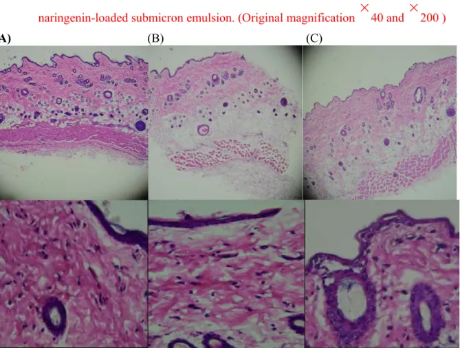
The rat skin irritation test was operated to evaluate the irritation caused by naringenin-loaded submicron emulsion, water (negative control) and the solution of formalin (positive control group). As compared to Fig. 2A of the negative control group, paraformaldehyde-treated skin showed collagen fiber swelling within dermis and edema in hypodermis as observed in Fig. 2B. Furthermore, slight damage and exfoliation of the stratum corneum of epidermis was observed. Non-significant edema and erythema was found in tested formulation (Fig. 2C) when compared to the negative control group, indicated that the drug-loaded micron emulsion had good biocompatibility with skin.
3.5. Stability
The selected naringenin-loaded formulation was subject to various thermodynamic
stability tests including centrifugation and three heating-cooling cycles tests. After these tests, no phase separation, liquefaction, creaming or precipitation was recorded, indicating that the selected formulation had good physical stability. This might be attributed to submicron emulsion with very low interfacial tension between water and oil phases, and small droplet size made the system thermodynamically stable (Azeem et al., 2009; Lawrence and Rees,
2000).
After 3 months of storage at 25 ℃ and 40℃ , the naringenin-loaded submicron emulsion had no obvious change, and no drug crystal was observed. The viscosity and droplet size showed non-significant difference. The residual drug contents of tested drug-loaded microemulsions at 25 ℃ and 40 ℃ storage were 103.04±4.35% and 91.65±3.10% respectively, indicating that the experimental naringenin-loaded submicron emulsion was
1 4
stable.
4. Conclusions
The naringenin deposition amounts in SC, epidermis, dermis and total skin were
significantly increased by using submicron emulsion as drug carrier. The drug-loaded submicron emulsion showed thermodynamic stability after centrifugation and heating-cooling cycle tests. The naringenin concentrations were 103.04±4.35% and 91.65±3.10% respectively, after 3 months of storage at 25℃ and 40℃. Moreover, naringenin-loaded submicron emulsion was less of an irritant than was the standard irritant group, indicating that the formulation can possibly be developed for a topical application system.
Conflict of Interests
The authors declare that there is no conflict of interest regarding the publication of this paper.
Acknowledgment
This work was supported by Grants from the National Science Council of Taiwan (NSC 102-2320-B-037-006-MY2 and NSC101-2320-B-037-33).
References
Azeem, A., Ahmad, F.J., Khar, R.K., Talegaonkar, S., 2009. Nanocarrier for the transdermal delivery of an antiparkinsonian drug. AAPS PharmSciTech 10, 1093-1103.
Baboota, S., Shakeel, F., Ahuja, A., Ali, J., Shafiq, S., 2007. Design, development and evaluation of novel nanoemulsion formulations for transdermal potential of celecoxib. Acta. Pharm. 57, 315-332.
1 5
Cavia-Saiz, M., Busto, M.D., Pilar-Izquierdo, M.C., Ortega, N., Perez-Mateos, M., Muniz, P., 2010. Antioxidant properties, radical scavenging activity and biomolecule protection capacity of flavonoid naringenin and its glycoside naringin: a comparative study. J. Sci. Food Agric. 90, 1238-1244.
Chung, H., Kim, T.W., Kwon, M., Kwon, I.C., Jeong, S.Y., 2001. Oil components modulate physical characteristics and function of the natural oil emulsions as drug or gene delivery system. J. Control. Release 71, 339-350.
De Paula, D., Martins, C.A., Bentley, M.V., 2008. Development and validation of HPLC method for imiquimod determination in skin penetration studies. Biomed. Chromatogr. 22, 1416-1423.
Dhawan, B., Aggarwal, G., Harikumar, S., 2014. Enhanced transdermal permeability of piroxicam through novel nanoemulgel formulation. Int. J. Pharm. Investig. 4, 65-76. El Maghraby, G.M., 2008. Transdermal delivery of hydrocortisone from eucalyptus oil
microemulsion: effects of cosurfactants. Int. J. Pharm. 355, 285-292.
El Maghraby, G.M., Arafa, M.F., Osman, M.A., 2014. Microemulsion for simultaneous transdermal delivery of benzocaine and indomethacin: in vitro and in vivo evaluation. Drug Dev. Ind. Pharm. 40, 1637-1644.
Felgines, C., Texier, O., Morand, C., Manach, C., Scalbert, A., Regerat, F., Remesy, C., 2000. Bioavailability of the flavanone naringenin and its glycosides in rats. Am. J. Physiol. Gastrointest. Liver Physiol. 279, G1148-1154.
Fouad, S.A., Basalious, E.B., El-Nabarawi, M.A., Tayel, S.A., 2013. Microemulsion and poloxamer microemulsion-based gel for sustained transdermal delivery of diclofenac epolamine using in-skin drug depot: in vitro/in vivo evaluation. Int. J. Pharm. 453, 569-578.
1 6
for dermal and transdermal drug delivery. J. Pharm. Sci. 97, 603-631.
Hsiu, S.L., Huang, T.Y., Hou, Y.C., Chin, D.H., Chao, P.D., 2002. Comparison of metabolic pharmacokinetics of naringin and naringenin in rabbits. Life Sci. 70, 1481-1489.
Huang, Y.B., Huang, C.T., Tsou, H.Y., Fu, L.T., Fu, Y.S., Tsai, Y.H., Wu, P.C., 2013. The transport effect of submicron emulsions on 5-flurouracil topical application. J. Microencapsul. 30, 425-431.
Kibbe, A.H., 2000. Handbook of pharmaceutical excipients. 3rd ed. London: Pharmaceutical Press.
Kitagawa, S., Tanaka, Y., Tanaka, M., Endo, K., Yoshii, A., 2009. Enhanced skin delivery of quercetin by microemulsion. J. Pharm. Pharmacol. 61, 855-860.
Kogan, A., Garti, N., 2006. Microemulsions as transdermal drug delivery vehicles. Adv. Colloid Interface Sci. 123-126, 369-385.
Lademann, J., Jacobi, U., Surber, C., Weigmann, H.J., Fluhr, J.W., 2009. The tape stripping procedure--evaluation of some critical parameters. Eur. J. Pharm. Biopharm.72, 317-323. Lawrence, M.J., Rees, G.D., 2000. Microemulsion-based media as novel drug delivery
systems. Adv. Drug Deliv. Rev. 45, 89-121.
Lewis, G.A., Mathieu, D., Phan-Tan-Luu, R., 1999. Pharmaceutical Experimental Design. Dekker, New York,.
Lin, C.F., Leu, Y.L., Al-Suwayeh, S.A., Ku, M.C., Hwang, T.L., Fang, J.Y., 2012. Anti-inflammatory activity and percutaneous absorption of quercetin and its polymethoxylated compound and glycosides: the relationships to chemical structures. Eur. J. Pharm. Sci. 47, 857-864.
Makraduli, L., Crcarevska, M.S., Geskovski, N., Dodov, M.G., Goracinova, K., 2013. Factorial design analysis and optimisation of alginate-Ca-chitosan microspheres. J. Microencapsul. 30, 81-92.
1 7
Mostafa, D.M., Ammar, N.M., Abd El-Alim, S.H., El-anssary, A.A., 2014. Transdermal microemulsions of Glycyrrhiza glabra L.: characterization, stability and evaluation of antioxidant potential. Drug delivery 21, 130-139.
Mutalik, S., Udupa, N., 2004. Glibenclamide transdermal patches: physicochemical, pharmacodynamic, and pharmacokinetic evaluations. J. Pharm. Sci. 93, 1577-1594. Pabari, R.M., Ramtoola, Z., 2012. Application of face centred central composite design to
optimise compression force and tablet diameter for the formulation of mechanically strong and fast disintegrating orodispersible tablets. Int. J. Pharm. 430, 18-25.
Peltola, S., Saarinen-Savolainen, P., Kiesvaara, J., Suhonen, T.M., Urtti, A., 2003. Microemulsions for topical delivery of estradiol. Int. J. Pharm. 254, 99-107.
Renugadevi, J., Prabu, S.M., 2009. Naringenin protects against cadmium-induced oxidative renal dysfunction in rats. Toxicology 256, 128-134.
Ropke, C.D., Kaneko, T.M., Rodrigues, R.M., da Silva, V.V., Barros, S., Sawada, T.C., Kato, M.J., Barros, S.B., 2002. Evaluation of percutaneous absorption of 4-nerolidylcathecol from four topical formulations. Int. J. Pharm. 249, 109-116.
Saija, A., Tomaino, A., Trombetta, D., Giacchi, M., De Pasquale, A., Bonina, F., 1998. Influence of different penetration enhancers on in vitro skin permeation and in vivo photoprotective effect of flavonoids. Int. J. Pharm. 175, 85-94.
Shulman, M., Cohen, M., Soto-Gutierrez, A., Yagi, H., Wang, H., Goldwasser, J., Lee-Parsons, C.W., Benny-Ratsaby, O., Yarmush, M.L., Nahmias, Y., 2011. Enhancement of naringenin bioavailability by complexation with hydroxypropyl-beta-cyclodextrin. Plos One 6, e18033.
Spernath, A., Aserin, A., 2006. Microemulsions as carriers for drugs and nutraceuticals. Adv. Colloid Interface Sci. 128-130, 47-64.
1 8
2007. Comparison of stratum corneum penetration and localization of a lipophilic model drug applied in an o/w microemulsion and an amphiphilic cream. Eur. J. Pharm. Biopharm. 67, 699-706.
Thitilertdecha, P., Guy, R.H., Rowan, M.G., 2014. Characterisation of polyphenolic compounds in Clerodendrum petasites S. Moore and their potential for topical delivery through the skin. J. Ethnopharmacol. 154, 400-407.
Tian, Q., Ren, F., Xu, Z., Xie, Y., Zhang, S., 2012. Preparation of high solubilizable microemulsion of naproxen and its solubilization mechanism. Int. J. Pharm. 426, 202-210.
Tsai, M.J., Fu, Y.S., Lin, Y.H., Huang, Y.B., Wu, P.C., 2014. The effect of nanoemulsion as a carrier of hydrophilic compound for transdermal delivery. Plos One 9, e102850.
Tsai, P.J., Huang, C.T., Lee, C.C., Li, C.L., Huang, Y.B., Tsai, Y.H., Wu, P.C., 2013. Isotretinoin oil-based capsule formulation optimization. TheScientificWorldJournal 2013, 856967.
Tsai, Y.H., Chang, J.T., Chang, J.S., Huang, C.T., Huang, Y.B., Wu, P.C., 2011. The effect of component of microemulsions on transdermal delivery of buspirone hydrochloride. J. Pharm. Sci. 100, 2358-2365.
Tsai, Y.H., Hsieh, Y.H., Huang, Y.B., Chang, J.S., Huang, C.T., Wu, P.C., 2010a. Microemulsions for intravesical delivery of gemcitabine. Chem. Pharm. Bull. 58, 1461-1465.
Tsai, Y.H., Lee, K.F., Huang, Y.B., Huang, C.T., Wu, P.C., 2010b. In vitro permeation and in vivo whitening effect of topical hesperetin microemulsion delivery system. Int. J. Pharm. 388, 257-262.
Vicentini, F.T., Simi, T.R., Del Ciampo, J.O., Wolga, N.O., Pitol, D.L., Iyomasa, M.M., Bentley, M.V., Fonseca, M.J., 2008. Quercetin in w/o microemulsion: in vitro and in vivo
1 9
skin penetration and efficacy against UVB-induced skin damages evaluated in vivo. Eur. J. Pharm. Biopharm. 69, 948-957.
Watkinson, R.M., Guy, R.H., Hadgraft, J., Lane, M.E., 2009. Optimisation of cosolvent concentration for topical drug delivery - II: influence of propylene glycol on ibuprofen permeation. Skin Pharmacol. Physiol. 22, 225-230.
2 0
Legends Table legends Table 1
The composition, physicochemical properties, and physical properties of model naringenin-loaded submicron emulsions provided mixture design.
Table 2
The permeability parameters of model naringenin-loaded submicron emulsions and control group (saturated aqueous solution naringenin).
Table 3
Regression coefficients and statistical analysis of dependent variables.
Figure legends
Fig. 1. Three dimensional response surface plots illustrating the effect of mixture surfactant
(X1), oil phase (X2) and aqueous phase containing 40% cosurfactant (X3) on the drug
deposition in skin of naringenin-loaded submicron emulsion formulations. A: stratum corneum layer; B: epidermis layer; C: dermis layer; D: total deposition in skin).
Fig. 2. Microscopic photos of rat skin after application of water (A), formalin (B), and tested
naringenin-loaded submicron emulsion. (Original magnification
40 and
200 )Table 1
The composition, physicochemical properties, and physical properties of model naringenin-loaded submicron emulsions provided mixture design.
X1 X2 X3 Size Viscosity (nm) (cps×103) F01 25 10 65 525.2 ± 44.5 194.8 ± 10.0 F02 25 24 51 149.7 ± 27.4 144.3 ± 3.7 F03 25 24 51 163.8 ± 14.4 142.1 ± 9.0 F04 31 25 44 150.8 ± 9.7 ND F05 29 17 54 90.0 ± 4.0 ND F06 35 10 55 98.2 ± 13.4 ND F07 35 22 43 103.7 ± 4.6 143.0 ± 5.8 F08 35 17 48 65.5 ± 3.0 191.2 ± 9.3 F09 25 10 65 504.7 ± 79.1 218.5 ± 13.5 F10 30 11 59 587.3 ± 210.3 568.8 ± 10.3
*The total amount of three variables of X1 (Tween 80/ Span 20 of 3/2), X2(IPM), and X3
(distilled water containing 40% PG) was 100%. X1+ X2+ X3=100%.
*ND: non-detect.
Table 2
The permeability parameters of model naringenin-loaded submicron emulsions and control group (saturated aqueous solution of naringenin).
Q24h SC Epidermis Dermis Total ER
(μg/cm2) (μg/cm2) (μg/cm2) (μg/cm2) (μg/cm2) F01 177.2 ± 37.0 1.2 ± 0.4 13.6 ± 3.3 1.6 ± 0.4 16.3 ± 3.7 6.3 F02 218.5 ± 45.7 2.2 ± 0.5 16.8 ± 5.2 2.3 ± 0.1 21.4 ± 5.5 8.3 F03 247.7 ± 52.4 1.9 ± 0.2 15.2 ± 2.8 2.3 ± 1.4 19.4 ± 4.4 7.5 F04 150.2 ± 21.1 2.1 ± 0.2 13.9 ± 1.0 1.8 ± 0.3 17.8 ± 1.3 6.9 F05 149.1 ± 28.8 1.6 ± 0.3 12.5 ± 2.7 0.9 ± 0.1 14.9 ± 2.6 5.8 F06 135.1 ± 24.8 1.6 ± 0.2 8.6 ± 1.0 1.3 ± 0.4 11.5 ± 0.6 4.5 F07 215.4 ± 13.2 1.7 ± 0.3 12.0 ± 0.8 1.0 ± 0.7 14.6 ± 1.6 9.4 F08 201.5 ± 38.3 1.6 ± 0.2 10.7 ± 1.8 1.5 ± 0.9 13.8 ± 2.6 5.3 F09 285.9 ± 138.7 1.9 ± 0.7 16.6 ± 5.4 3.7 ± 3.8 22.2 ± 1.0 8.6 F10 200.8 ± 36.0 2.0 ± 0.6 9.2 ± 3.9 1.3 ± 0.6 12.5 ± 4.6 4.9 C 4.8 2.7 0.5 ± 0.1 1.8 ± 0.5 0.3 ± 0.1 2.6 ± 0.7 1.0 *SC: stratum corneum
*ER=enhancement ratio= skin deposition of formulation/ skin deposition of saturated aqueous solution
Table 3
Regression coefficients and statistical analysis of dependent variables.
Regression Stratum Epidermis Dermis Total skin
coefficient corneum
Coefficient Coefficient Coefficient Coefficient
Estimate Estimate Estimate Estimate
b1 (X1) -2.06 2.27 -0.05 3.67 b2 (X2) 3.68 19.02 2.50 23.87 b3 (X3) 1.33 12.67 1.59 15.77 b12 (X1X2) 1.59 b13 (X1X3) 7.32 b23 (X2X3) -2.54 b123 (X1X2X3) Model (P value) 0.0211 0.0002 0.0324 0.0001
Lack of Fit (P value) 0.1522 0.8227 0.6296 0.9457
Stratum corneum
ign-Expert?Software ponent Coding: Actual al
5.7653 .05853 = A: T80/S20 = B: IPM = C: 40% PG sign-Expert?Software mponent Coding: Actual I 20.6535 6.62675 = A: T80/S20 = B: IPM = C: 40% PG
Fig. 1. Three dimensional response surface plots illustrating the effect of mixture surfactant
(X1), oil phase (X2) and aqueous phase containing 40% cosurfactant (X3) on the
drug deposition in skin of naringenin-loaded submicron emulsion formulations. A: stratum corneum layer; B: epidermis layer; C: dermis layer; D: total deposition in skin).
Design-Expert?Software Component Coding: Actual Original Scale SC 2.75139 30 0.715062 3 X1 = A: T80/S20 25 X2 = B: IPMX3 = C: 40% PG 2.5 To ta lsk in de po sit io n 20 2 St ra tu m co rn eu m 15 1.5 10 1 5 0.5 X1(1.0) X (0.0) X1(1.0) X (0.0) 2 2 X3(1.0) X3(1.0) X3(040.0) X3(0.0) X1(0.0) X1(0.0) X2(1.0) X (1.0) 2 Design-Expert?Software
Component Coding: Actual Original Scale Dermis 3.79015 22 0.360123 20 4 X1 = A: T80/S20 18 X2 = B: IPM X3 = C: 40% PG 3 16 Ep id er m is 14 D er m is 12 2 10 8 1 6 0 X1(1.0) X2(0.0) X1(1.0) X2(0.0) X (1.0) X (1.0) 3 3 X3(0.0) X1(0.0) X3(0.0) X1(0.0)
X2(1.0)
Figure-2
Fig. 2. Microscopic photos of rat skin after application of water (A), formalin (B), and tested
naringenin-loaded submicron emulsion. (Original magnification