行政院國家科學委員會專題研究計畫 期中進度報告
以基因分析評估腫瘤血管內皮細胞基因的表達:從鼷鼠到人
類(2/3)
計畫類別: 個別型計畫 計畫編號: NSC91-2314-B-002-312-執行期間: 91 年 08 月 01 日至 92 年 07 月 31 日 執行單位: 國立臺灣大學醫學院婦產科 計畫主持人: 謝豐舟 計畫參與人員: 鄭文芳 報告類型: 精簡報告 處理方式: 本計畫可公開查詢中
華
民
國 92 年 5 月 27 日
一、計畫中文摘要 血管新生-從已存在的血管生長出來的微血管-對胚胎的發育、器官的形 成、組織的重建和重建是經不可或缺的。血管新生已經被證實對腫瘤生長和轉 移的發生和進展有所貢獻。抗血管新生被認為是抵抗腫瘤細胞抗藥性的一種新 的癌症治療。然而許多關於腫瘤血管的問題仍無法回答。最近人類大腸直腸癌 的腫瘤血管內皮細胞被發現表達一些和正常血管內皮細胞不相同的基因。看來 在腫瘤組織中血管的內皮細胞和正常組織中血管的內皮細胞其性質並不相同。 因此我們提出這個兩年度的計畫來回答一些有趣的問題。首先,在其他腫瘤中 的血管內皮細胞其基因的表達是否和正常組織中血管的內皮細胞並不相同。其 次,不同器官所產生的不同腫瘤的血管內皮細胞其基因的表達是否會相同。我 們將利用在鼷鼠動物模式的各個細胞株以分示法來評估腫瘤血管內皮細胞基因 表達的差異。接著我們將進一步評估在鼷鼠動物模式所觀察到的腫瘤血管內皮 細胞基因表達的差異,是否也可以在人類腫瘤被觀察到。最後我們對所找到的 基因做功能的分析。這些問題的回答將對了解不同器官的腫瘤血管新生和針對 特異腫瘤的抑制血管新生這個新的的治療方式具有決定性地衝擊。 二、計畫英文摘要
Angiogenesis, the outgrowth of new capillaries from pre-existing vessels, is essential for embryonic development, organ formation, tissue regeneration, and remodeling. Angiogenesis has been proved to contribute to the development and progression of tumor growth and metastasis. Anti-angiogenesis was regarded to be a new cancer therapy resistant to drug resistance of tumor cells. However, many basic questions about tumor vessels remain unanswered. Recently tumor endothelium of human colorectal cancer has been found to express some different genes than normal
endothelium does. It seems that endothelium that lines blood vessels in tumors qualitatively different from endothelium in vessels of normal tissue. So we propose this two-year projects to answer some interesting questions. First, if the gene expressions of tumor endothelium are different from those of normal endothelium in the other different tumors. Second, if the gene expressions of tumor endothelium are the same in different tumors from different organs. We will utilize the tumor cells lines in the murine model to evaluate the differences of gene expressions in the tumor endothelium by differential expression. Then we will further evaluate if the differences of gene expressions that we observed in murine model can also be observed in human tumors. Finally, we will try to evaluate the function of these observed genes from tumor endothelium. The answers to these questions critically impact the potential for understanding the tumor angiogenesis in different organ and for new therapeutic approaches to inhibit angiogenesis in a tumor-specific manner.
三、研究計畫之原由及目的:
Tumors require a blood supply for expansive growth (1–3), an observation that has stimulated a profusion of research on tumor angiogenesis. However, several basic questions about tumor vessels remain unanswered. For example, is endothelium that lines blood vessels in tumors qualitatively different from endothelium in vessels of normal tissue ? What is the relation of tumor angiogenesis to angiogenesis associated with wound healing or other physiological processes? The answers to these questions critically impact the potential for new therapeutic approaches to inhibit angiogenesis in a tumorspecific manner.
To determine if tumor-specific endothelial markers exist, we will compare gene expression profiles in endothelium derived from normal and tumor tissue.
incidence, tends to grow slowly, and is often resistant to chemotherapeutic drugs. Importantly, the progressive growth of this tumor type appears to be angiogenesis-dependent (4).
Global analysis of gene expression in tumor and normal endothelium is difficult because (i) the endothelium is enmeshed in a complex tissue consisting of vessel wall components, stromal cells, and epithelial cells, and (ii) only a small fraction of the cells within these tissues are endothelial. Thus, we needed to develop methods for isolating highly purified endothelial cells (ECs) and for evaluating gene expression profiles from relatively few cells.
To overcome the first obstacle, we attempted to purify ECs from dispersed human colorectal tissue using CD31, an endothelial marker commonly used for this purpose (5–8). This resulted in a substantial enrichment of ECs but also in contamination of the preparations by hematopoietic cells, most likely due to expression of CD31 by macrophages (9). We therefore purified ECs from human tissues using P1H12, a recently described marker for ECs (10). Unlike CD31, CD34, and VE-cadherin, P1H12 specifically localized to ECs of all vessels including microvessels of normal and cancerous colorectal tissue (Fig. 1A). Our purification protocol also optimized the detachment of ECs from neighboring cells, leaving cell surface proteins intact, and included positive and negative affinity purifications using a mixture of antibodies (Fig. 1B). The ECs purified from normal colorectal mucosa and colorectal cancers were essentially free of epithelial and hematopoietic cells as judged by reverse transcription –polymerase chain reaction (RTPCR) (Fig. 1C) and subsequent gene expression analysis (see below).
To evaluate modification of the serial analysis of gene expression (SAGE) technique (11). SAGE associates individual mRNA transcripts with 14–base pair (bp) tags derived from a specific position near their 39 termini (12). The abundance of
each tag provides a quantitative measure of the transcript level present within the mRNA population studied. SAGE is not dependent on preexisting databases of expressed genes and therefore provides an unbiased view of gene expression profiles. This feature is particularly important in the analysis of cells that constitute only a small fraction of the tissue under study, as transcripts from these cells are unlikely to be well represented in extant expressed sequence tag (EST) databases. generated a SAGE library of ;96,000 N.T.
四、研究方法、進行步驟及執行進度: Murine part
1. Tumor cell line
The production and maintenance of TC-1 cells has been described previously [Lin, 1996 #175]. HPV-16 E6, E7 and ras oncogene were used to transform primary lung epithelial cells of C57BL/6 mice. This tumorigenic cell line was named TC-1. 2. Isolation of tumor endothelial cells
C57BL/6 mice (five per group) were injected with 5x10⁴ TC-1 tumor cells will be injected subcutaneously in the right hind leg or intravenously via tail vein. Mice will be were sacrificed. The subcutaneous tumors and lungs will be explanted on 28 days after tumor challenge.
After removal of lungs or subcutaneous tumors, these tumor tissues will be minsed and will be digested with collagenase A and trypsin. The enzyme-treate tumor tissues will be filtered with 25 µm filters and Percoll gradient solution will be performed to get rid of RBC. The remaining cells were stained with VCAM-1 antibody, purified with anti-rat immunoglobulin G (IgG)–coupled magnetic beads.
The isolation of normal endothelial cells will be performed as the previous description of tumor endothelial cells. Lung tissues and aorta of the naive mice will be collected and the endothelial cells of the lungs and aortas will be further isolated as normal endothelial cells.
4. Mice
Six- to eight-week-old female C57BL/6 mice were purchased from the National Taiwan University Hospital (Taipei, Taiwan) and bred in the animal facility of the National Taiwan University Hospital (Taipei, Taiwan). All animal procedures were performed according to approved protocols and in accordance with recommendations for the proper use and care of laboratory animals.
Human part
1. Isolation of normal and tumor endothelial cells
Human gastric and hepatic tissues were obtained <30 min after surgical removal. Normal tissue was taken from regions 1.0 cm away from bulk tumor tissue that had clearly defined margins. For normal tissue, sheets of epithelial cells were removed with a glass slide after treatment with 5 mM dithiothreitol, then 10 mM EDTA, leaving an intact lamina propria for subsequent processing. After a 2-hour incubation in collagenase at 37°C, cells were filtered sequentially through 400-,100-, 50-, and 25-mm mesh and centrifuged through a 30% preformed Percoll gradient to sediment red blood cells. Epithelial cells (Epithelial Fraction), which were found to nonspecifically bind magnetic beads, were removed with Dynabeads coupled to BerEP4. Subsequently, macrophages and other leukocytes (Hematopoietic Fraction) were removed with a mixture of beads coupled to anti-CD45, anti-CD14, and anti-CD64 (Dynal). The remaining cells were stained with P1H12 antibody, purified
with anti-mouse immunoglobulin G (IgG)–coupled magnetic beads.
五、結果與討論
Isolation and collection of tumor and normal endothelial cells from cancer patients Human gastric tissues were obtained <30 min after surgical removal. Normal tissue was taken from regions 1.0 cm away from bulk tumor tissue that had clearly defined margins. For normal tissue, sheets of epithelial cells were removed with a glass slide after treatment with 5 mM dithiothreitol, then 10 mM EDTA, leaving an intact lamina propria for subsequent processing. After a 2-hour incubation in collagenase at 37°C, cells were filtered sequentially through 400-,100-, 50-, and 25-mm mesh and centrifuged through a 30% preformed Percoll gradient to sediment red blood cells. Epithelial cells (Epithelial Fraction), which were found to nonspecifically bind magnetic beads, were removed with Dynabeads coupled to BerEP4. Subsequently, macrophages and other leukocytes (Hematopoietic Fraction) were removed with a mixture of beads coupled to anti-CD45, anti-CD14, and anti-CD64 (Dynal). The remaining cells were stained with P1H12 antibody, purified with anti-mouse immunoglobulin G (IgG)–coupled magnetic beads.
Tumor and normal endothelial cells were collected from 12 gastric cancer patients. These isolated tumor and normal endothelial cells were frozen in –700C refrigerator for further experiments. The identification and characterization of the magnetic beads-isolated cells were further stained with anti-CD34 antibody to clarify that these isolated cells were endothelial cells ( Figure 1).
Isolation and collection of tumor and normal endothelial cells from murin tumor and normal tissues
C57BL/6 mice (five per group) were injected with 5x10⁴ TC-1 tumor cells will be injected subcutaneously in the right hind leg or intravenously via tail vein. Mice will be were sacrificed. The subcutaneous tumors and lungs will be explanted on 28 days after tumor challenge.
After removal of lungs or subcutaneous tumors, these tumor tissues will be minsed and will be digested with collagenase A and trypsin. The enzyme-treate tumor tissues will be filtered with 25 µm filters and Percoll gradient solution will be performed to get rid of RBC. The remaining cells were stained with VCAM-1 antibody, purified with anti-rat immunoglobulin G (IgG)–coupled magnetic beads.
The isolation of normal endothelial cells will be performed as the previous description of tumor endothelial cells. Lung tissues and aorta of the naive mice will be collected and the endothelial cells of the lungs and aortas will be further isolated as normal endothelial cells. The identification and characterization of the magnetic beads-isolated endothelial cells were further stained with anti-CD34 antibody to clarify that these isolated cells were endothelial cells.
六、計畫成果自評 我們將人類及鼷鼠腫瘤中的血管內皮細胞分離出來。我們將進一步利 用分示法分析腫瘤組織內血管內皮細胞基因表達與正常組織內血管內皮細胞基 因表達的差異。接著尋找人類血管內皮細胞中與血管新生相關的基因,比較人類 及鼷鼠調控血管新生基因的相同處和差異處。這將幫助我們了解調控腫瘤血管新 生的基因的功能,對未來以抗腫瘤血管新生作為治療癌症提供新的治療方針。 REFERENCES
1. Pfleiderer, A. 1984. Diagnosis and staging of ovarian cancer. J Cancer Res Clin Oncol 107:81-8.
2. Tarone, R. E., and K. C. Chu. 2000. Age-period-cohort analyses of breast-, ovarian-, endometrial- and cervical-cancer mortality rates for Caucasian women in the USA [In Process Citation]. J Epidemiol Biostat 5:221-31.
3. Gonzalez-Diego, P., G. Lopez-Abente, M. Pollan, et al. 2000. Time trends in ovarian cancer mortality in europe (1955-1993). effect Of age, birth cohort and period of death [In Process Citation]. Eur J Cancer 36:1816-24.
4. Parkin, D. M., P. Pisani, and J. Ferlay. 1993. Estimates of the worldwide incidence of eighteen major cancers in 1985. Int J Cancer 54:594-606.
5. Beard, C. M., L. C. Hartmann, E. J. Atkinson, et al. 2000. The epidemiology of ovarian cancer: a population-based study in Olmsted County, Minnesota, 1935-1991. Ann Epidemiol 10:14-23.
6.Boyle, P., P. Maisonneuve, and P. Autier. 2000. Update on cancer control in women. Int J Gynaecol Obstet 70:263-303.
7. Holschneider, C. H., and J. S. Berek. 2000. Ovarian cancer: epidemiology, biology, and prognostic factors. Semin Surg Oncol 19:3-10.
8. Chen, C. A., W. F. Cheng, C. N. Lee, et al. 1999. Serum vascular endothelial growth factor in epithelial ovarian neoplasms: correlation with patient survival. Gynecol Oncol 74:235-40.
9. Papadimitriou, C. A., L. A. Moulopoulos, G. Vlahos, Z. et al. 2000. Paclitaxel, cisplatin, and epirubicin first-line chemotherapy in stage III and IV ovarian carcinoma: long-term results of a phase II study. Cancer 89:1547-54.
10. Berek, J. S., K. Bertelsen, A. du Bois, et al. 2000. [Epithelial ovarian cancer (advanced stage): consensus conference (1998)]. Gynecol Obstet Fertil 28:576-83. 11. Joly, F., J. F. Heron, P. Kerbrat, et al. 2000. High-dose platinum versus standard
dose in advanced ovarian carcinoma: a randomized trial from the Gynecologic Cooperative Group of the French Comprehensive Cancer Centers (FNCLCC).
Gynecol Oncol 78:361-8.
12. Rofstad, E. K. 2000. Microenvironment-induced cancer metastasis. Int J Radiat Biol 76:589-605.
13. Fidler, I. J. 2000. Angiogenesis and cancer metastasis. Cancer J Sci Am 6 Suppl 2:S134-41.
14. Fidler, I. J., and R. Radinsky. 1996. Search for genes that suppress cancer metastasis [editorial; comment]. J Natl Cancer Inst 88:1700-3.
15. Roberts, D. D. 1996. Regulation of tumor growth and metastasis by thrombospondin-1. Faseb J 10:1183-91.
16. Bao, L., M. Loda, P. A. Janmey, et al. 1996. Thymosin beta 15: a novel regulator of tumor cell motility upregulated in metastatic prostate cancer [see comments]. Nat Med 2:1322-8.
17. Habets, G. G., E. H. Scholtes, D. Zuydgeest, et al. 1994. Identification of an invasion-inducing gene, Tiam-1, that encodes a protein with homology to GDP-GTP exchangers for Rho-like proteins. Cell 77:537-49.
18. Roby, K. F., C. C. Taylor, J. P. Sweetwood, et al. 2000. Development of a syngeneic mouse model for events related to ovarian cancer. Carcinogenesis 21:585-91.
19. Fambrough, D., K. McClure, A. Kazlauskas, et al. 1999. Diverse signaling pathways activated by growth factor receptors induce broadly overlapping, rather than independent, sets of genes [see comments]. Cell 97:727-41.
20. Clark, E. A., T. R. Golub, E. S. Lander, et al. 2000. Genomic analysis of metastasis reveals an essential role for RhoC [see comments]. Nature 406:532-5. 21. Zuo, L., C. K. Ogle, J. E. Fischer, et al. 1997. mRNA differential display of
colonic mucosa cells in ulcerative colitis. J Surg Res 69:119-27.
beta-induced phosphorylation of Smad3 is required for growth inhibition and transcriptional induction in epithelial cells. Proc Natl Acad Sci U S A 94:10669-74. 23. Yebra, M., G. C. N. Parry, S. Stromblad, et al. 1996. Requirement of
receptor-bound urokinase-type plasminogen activator for integrin alphavbeta5-directed cell migration. J Biol Chem 271:29393-9.
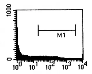
Figure Legends
Figure1. C57BL/6 mice (five per group) were injected with 5x10⁴ TC-1 tumor cells will be injected intravenously via tail vein. Mice will be were sacrificed. on 28 days after tumor injection. After removal of lungs tumors, these tumor tissues will be minsed and will be digested with collagenase A and trypsin. The enzyme-treate tumor tissues will be filtered with 25 µm filters and Percoll gradient solution will be performed to get rid of RBC. The remaining cells were stained with VCAM-1 antibody, purified with anti-rat immunoglobulin G (IgG)–coupled magnetic beads. (A) Isolated cells from tumor tissues showed positive CD144 surface marker expression. (B) Isolated cells from spleen showed negative CD144 surface marker expression.