行政院國家科學委員會專題研究計畫 期中進度報告
雷射捕陷結晶法之研究(2/3)
期中進度報告(精簡版)
計 畫 類 別 : 個別型 計 畫 編 號 : NSC 98-2113-M-009-001- 執 行 期 間 : 98 年 04 月 01 日至 99 年 03 月 31 日 執 行 單 位 : 國立交通大學應用化學系(所) 計 畫 主 持 人 : 增原宏 報 告 附 件 : 出席國際會議研究心得報告及發表論文 處 理 方 式 : 本計畫可公開查詢中 華 民 國 99 年 01 月 20 日
行政院國家科學委員會補助專題研究計畫
□ 成 果 報 告
■期中進度報告
雷射捕陷結晶法之研究
計畫類別:■ 個別型計畫 □ 整合型計畫
計畫編號:NSC 98-2113-M-009-001-
執行期間:98 年 4 月 1 日至 99 年 3 月 31 日
計畫主持人:增原宏
共同主持人:
計畫參與人員: Usman ANWAR
成果報告類型(依經費核定清單規定繳交):■精簡報告 □完整報告
本成果報告包括以下應繳交之附件:
■赴國外出差或研習心得報告一份
□赴大陸地區出差或研習心得報告一份
□出席國際學術會議心得報告及發表之論文各一份
□國際合作研究計畫國外研究報告書一份
處理方式:除產學合作研究計畫、提升產業技術及人才培育研究計畫、
列管計畫及下列情形者外,得立即公開查詢
□涉及專利或其他智慧財產權,□一年□二年後可公開查詢
執行單位:國立交通大學應用化學系
國科會専題研究計劃成果報告書(増原 宏)
目錄
中、英文摘要 I 報告內容 I-1 前言 1 I-2 研究目的 1 I-3 文獻探討 2 I-4 研究方法 2 I-5 結果與討論 3 II 計畫成果自評 5 附錄 I 表及圖 6 附錄 II 計画中獲補助国外或大陸地区差旅費 10附錄 III Publication list 11
国科会専題研究計画成果報告書(増原 宏)
英文摘要
We have started to elucidate underlying molecular dynamics and mechanism of ”Laser Trapping Crystallization” in a new laboratory of Tin Ka Ping Photonics Center.. Raman microspectroscopy is being developed to monitor crystal nucleation and its growth induced by laser trapping. Polymorph control of glycine crystal is successfully demonstrated for the first time, and its mechanism is considered in view of photon pressure and temperature, both of which are due to its irradiation laser power. The new results and related works have been summarized as original 4 papers and 6 invited lectures.
中文關鍵詞
雷射捕陷,結晶,金奈米粒子,胺基酸,晶型結構控制
英文關鍵詞
I.
報告內容
I-1. 前言
Laser has high potential in advancing chemical research by realizing new spectroscopy, analysis, reaction, and fabrication, while their spatial resolution was limited to light wavelength. We have utilized various lasers and microscopes, developed new spectroscopy and imaging methods with nm resolution, explored novel nm chemical phenomena, elucidated their mechanism and dynamics, and extended the studies to material and bio applications. This achievement was already summarized as follows.
Laser Nano Spectroscopy and Nano Photochemistry Laser Nano Ablation: Dynamics and Bio Application
Laser Nano Manipulation and Chemistry of Photon Pressure
Most of new interesting phenomena which we have found are characteristic of intense laser irradiation into small volume and not observed under conventional conditions of laser spectroscopy and photochemistry. Among them we have chosen “Laser Trapping Crystallization” as a subject for this NSC project and are extending it to an original work in NCTU. This phenomenon is induced by focusing near infrared CW laser into concentrated solution of molecules. By increasing laser intensity, nm-sized molecular aggregates can be trapped at the focal point even at room temperature. When they are gathered, their effective polarizability is increased, so that the trapping is nonlinearly enhanced, forming larger assemblies. Eventually they should form crystals, which has been recognized as an important milestone experiment and tried by many physicists and optical scientists. However no successful report has been given as far as we know. Quite recently we have demonstrated the trapping and crystallization of an amino acid in 2007 (Chem. Lett., 2007, Vol.36, No.12, pp.1480-1481), which is the first report in the relevant works. Under these backgrounds we planned to elucidate underlying molecular dynamics and mechanism of the “Laser Trapping Crystallization”. Last October installations of lasers, microscopes, and related devices have been finalized in a new laboratory in the new building of Tin Ka Ping Photonics Center. Now we have started our research under nice conditions.
I-2. 研究目的
The initial gathering of molecules and clusters is very important for crystallization and will be followed spectroscopically in real time under the irradiation condition of the power of subW. Weakly associated molecules and nanocrystals should be discriminated by fluorescence spectroscopy, and assembling dynamics will be followed by fluorescence correlation spectroscopy. More structural information on gathered matters can be obtained by introducing
Raman microscopy. Polarized laser beam prefers oriented assembling of anisotropic clusters, which will be very useful to create crystals with different phases. What kinds of cluster structures are preferred, what is their size, how they fluctuate, and how they become nucleus? To control trapping and crystallization, suitable optical conditions and nice combinations of molecules and solvents should be searched and theoretically interpreted. The various molecules are examined for understanding molecular dynamics and mechanism of the crystallization phenomenon.
I-3. 文獻探討
(1) H. Adachi, K. Takano, Y. Hosokawa, T. Inoue, Y. Mori, H. Matsumura, M. Yoshimura, Y. Tsunaka, M. Morikawa, S. Kanaya, H. Masuhara, Y. Kai and T. Sasaki, Jpn. J. Appl. Phys., Vol.42, Part 2, No.7B, pp.L 798-L 800 (2003)
(2) Y. Hosokawa, H. Adachi, M. Yoshimura, Y. Mori, T. Sasaki, and H. Masuhara, Crystal Growth & Design, Vol.5, No.3, pp.861-863 (2005)
(3) J. Borowicz, J. Hotta, K. Sasaki, and H. Masuhara, J. Phys. Chem. B, Vol.101, No.31, pp.5900-5904 (1997)
(4) J. Hotta, K. Sasaki, H. Masuhara, and Y. Morishima, J. Phys. Chem. B, Vol.102, No.40, pp.7687-7690 (1998)
(5) T. Smith, J. Hotta, K. Sasaki, H. Masuhara, and Y. Ito, J. Phys. Chem. B, Vol.103, No.10, pp.1660-1663 (1999)
(6) C. Hosokawa, H. Yoshikawa, and H. Masuhara, Phys. Rev. E, Vol.70, pp.061410_1-061410_7 (2004); ibid., Vol.72, pp.021408_1-021408_7 (2005)
(7) K. Sasaki, M. Tsukima, and H. Masuhara, Appl. Phys. Lett., Vol.71, No.1, pp.37-39 (1997)
(8) H. Yoshikawa, T. Matsui, and H. Masuhara, Phys. Rev. E, Vol.70, pp.061406_1-061406_6
(9) S. Ito, H. Yoshikawa, and H. Masuhara, Proc. SPIE, Vol.4977, pp.623-631 (2003)
(10) H. Masuhara and F.C. De Schryver, "Organic Mesoscopic Chemistry" (IUPAC 21st century chemistry monograph), Blackwell Science (1999)
(11) H. Masuhara, H. Nakanishi, and K. Sasaki, "Single Organic Nanopariticles", Springer (2003)
(12) H. Masuhara and S. Kawata Ed., "Nanophotonics: Integrating Photochemistry, Optics and Nano/Bio Materials Studies", Elsevier (2004)
(13) H, Masuhara, S. Kawata, and F. Tokunaga, “Nano Biophotonics”, Elsevier (2007)
(14) T. Sugiyama, T. Adachi, and H. Masuhara, Chem. Lett., Vol.36, pp.1480-1481 (2007) (15) T. Sugiyama, T. Adachi, and H. Masuhara, Chem. Lett., Vol.38, pp.482-483 (2009)
solution. The concentration, temperature, shape and volume of the solution will be adjusted, while selection of molecules and solvents are critical. First an intense CW near infrared laser is installed. As the initially formed nucleus should be very small, single molecular level detection is necessary, and extreme optical arrangement should be set up under a microscope. The planned measurements are as follows.
(a) Fluorescence and fluorescence correlation spectroscopy of molecular assembling under laser trapping
(b) Dynamic interference measurement of solution surface under laser trapping (c) Imaging of crystal growth realized by laser trapping.
I-5. 結果與討論
1. New laboratory in Tin Ka Ping Photonics Center
In April 2008 we started our research activity in NCTU in Science building I by borrowing a corner of a previous student experimental room from collage of Science. We used this space as a tentative staff and student office space and as an experimental room after renovation. In September 2009, a construction of Tin Ka Pin Photonics Center which was allocated as our permanent office and experimental space was finally finished. We moved in sixth floor of this building on late October. Now we have two faculty office rooms (612 and 613, for Miura and Masuhara, respectively) and one experiment/student space (room 611) which is used as a clean room for laser/microscope experiments, the student office space, and a preparation room for experimental samples. We have moved and rearranged the experimental equipments to the new experimental room and, in addition, we installed additional experimental equipments such as femtosecond regenerative amplifier laser system. We have finished preparation of the new laboratory within 2009 and have started research works with new environment.
2. Raman Spectroscopy for Laser Trapping Characterization of Organic Compunds
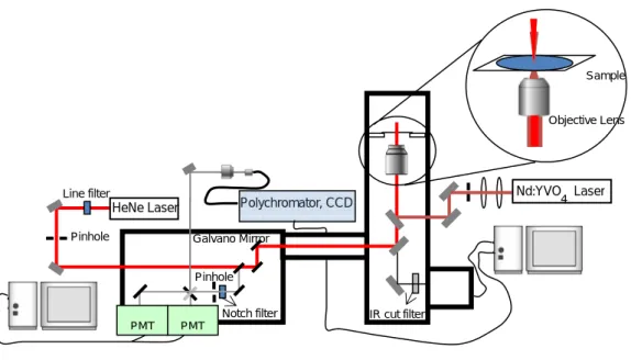
The intended purpose of this research topic is to develop a high-performance method to observe the early nucleation process of the laser-induced crystallization. To this purpose a fast and sensitive technique is required to monitor the aggregation and nucleation process at the focal spot under the microscope. We are therefore developing confocal Raman microspectroscopy, observing the fluctuation Raman bands with spatial resolution to monitor the process of the laser-induced and trapping crystallization. The instrument employed is a confocal microscope [Fig. 1]. The sample is illuminated using a 632.8 nm line from a He–Ne laser through an objective lens. Raman scattering light in backward direction is collected by
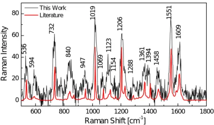
the objective lens, it is passed through a notch filter in order to cut Rayleigh scattering light, and then it is detected by a polychromator coupled with an electrically cooled charge-coupled device (CCD) detector. A single grating (600 lines/cm) is used for the spectral dispersion of the Raman bands. Figure 2 shows the Raman spectrum of indene, a standard sample, in the region of 500–1800 cm-1 with spectral resolution being 2-3 cm-1, which agreed well with the literature. Though the signal to noise remains to be improved, this preliminary result confirms that our confocal Raman microscopy setup is technically suitable for the detection of the Raman spectrum under trapping condition.
Important future development in the experimental setup is to monitor the evolution of the Raman bands at and around a small focal volume of the 1064 nm trapping laser. Thus, the Raman microspectroscopy will provide much structural and dynamical information on the process of the laser-induced crystallization.
Fig. 1. Schematic drawing of Raman microspectroscopy for laser-trapping crystallization
IR cut filter
HeNe Laser Polychromator, CCD
Pinhole Line filter Notch filter Sample Galvano Mirror Pinhole Nd:YVO Laser 4 PMT PMT Objective Lens
600 800 1000 1200 1400 1600 1800 0 20 40 60 80 Raman Shift [cm-1] This Work Literature 594 84 0 947 1154 1069 1123 1019 732 536 1288 13 61 13 94 145 8 1551 1206 Raman Intens it y 1609
Fig. 2. Raman spectrum of indene observed by our Raman microspectroscopy.
3. Polymorph Control of Glycine Crystal by Laser Trapping Characterization
We have already succeeded in the glycine crystallization spatiotemporally in the supersaturated D2O solution. In this laser trapping crystallization, the liquid-like clusters of
glycine formed in the supersaturated solution are gathered in a focal point under the photon pressure, and the local association of the clusters is enhanced. The molecules in the association should be reoriented and reorganized, eventually leading to the nucleation. Separately, we have reported that the growth rate and its direction of a spontaneously produced glycine crystal in D2O is arbitrarily controlled by trapping the glycine clusters near
the crystal. This means high concentration regions of the clusters are formed. Next step of laser trapping crystallization study is on possible polymorph control.
Control of crystal polymorph has received much attention in many fields of molecular, material, biological, and chemical sciences, since the crystal polymorphs show different physical and chemical properties such as melting point, solubility, optical property, and so on. Glycine has been employed as a representative compound in many researches on a polymorphic crystallization, and three polymorphs are known. α-Form is prepared by the spontaneous crystallization from the neutral aqueous solution, although it is not the most stable polymorph among those forms at ordinary temperatures and pressures. β-Form is
present just under a special condition, and rapidly transforms into the α-one in air or/and water. γ-Form is prepared only under comparatively severe experimental conditions (e.g. high
pressure, high acidic/basic, or high supersaturated) or by adding some additive salts into the aqueous solution, despite the fact that it is the most thermodynamically stable among those three polymorphs.
In collaboration with Prof. T. Sugiyama, Dr. T. Rungusimanon, and Mr. K. Yuyama of Nara Institute of Science and Technology, we have obtained the first result on crystal
polymorph control by photon pressure of a focused CW NIR laser beam. Except under the severe conditions, glycine tends to crystallize to the γ-form from the high supersaturated solution via quite slow evaporation or cooling, so that the high supersaturated spot of the molecules produced by photon pressure at high laser power may lead to the γ-form preparation.
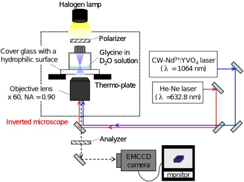
Figure 3 shows a schematic illustration of the optical trapping system in this experiment. In order to avoid the temperature elevation during laser irradiation, D2O was used as a solvent.
Upon focusing a linearly polarized CW 1064-nm laser beam at the air/solution interface of the supersaturated glycine/D2O solution, one crystal was prepared and observed at the focal spot
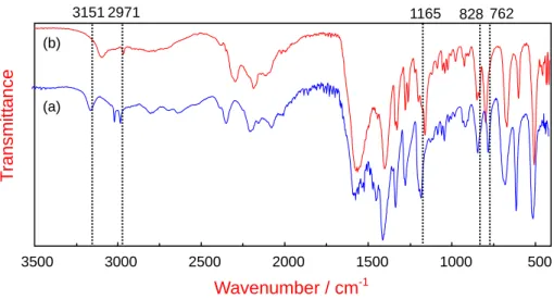
within a few tens of seconds as in previous report. The crystallization was observed in all samples that we tried in this experiment. After one crystal grew up to a few mm in size by aging for 3 h in the solution, it was taken out from the sample bottle and examined by FTIR measurement. Always only one large crystal was prepared in the center of the bottom near the focal point. By measuring the FTIR spectrum of each large crystal, we found that all of the obtained spectra can be classified into two types [Fig. 4] and no mixture of these two spectra was formed.
Fig. 3. A schematic illustration of the optical trapping system of the photon pressure-induced
crystallization from a supersaturated glycine/D2O solution. Thermo-plate Objective lens x 60, NA = 0.90 CW-Nd3+:YVO 4laser (λ = 1064 nm) He-Ne laser (λ =632.8 nm) Cover glass with a
hydrophilic surface monitor Halogen lamp EMCCD camera Glycine in D2O solution Polarizer Analyzer Inverted microscope Thermo-plate Objective lens x 60, NA = 0.90 CW-Nd3+:YVO 4laser (λ = 1064 nm) He-Ne laser (λ =632.8 nm) Cover glass with a
hydrophilic surface monitor Halogen lamp EMCCD camera EMCCD camera Glycine in D2O solution Polarizer Analyzer Inverted microscope
The H atoms of an amino group of glycine in heavy water are replaced by D atoms, so that the vibrational frequencies of N-deuterated glycine (ND3+CH2COO-, glycine-d3) are
important which was reported by Suzuki et al., in 1963. They pointed out that the FTIR spectrum of α-form showed the sharp peaks of CH2 stretching around 3070-2940 cm-1,
whereas that of glycine appeared to be a broad region. In addition, the peaks shifted from the regions at 1527-1502 and 1131-1111 to 1188-1166 and 822-763 cm-1, respectively, which showed the change in vibrational mode of NH3 to ND3 deformation and rocking, respectively.
Taking this into consideration, Figure 2a shows the sharp peaks at 3151-2971, 1180-1165, and 828-762 cm-1, all of which are consistent with the previous reports. It implies that glycine was changed into glycine-d3 in D2O and its polymorph was ascribed to the α-form. In addition, the
crystal polymorph was also confirmed by single crystal X-ray crystallographic analysis (see a further discussion below). Indeed, the glycine crystal spontaneously obtained from the supersaturated glycine/D2O solution after 1-2 days also showed the FTIR bands as same as
those in Fig. 4a.
It was reported that when keeping α-form glycine crystal left in the solution for a long period, its polymorph gradually changes to γ-form one, which is the most thermodynamically stable phase, namely the solution mediated phase transformation. Therefore, in order to investigate whether or not the transformation occurred while the formed crystal is kept in the solution, we measured temporal changes of the FTIR spectrum of the α-form crystal spontaneously formed in the solution. We found that no change in the FTIR spectrum was identified until 4 days, while after that it changed to a spectrum remaining characteristics of
Wavenumber / cm-1 T ransmitt ance 3500 3000 2500 2000 1500 1000 500 (a) (b) 3151 2971 1165 828 762
Fig. 4. FTIR spectra of glycine crystals prepared by applying the photon pressure of a focused
CW NIR laser beam. Obtained spectra a) and (b) are assigned to α- and γ-forms of glycine-d3, respectively.
the α-form and having some partial peak shifts. This indicates that the crystal polymorph started transforming from the α-form to the γ-one after 4 days. On the basis of the results and considering carefully our experimental conditions, it is considered that the solution mediated transformation did not take place during 3 h before FTIR measurement. Furthermore, if the transformation had occurred within such a short time, we would have obtained only the
γ-form. After aging for 1 week, the transformation from the α-form to the γ-one was almost
finished, giving a same FTIR spectrum as shown in Fig. 4b. In other words, Fig. 4b, which shows a low frequency shifted CH2 stretching at 3094-2971, ND3 deformation at 1167-1153,
and ND3 rocking at 824-787 cm-1, can be ascribed to the characteristic peak of the γ-form of
glycine-d3. Consequently, we conclude that both the α- and γ-forms were produced by the
photon pressure, not via the solution mediated transformation.
To clarify the precise structures of two polymorphs, single crystal X-ray crystallographic analysis was carried out. The results revealed that two polymorphs of glycine prepared by the photon pressure were certainly ascribed to the α- and γ-forms, respectively, which are consistent with the results of FTIR measurement as described above. The crystal structure of the α-form is monoclinic, and belongs to space group P21/n. For the γ-form, the
crystal structure is trigonal and has a chiral space group (P31). The crystal parameters of the
two polymorphs of deuterated glycine are quite similar to those prepared from H2O.
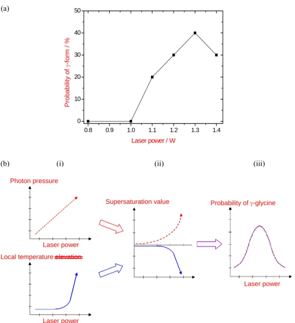
As described above, glycine tends to crystallize to the γ-form from the high supersaturated solution. As laser power increases, the concentration of the liquid-like clusters in the focal spot should be higher due to deeper optical potential. Therefore, in order to investigate how the laser power affects the probability of γ-form preparation, FTIR measurement was carried out for the crystals prepared with the power varying from 0.8 to 1.4 W. The probability was investigated for 10 samples at each laser power and the results are shown in Figure 5a. In the case of the laser power lower than 1.0 W, only the α-form was obtained. While the laser power
became higher, the probability gradually became higher and reached to the max of 40% at 1.3 W. However, it decreased to 20% at 1.4 W, which is the maximum laser power in this experiment. Why the probability does not reach to 100% is explained as follows: The concentration of glycine liquid-like clusters in the focal spot increases from time to time by laser irradiation. At first, a certain supersaturation, where the nucleation to α-form occurs as in bulk solution, is attained. As the nucleation is a statistical phenomenon, the concentration sometimes increases faster before the α-form nucleus is produced and possibly comes to higher region, where γ-form can be nucleated. Therefore, 40% of the probability of γ-glycine was obtained under the present condition.
The generation of γ-form can possibly be explained by two reciprocal laser power dependent effects; photon pressure and local temperature elevation in the focal spot. As illustrated in Figure 5b, the interaction of a focused laser beam and the liquid-like cluster induces photon pressure, which force is proportional to the laser power. Simultaneously, the laser beam causes a local temperature elevation
Fig. 5. (a) Laser power dependence of the preparation probability of γ-glycine induced by the photon
pressure of the linearly polarized CW NIR laser beam and (b) a bell-shaped curve of the multiplication of two effects; photon pressure and local temperature elevation.
(a)
(b) (i) (ii) (iii)
Probability of γ-glycine
Laser power Photon pressure
Laser power Local temperature elevation
Laser power Supersaturation value Laser power 0.8 0.9 1.0 1.1 1.2 1.3 1.4 0 10 20 30 40 50 Prob ability of γ -f orm / % Laser power / W
mainly due to the absorption of 1064 nm-photon by glycine molecule itself, since it has a larger absorption coefficient at 1064 nm than D2O (Figure 5b (i)). The molecular concentration of glycine
liquid-like clusters is increased with time, and larger associates receive deeper trapping potential. Thus, nonlinear increase of glycine clusters is realized. These glycine clusters are responsible to laser-induced heating. Thus, the temperature elevation is nonlinearly enhanced with the laser power. The elevation becomes apparent relatively at the high laser power region, and overcomes the dissipation.
Next, we consider the changes of the supersaturation value with respect to the photon pressure and the local temperature elevation as illustrated in Figure 5b (ii), as the polymorph of glycine depends on the supersaturation degree on the nucleation as described above. Since the photon pressure is enhanced more due to the gathering of the liquid-like clusters, the supersaturation value in the focal spot nonlinearly increases with the laser power, leading to a higher probability of γ-glycine formation. On the contrary, the supersaturation value is reduced by the local temperature elevation, and its tendency suddenly becomes large above a certain laser power where the thermal dissipation is overcome by input laser power. Thus, the probability of γ-form preparation is considered to be represented by the multiplication of these two factors, consequently a bell-shaped curve as illustrated in Figure 5b (iii) is obtained. This explanation supports the present result showing in Figure 5a.
In summary, we have succeeded in the crystallization of glycine in D2O by photon pressure of a
focused CW NIR laser beam, and in controlling the crystal polymorph just by changing the laser power. Interestingly, only one crystal with the polymorph control was spatiotemporally obtained just at the focal spot within a few tens of seconds. The control mechanism was proposed in view of the change of supersaturation degree in the focal spot, and discussed on the basis of photon pressure and temperature elevation depending on the laser power. In spite of the fact that the spontaneous crystallization of glycine is controlled kinetically rather than thermodynamically, giving the α-form, we have found that the γ-form glycine crystal was achieved by high photon pressure. The present work clearly shows that photon pressure enables us to control the polymorph. Other parameters probably reflecting the crystal polymorph, e.g. concentration, temperature, solvent, laser polarization, and so on, are under investigation and will be reported near future.
II. 計畫成果自評
In our Laser Bio/Nano Science Laboratory we have settled the lasers, microscopes, and related instruments, and have developed some microscopy and imaging methods. Last October we moved to the “final” destination of our office and experimental space in Tin Ka Ping Photonics Center where we now work comfortably. Some promising experimental data for conducting Laser Trapping Crystallization studies have been obtained and presented in conferences and symposia, and particularly Raman microspectroscopic analysis and and crystal polymorph control are quite important will be a milestone of our research project. In addition, amplified femtosecond laser system was installed recently. It should accelerate the research on Laser Tsunami Crystallization study, which will be coupled with the present subject.
We have published 4 papers in international journals and presented 6 invited lectures in international conferences in 2009 period. Further more we organized The 1st NCTU-NAIST (Nara Institute of Science and Technology) Workshop on Molecular/Nano Science 2009 in collaboration with many colleagues in NCTU. Some of our scientific results on Laser Trapping Crystallization were regarded as new interesting and original trials from this laboratory and we received very positive responses. Also our activity is being accepted well, one of which examples is the publication of Hiroshi Masuhara Festschrift in J. Phys Chem. C from American Chemical Society.
Thus we think that our efforts in the last year are very efficient and conduction of the research in this laboratory is successful.
錄
附 I. 表及圖
Fig. 1. Schematic drawing of Raman microspectroscopy for laser-trapping crystallization
IR cut filter
HeNe Laser Polychromator, CCD
Pinhole Line filter Notch filter Sample Galvano Mirror Pinhole Nd:YVO Laser 4 PMT PMT Objective Lens
Fig. 2. (a) Laser power dependence of the preparation probability of γ-glycine induced by the photon
pressure of the linearly polarized CW NIR laser beam and (b) a bell-shaped curve of the multiplication of two effects; photon pressure and local temperature elevation.
(a)
(b) (i) (ii) (iii)
Probability of γ-glycine
Laser power Photon pressure
Laser power Local temperature elevation
Laser power Supersaturation value 0.8 0.9 1.0 1.1 1.2 1.3 1.4 0 10 20 30 40 50 Prob ability of γ -f orm / % Laser power / W
附錄 III. Publication list
Journal papers
[1] “Nanosecond laser preparation of C60 aqueous nanocolloids”
Teruki Sugiyama, Sen-ichi Ryo, Isamu Oh, Tsuyoshi Asahi, Hiroshi Masuhara
J. Photochem. Photobiol. A: Chem., Vol. 207, No. 1, pp.7-12 (2009).
[2] “Comparative investigation of ultrafast photoinduced processes in salicylidene-aminopyridine in solution and solid state”
Michel Sliwa, Nicolas Mouton, Cyril Ruckebusch, Stephane Aloise, Olivier Poizat, Guy Buntin, Remi Metivier, Keitaro Nakatani, Hiroshi Masuhara, Tsuyoshi Asahi
J. Phys. Chem. C, Vol. 113, No. 27, pp.11959–11968 (2009).
[3] “Crystal growth of glycine controlled by a focused cw near-infrared laser beam” Teruki Sugiyama, Takuji Adachi, and Hiroshi Masuhara
Chem. Lett., Vol. 38, No.5, pp.482-483 (2009).
[4] “Blinking photoluminescence properties of single TiO2 nanodiscs: interfacial electron transfer
dynamics”
Ki-Seok Jeon, Seung-Do Oh, Yung Doug Suh, Hiroyuki Yoshikawa, Hiroshi Masuhara and Minjoong Yoon
Phys. Chem. Chem. Phys., Vol. 11, No. 3, pp.534-542 (2009).
Books, Book chapters
[1] 光科学研究の最前線 2 (Tokyo, 強光子場科学研究懇談会, 2009) 加藤 義章、増原 宏、他(Eds.) [2] “レーザーナノ化学”, pp. 178. 増原 宏 In 光科学研究の最前線 2 (Tokyo, 強光子場科学研究懇談会, 2009) edited by加藤 義章、増 原 宏、他
[3] Molecular Nano Dynamics, Vol. 1: Spectroscopic Methods and Nanostructures (Berlin, Wiley-VCH, 2009)
[4] Molecular Nano Dynamics, Vol.2: Active Surfaces, Single Crystals and Single Biocells (Berlin, Wiley-VCH, 2009)
Fukumura, Masahiro Irie, Yasuhiro Iwasawa, Hiroshi Masuhara, and Kohei Uosaki (Eds.)
[5] “Femtosecond laser tsunami processing and light scattering spectroscopic imaging of single animal cells”, pp. 547-570.
Hiroshi Masuhara, Yoichiroh Hosokawa, Takayuki Uwada, Guillaume Louit, Tsuyoshi Asahi
In Molecular Nano Dynamics Volume 2 (Berlin, Wiley-VCH, 2009) edited by Hiroshi Fukumura, Masahiro Irie, Yasuhiro Iwasawa, Hiroshi Masuhara, and Kohei Uosaki.
[6] 反応すれば形が変わるナノの世界 ~細胞から結晶まで~ (Tokyo, Kuba Pro., 2009) 増原 宏 (Ed.)
International conferences, workshop, and seminar (invited)
Title Spectroscopy, Photochemistry, and Fabrication of Single Nanocrystals Author(s) Hiroshi Masuhara* Invited
Conference 2009 RCAS Taiwan-Japan Workshop on Single Molecule/Confocal Microscopy
1
Place, date Oct. 15, 2009, Taipei, Taiwan
Title Spectroscopic and Imaging Study on Laser Trapping Dynamics and Crystallization of Amino Acids and Proteins in Solution
Author(s) Hiroshi Masuhara,* Teruki Sugiyama, Ken-ichi Yuyama, Thitiporn Rungsimanon, Takayuki Uwada, and Atsushi Miura Invited
Conference The 11th International Conference on Organic Nonlinear Optics (ICONO'11)
2
Place, date September 20-25, 2009, Beijing, China
Title Crystallization and Crystal Growth of Amino Acids in Solution by Photon Pressure of a Focused Cw Near Infrared Laser Beam
Author(s) Hiroshi Masuhara,* Teruki Sugiyama, Kenichi Yuyama, Thitiporn Rungsimanon Invited
Conference ICP2009. XXIV International Conference on Photochemistry 3
Place, date 19 - 24 July 2009, Toledo, Spain Title Laser and Organic Nanoparticles Author(s) Hiroshi Masuhara Plenary
Conference International Conference Organic nanophotonics (ICON2009) 4
Title Femtosecond “Laser Tsunami” Mamipulation for Single Living Cells in Solution
Author(s) Hiroshi Masuhara* and Yoichiroh Hosokawa Invited Conference
Spring Annual Meeting of the Korean Chemical Society, A Special Symposium of Physical Chemistry Division “Physical Chemistry for Biological Application”
5
Place, date 2009 April 17, Seoul, Korea
Title Laser Trapping Spectroscopy and Crystallization in Solution Author(s) Hiroshi Masuhara Invited
Conference Asian Academy of Sciencec by JSPS and KOSEF 6
附錄 IV. 獎項獲得
2009
Hiroshi Masuhara Festscrift on The Journal of Physical Chemistry C, Volume 113, Issue 27. - Exploration with Lasers into New Areas of Molecular Photoscience -
Website
國立交通大學出國報告書
98 年 11 月 20 日 職稱 講座教授 報告人姓名 増原 宏 申請單位 (學生請加註系級) 應用化學系 電話 Ext. 56593 出國類別 □考察 ■訪問 □進修 □研究 ■國際會議 ■其他:Collaboration experiment 會議/出國計畫名稱 出國期間 自98 年 11 月 5 日至 98 年 11 月 11 日 出國地點 日本 奈良、東京 出國目的/發表論 文題目Collaboration on “Formation of millimeter scale liquid-like domain of glycine by a focused laser beam” and invited presentation on “Laser-induced crystallization ad crystal growth of amino acids and proteins in solution” in 11th Japan-Belgium Symposium on Polymer Science (Nov. 8-11, 2009, Tokyo)
金額 69,298.- 經費來源 (校內會計編 號) 98N035 雷射捕陥結晶法之研究 報告內容應包括下列各項: 一、 經過
November 5-7; Left Hsinchu and arrived at Nara Institute of Science and Technology (NAIST), and extended collaboration experiment with Associate Professor Teruki Sugiyama, Mr. Kei Ishiguro, and Mr. Ken-ichi Yuyama. November 8-10; Attended the 11th Japan-Belgium Symposium on Polymer Science in Tokyo Institute of
Technology, presented an invited paper on “Laser–induced Crystallization and Crystal Growth of Amino Acids and Proteins in Solution”, and discussed on the relevant topics with Japanese and Belgian scientists. Also chaired the 1st morning session of Nov. 10.
November 11; Left Tokyo and arrived back at Hsinchu
二、 心得(可含照片) (1) Collaboration experiment
In this collaboration, we have succeeded in the formation of a mm-scale liquid-like cluster of glycine by focusing a laser beam at a different focal position, a glass/solution interface of the solution. We examine and consider the formation process of the large cluster on the basis of the results of direct observation and surface height measurement.
A 40 µl portion of glycine D2O solution (3.6 M) was put on a cover glass with a highly hydrophilic surface,
where a thin solution layer with 120-160 µm thickness was prepared. A near-infrared CW laser beam of the linear polarization (λ = 1064 nm) with varying the intensity from 0.7 to 1.4 W was focused at a glass/solution interface via an objective lens (60× magnification, NA=0.90). The surrounding area at the focal spot was directly observed under halogen light irradiation, and the images were captured from the obliquely upward of a sample using a CCD camera. The temporal change of the solution surface height during 10 min-laser irradiation was monitored by a laser confocal displacement meter every 50 ms.
Before laser irradiation, only the top of the objective lens was observed as a dark disk through a petri dish and the solution in Fig. 1a (1). After 30 sec-laser irradiation, we clearly identified about 2 mm-sized and circular area (white dashed line) around the focal spot by CCD camera as shown in Fig. 1a (2), which had higher refractive index than that of the surrounding solution. For further laser irradiation, the area gradually became larger until 210 sec, and eventually it grew to the size with a few mm in diameter, as could be seen even by the naked eye as in Fig. 1a (3). However, at 225 sec the area suddenly disappeared in Figure 1a (4). During the area formation, temporal change of the solution surface height simultaneously measured. The result in Fig. 1b shows that the solution surface elevation is followed with the area growth observed directly. Surprisingly the height became higher than the initial position. We consider that this area with a high refractive index is produced by gathering the small liquid-like cluster. As far as we know this is the first demonstration of the formation of the mm-scale liquid-like cluster, which is much larger than the focal spot. Note that the focal point was moved to the surface of the large cluster after it formed, the crystallization was immediately induced. Thus, the large cluster is expected to be the precursor of the crystal, and the understanding will lead us the elucidation of nucleation and crystallization process. 0 sec 30 sec (1) (2) 1 mm 210 sec 225 sec (3) (4) 0 100 200 300 400 500 600 0 50 100 150 200 250 S o lu ti o n s u rf ac e hei g h t ( u m ) Time (sec) (1) (2) (3) (4)
(a)
(b)
Fig.1 (a) CCD camera images around the focal spot.
(b) Temporal change of the solution surface height during laser irradiation. The images shown in Fig.1(a) were captured at each point in the graph.
(2) Invited paper and chair
The content of my lecture is concerned with new frontiers in molecular science opened by intense laser irradiation, particularly with laser-induced crystallization and crystal growth of molecules in solution. I talked as follows.
In 2002 we reported for the first time lysozyme crystallization triggered by femtosecond laser ablation of its saturated aqueous solution. The ablation induces shockwave propagation, and generation and collapse of tiny bubbles, leading to local convection in solution, and thus we call this phenomenon “Laser Tsunami
Now we have been extending the studies on the underlying dynamics and mechanism by time-resolved spectroscopy and imaging.
In 2007 we succeeded in crystallization of glycine by photon pressure of a focused CW near IR laser beam and now we call this crystallizaiton “Laser Trapping Crystallization” and explore the relevant behaviors. An intense CW laser beam of 1064 nm was introduced into an inverted microscope and focused into a thin film of super-saturated heavy water solution of glycine with an objective lens of 40 x magnification and NA of 0.9. The irradiation position is important and single crystal formation was observed only upon focusing the beam at an air/solution interface. The crystal grew up to a few tens μm and dependences of the crystallization on laser power and laser polarization are now being examined.
When laser irradiation was performed at a solution/glass interface of the glycine solution, no crystallization was observed, and instead millimeter-sized liquid-like domain was formed. It is surprising to see that the size of the domain is extremely larger than the focal point, while the crystallization through this domain was also confirmed.
In addition it was demonstrated that growth of a spontaneously prepared (not by laser) glycine crystal can be controlled by irradiation at the solution/glass interface with the trapping laser.
These results show high potential of laser trapping method in molecular, supramolecular, and polymer sciences, while we expect our approach will contribute to understand crystallization mechanism of amino acids and proteins in solution.
Also I chaired two speakers ; Th. Verbiest (Belgium) and H. Takezoe (Japan).
三、 考察參觀活動(無是項活動者,或前已敘述者可省略此項) None
四、 建議
The collaboration topic is extremely unique and will give a strong impact upon different two research fields of laser trapping and crystallization mechanism. Systematic and detailed study is necessary and will be developed in NCTU and NAIST.
The intimate relation between Japan and Belgium has been grown up since 1982. Under this situation I have participated from NCTU and discussed on many topics of mutual interests with Japanese and Belgian scientists. I am sure my presence will contribute to setup of future collaboration of NCTU with Japanese and Belgian Universities.
五、 攜回資料名稱及內容
A copy of program and some information of 11th Japan-Belgium Symposium on Polymer Science
六、其他 None
Eleventh Japan - Belgium Symposium on Polymer Science
November 8-11, 2009
Tokyo Institute of Technology
Eleventh Japan - Belgium Symposium
on
Polymer Science
November 8 -11, 2009
Tokyo Institute of Technology,
Tokyo, Japan
Organized by
International Research Center of Macromolecular Science,
Tokyo Institute of Technology
History of Japan – Belgium Symposium on Polymer Science
This series of symposia was organized on the original initiative of Professor G. Smets (Katholieke Universiteit Leuven) and Professor K. Hayashi (Osaka University)
Year Place Chairman
1st 1982 Osaka (Japan) Prof. K. Hayashi
2nd 1984 Brussels (Belgium) Prof. G. Smets
3rd 1986 Sapporo (Japan) Prof. J. Sohma
4th 1989 Brussels (Belgium) Prof. M. Van Beylen
5th 1991 Sendai (Japan) Prof. M. Matsuda
6th 1994 Namur (Belgium) Prof. J.-M. André
7th 1997 Hayama (Japan) Prof. M. Irie
8th 2000 Ghent (Belgium) Prof. E. Goethals
9th 2003 Suita (Japan) Prof. A. Matsumoto
10th 2006 Liege (Belgium) Prof. R. Jérôme
Eleventh Japan - Belgium Symposium on Polymer Science
November 8 (Sunday)
17:00 – 18:00 Registration (Hotel Princess Garden) 18:00 – 20:00 Welcome mixer (Hotel Princess Garden)
November 9 (Monday)
9:00 - 9:30 Registration (Tokyo Tech Front, Royal-Blue Hall) 9:30 – 9:45 Opening Remarks and Addresses
Prof. Ken Okazaki (Dean, Graduate School of Engineering, TIT) Prof. Mitsuo Sawamoto (President, Society of Polymer Science, Japan)
Session 1: Chairperson: Prof.’s Sawamoto/Goethals
9:45 – 10:15
Chip calorimetry for the investigation of transformations in thin polymer films
N. Gotzen, G. Van Assche, B. Van Mele* (VUB) 10:15 -10:45
Organic nano-electronics based on polymer nano-sheet assemblies
T. Miyashita (Tohoku U.)
10:45 – 11:15 Coffee Break
Session 2: Chairperson: Prof.’s Miyashita/Berghmans
11: 15 – 11: 45
Co-continuous polymer blend morphology tailored by nanofillers : from nanostructures to mechanical performances
O. Persenaire, J.-M. Raquez, L. Bonnaud, Ph. Dubois* (UMH) 11:45 – 12:15
Fabrication of tissues chips using a layer-by-layer technique
M. Matsusaki, K. Kadowaki, K. Sakaue, M. Akashi* (Osaka U.)
Session 3: Chairperson: Prof.’s Matsumoto/Dubois
13: 30 – 14:00
Cobalt-mediated radical coupling (CMRC); a powerful route to the preparation of novel ABA triblock copolymers
C. Detrembleur*, C. Jérôme, A. Debuigne (ULg) 14:00 – 14:30
Metal catalysis and precision synthesis in living radical polymerization
M. Sawamoto (Kyoto U.) 14:30 – 15:00
'Clicked' polymeric microcapsules and beads: azide-alkyne versus thiol-ene and thiol-yne approach
F. Du Prez*, W. Van Camp, A. Prasath, T. Dispinar, T. Gokmen, B. G. De Geest (RUG) 15:00 -15:30
Topological polymer chemistry in pursuit of elusive polymer rings
Y. Tezuka*, T. Yamamoto (TIT)
15:30 – 16:00 Coffee Break
Session 4: Chairperson: Prof.’s Akashi/Du Prez
16:00 – 16:30
Nanopatterned smart polymer surfaces
A. Jonas (UCL) 16:30 – 17:00
Precision synthesis of condensation polymers and π-conjugated polymers
T. Yokozawa (Kanagawa U.) 17:00 – 17:30
Smart drug delivery systems based on pH-sensitive block copolymers
Ch. Jerome* (ULg) 17:30 – 18:00
Structure-property relationship of stereoblock polylactides with controlled sequences
Y. Kimura (KIT) 18:00 – 18:30
Multifunctional nanostructures for controlled and targeted drug delivery
November 10 (Tuesday)
Session 5: Chairperson: Prof.’s Masuhara/Hofkens
9:30 – 10:00
Magneto-optical properties of polymers and nanoparticle/polymer blends
Th. Verbiest (KUL) 10:00 – 10:30
Liquid crystal polymers for photonic devices
H. Takezoe (TIT)
10:30 – 11:00 Coffee Break
Session 6: Chairperson: Prof.’s Vacha(TIT)/Lazzaroni
11:00 – 11:30
Visualizing dynamics of polymers by single molecule fluorescence microscopy
E. Braeken, A. Deres, H. Ujii, J. Hofkens* (KUL) 11:30 – 12:00
Laser-induced crystallization and crystal growth of amino acids and proteins in solution
H. Masuhara*, T. Sugiyama, K. Yuyama, Th. Rungamanon, A. Miura, T. Uwada, A. Usman (NAIST, NCTU)
12:00 -12:30
Theoretical modeling of structure and properties of extended systems – unraveling the structure of polymers by vibrational Raman optical activity spectroscopy
B. Champagne (FUNDP)
12:30 – 14:00 Lunch (Conference Room-L, Tokyo Tech Front , 2F)
Session 7: Chairperson: Prof.’s Iwai/Jonas
14: 00 – 14:30
Organic semiconducting nanostructures from conjugated polymer self-assembly
R. Lazzaroni (UMH) 14:30 – 15:00
New functions related to the helical structures of substituted polyacetylenes prepared using a [Rh(nbd)Cl]2 catalyst
15:00 – 15:30
The lifetime of organic photovoltaic cells: A critical problem with a strong need for a fundamental solution
D. Vanderzande*, B. Campo, S. Bertho, L. Lutsen, J. Manca, T. Cleij, T. Aernouts, A. Hadipour (Uhasselt)
15:30 – 16:00 Coffee Break
Session 8: Chairperson: Prof.’s Tabata/Vanderzande
16:00 – 16:30
Fluorescence label studies of thermo-responsive copolymers in aqueous solution
K. Iwai (Nara Women’s U.) 16:30 – 17:00
Use of metal-ligands interactions to control the self-assembly of block copolymers
J.-F. Gohy (UCL) 17:00 – 17:30
Construction of crosslink-system-materials consisting of designed network-polymer-precursor modules
A. Matsumoto (Kansai U.) 17:30 – 18:00
Polymers as versatile materials for biomedical applications
P. Dubruel*, S. van Vlierberghe, Tim Desmet, E. Schacht (RUG)
19:00 – 21:00 Banquet (Japanese Restaurant, “Botan”)
November 11 (Wednesday)
Session 9: Chairperson: Prof.’s Saruyama/Van Mele
9:30 – 10:00
Selective removal of VOCs by polymer membranes containing ionic liquid
T. Uragami (Kansai U.) 10:00 – 10:30
Development of POSS containing block copolymers for direct patterning templates
10:30 – 11:00 Coffee Break
Session 10: Chairperson: Prof.’s Uragami/ Jérôme
11:00 – 11:30
Application of the temperature modulation method to polymers
Y. Saruyama (KIT) 11:30 – 12:00
Chemorheology of thermosetting polymer systems studied by RheoDSC - a novel technique for simultaneous rheological and calorimetric analysis
G. Van Assche*, Ch. Block, V. Janssens, P. Van Puyvelde, B. Van Mele (VUB)
12:00 – 12:15 Closing Remarks
Prof. Bruno Van Mele (Vrije Universiteit Brussel)