Polymethylmethacrylate (PMMA) has been extensively used as bone cement in orthopedic procedures. Adverse circulatory and pulmonary events associated with hip arthroplasty using PMMA bone cement have been documented. Percutaneous vertebroplasty is an image-guided procedure in which the bone cement is injected into the compression fracture due to osteoporosis or neoplasm. We experienced a case of sudden tachypnea associated with pulmonary cement embolism in an adult male who underwent percutaneous vertebroplasty.
Key words: Embolism, Percutaneous Vertebroplasty, Polymethylmethacrylate
Percutaneous vertebroplasty involves an injection of polymethylmethacrylate (PMMA) into a diseased vertebral body for partial vertebral body remodeling and lumbar pain relief. Complications are infrequent, and consist of local processes such as infections and bone cement leakage into the spinal canal or the perivertebral venous system [1, 2]. Other complications are usually related to the initial vertebral disease rather than to the vertebroplasty technique itself [3]. We report a case of symptomatic pulmonary embolism caused by cement after percutaneous vertebroplasty.
CASE REPORT
A 69 year-old man suffering from persistent, severe lower back pain due to osteoporosis with compression fractures of L3 and L4 was admitted for L3 and L4 percutaneous vertebroplasty. All laboratory data were within normal limits. Verteroplasty was performed under local anesthesia and conscious sedation. Prophylactic antibiotic therapy was given. Vertebroplasty was performed by a unilateral transpedicular approach at L3 and bilateral transpedicular approach at L4 with two 10-gauge needles in each vertebral body. Methylmethacrylate cement (Simplex P; Howmedica, International, Ireland) was prepared at room temperature (20 degree) and opacified with sulfate power, and 6mL was then injected under lateral fluoroscopic guidance (GE; Compact 7700DF-151R Mobile Digital C-arm, 512 × 512) into each vertebral body. Tachypnea started immediately after percutaneous vertebroplasty of L3 and L4. Dyspnea did not develop until the second postoperative day; pulmonary embolism caused by PMMA was detected on the chest radiography. The chest radiograph shows multiple high-density tubular opacities outlining pulmonary vessels (Fig. 1). An Reprint requests to: Dr. Shaw-Nan Jean
Department of Radiology, DA Chien General Hospital. No. 6, Shinguang Street, Miaoli 360, Taiwan, R.O.C
Pulmonary Embolism of
Polymethylmethacrylate After Percutaneous
Vertebroplasty : A case report
Shaw-NaN JeaN1 YuNg-FaNg CheN2 Jui-FeN CheN2 SheN-JYe CheN3 Tao-ChYi Lo4 Su-JaNe hwaNg4
Department of Radiology1, Da-Chen General Hospital Department of Radiology2, China Medical College Hospital Department of Radiology3, Ching Chiuan Hsopital
immediate thoracic CT scan, with pre-contrast and post-contrast soft tissue window settings, reveals much linear high-density intraluminal cement in the right atrium (Fig 2 a). There is high-density intra-luminal bone cement outlining several pulmonary arteries and the segmental arteries of the superior and lower lobes (Fig 2 b). The CT scan with bone settings at the level of vertebroplasty (L3) shows cement in right latero-vertebral vein draining into the vena cava (Fig 3). The patient was treated with oxygen therapy with a regression of dyspnea. Since the day after the procedure, the patient had been admitted for total 5 times for mild dyspnea and chest discomfort and was treated by supplemental oxygen inhalation and bronchodilator agents for 3 or 4 days during each admission.
DISCCUSSION
Polymethylmethacrylate bone cement prepared in a liquid methylmethacrylate monomer is widely used to anchor prostheses in joint replacement surgery. Although multiple adverse effects have been documented to be associated with the use of PMMS cement in hip arthroplasty [4], untoward cardiovascular or pulmonary effects have been rarely reported in percutaneous vertebroplasty [5, 6]. The vertebral body is highly vascularized with intraosseous vertebral veins, making a freely communicating
valveless network with paravertebral and extradural venous plexus [7]. The osteoporotic compression may destroy the venous drainage, allowing a direct shunt of the bone cement fragment into the vein and subsequently into pulmonary circulation. Although percutaneous vertebroplasty is considered only minimally invasive, complications can occur during the procedure. Bone cement leaks into the external vertebral venous plexuses have been frequently
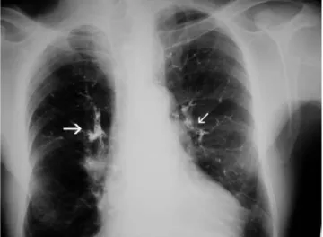
Figure 1. Chest radiography showed multiple
high-density tubular opacities outlining pulmonary vessels (arrows).
2a 2b
Figure 2. a.Pre-contrast Chest CT scan with soft tissue window settings demonstrated linear high-density intraluminal
cement (black arrow) outlining the right cardiac chamber. b. Post-contrast Chest CT scan with soft-tissue window showed high-density intraluminal cement (white arrow) outlining the pulmonary artery and its branchings.
described. There is also a potential risk for bone cement migration into the inferior vena cava [3] and pulmonary embolism. Jensen et al [8] reported two patients with a presumed embolization to the lungs that did not produce respiratory changes. Venous leaks can also occur toward the internal vertebral venous plexuses resulting in cord nerve root compression [2]. The great majority of these leaks are asymmetric. Such leaks can be explained by the fluid consistency of the PMMA at the moment of injection. More extensive leaks may occur if the needle is positioned incorrectly, especially if the tip lies in a basivertebral vein. Gangi et al [9] recommended that the technique be performed under both CT and fluoroscopic guidance to verify needle positioning.
The pulmonary embolism in our patient may have been caused by insufficient polymerization of the PMMS at the time of injection, which allowed migration into the inferior vena cava, right cardiac chamber and pulmonary arteries. For this reason, the acrylic cement must be mixed to the consistency of paste (i.e. a stage of advanced polymerization) before the injection so that several difficult- to-evaluate factors that affect the polymerization rate (i.e. room temperature) can be better controlled. Good-quality lateral fluoroscopy is essential during injection. Detection of even minimal cement leakage into the perivertebral veins or behind the line of the posterior wall should be an indication to halt the procedure
immediately. In our case, pulmonary embolism occurred because perivertebral venous migration was not recognized at the time of vertebroplasty. The bone cement was opacified only with barium. Jensen et al [8] recommend a barium/ tungsten combination for adequate visualization of needle positioning and venous flow during fluoroscopy. Recently, Padovani et al [10] reported a case of symptomatic pulmonary embolism caused by PMMA that resulted in pulmonary infarction. Three of the 27 patients with a malignant osteolytic spinal tumor in the current study experienced pulmonary embolism caused by PMMA after percutaneous vertebroplasty (total of 72 vertebrae).
The value of performing a prior vertebral venography through the vertebroplasty needle is controversial. Jensen et al [8] found vertebral venography to be of great use because it confirmed the needle position in the trabecular space and outlined the venous outflow pattern, indicating where to look for PMMA migration, so we did perform the vertebral venography before the percutaneous vertebroplasty. Some European authors [3, 9] have not performed vertebrography prior to vertebroplasty. Results appear doubtful to them because iodinated contrast agents have different physiochemical properties than PMMA, and nearly always drain into the venous plexuses. In addition, persistence of intravertebral opacification could interfere with the procedure of vertebroplasty.
The chest radiographs were checked routinely after vertebroplasty to allow detection of pulmonary embolism caused by PMMA at the early stage. The clinical manifestations of pulmonary embolism include sudden dyspnea, tachypnea, cyanosis, precordial or substernal oppressive pain, tachycardia, and hypotension. The presence of these symptoms following the procedure would suggest pulmonary embolism, and the chest radiography should then be taken immediately as in our case. If a patient reports the chemical odor of the PMMS solvent, pulmonary embolism should also be considered.
Supportive treatment for acute embolism should include rest, analgesics for pain, and oxygen as indicated. Pulmonary embolectomy is the more therapeutic way for critical patients. Tozzi et al [11] reported one case in which this rare and dreadful complication was successfully treated by pulmonary embolectomy. However, Yoo et al [12] reported a case of acute respiratory distress syndrome following percutaneous vertebroplasty treated with pulmonary embolectomy, while the subject died on the tenth day following this procedure. Therefore conservative treatment rather than surgical removal of the bone
Figure 3. CT scan with bone settings at the level of
vertebroplasty (L4) showed cement in right latero-vertebral vein (arrow) draining in the vena cava and epidural leak (small arrow).
cement entrapped in pulmonary circulation may be recommended, except that the obstruction is extensive enough to cause immediate cardio-circulatory changes. In our case, the patient was treated with oxygen therapy and stable regression of the symptoms was observed. In the absence of contraindications, anticoagulant therapy with heparin should be instituted immediately to obviate the progression of pulmonary infarction. Ventilation perfusion scanning is useful for demonstration and quantification of the perfusion defect, allowing repeated evaluation during patient follow-up course (5, 10).
In conclusion, a case of pulmonary embolism caused by PMMA migration after percutaneous vertebroplasty was reported. Proper techniques can minimize the risk of pulmonary embolism caused by PMMA during the percutaneous vertebroplasty. ◆
REFERENCES
1. Cotton A, Dewatre F, Cortet B et al. Percutaneous vertebroplasty for osteolytic metastases and myeloma: effects of the percentage of lesion filling and the leakage of Methylmethacrylate at clinical follow-up. Radiology 1996; 200: 525-530
2. Chiras J, Depriester C, Weill A, Sola-Martiner MT, Deramond H. Vertebroplasties percutanees. AJNR 1997; 24: 45-59
3. Weill A, Chiras J, Simon JM, Rose M, Sola-Martinex T, Enkaoua E. Spinal metastases: indications for and results of percutaneous injection of Acrylic surgical cement. Radiology 1996; 99: 241-247
4. Fallon KM, Fuller JG, Morley-Forster P. Fat embolization and fatal cardiac arrest during hip arthroplasty with Methylmethacrylate. Can J Anaesth 2001; 48: 626-629
5. Padovani B, Kasriel O, Brunner P, et al. Pulmonary embolism caused by Acrylic cement: a rare complication of percutaneous vertebroplasty. AJNR 1999; 20: 375-377
6. Vasconcelos C, Gailloud P, Martin JB, et al. Transient arterial hypotension induced Polymethylmethacrylate injection during percutaneous vertebroplasty. Radiology 2001; 12: 1001-1002
7. Gershater R, St. Louis EL. Lumbar epidural venography: Review of 1,200 cases. Radiology 1997; 57: 511-518
8. Jensen ME, Avery JE, Mathis JM, Kallmes DF, Cloft HJ, Dio JE. Percutaneous Polymethylmethacrylate vertebroplasty in the treatment of osteoporotic vertebral body compression fractures: technical aspects. AJNR 1997; 18:1897-1904
9. Gangi A, Kastler BA, Dietemann JL. Percutaneous vertebroplasty guided by a combination of CT and fluoroscopy. AJNR 1994; 15: 83-86
10. Jee SJ, Sang HL, Sang KJ, et al. Pulmonary embolism of Polymethymethacrylate after percutaneous vertebroplasty: a report of three cases. SPINE 2002; 27: E 416-418
11. Piergiorgio T, Yasmine A, Antonio FC, et al. Management of pulmonary embolism during acrylic vertebroplasty. Ann Thorac Surg 2002; 74: 1706-1708 12. Kyung YY, Seong W, et al. Acute respiratory distress
syndrome associated with pulmonary cement embolism following percutaneous vertebroplasty with Polymethylmethacrylate . Spine 2004; 29: E 294-297