MALDI MS Analysis of Oligonucleotides: Desalting
by Functional Magnetite Beads Using
Microwave-Assisted Extraction
Wei-Yu Chen†and Yu-Chie Chen*,†,‡Department of Applied Chemistry and Institute of Molecular Science, National Chiao Tung University, Hsinchu 300, Taiwan
The presence of alkali cation adductions of oligonucle-otides commonly deteriorates matrix-assisted laser de-sorption/ionization (MALDI) mass spectra. Thus, desalt-ing is required for oligonucleotide samples prior to MALDI MS analysis in order to prevent the mass spectra from developing poor quality. In this paper, we demon-strate a new approach to extract traces of oligonucleotides from aqueous solutions containing high concentrations of salts using microwave-assisted extraction. The C18-presenting magnetite beads, capable of absorbing micro-wave irradiation, are used as affinity probes for oligonu-cleotides with the addition of triethylammonium acetate as the counterions. This new microwave-assisted extrac-tion approach using magnetite beads as the trapping agents and as microwave-absorbers has been demon-strated to be very effective in the selective binding of oligonucleotides from aqueous solutions. The extraction of oligonucleotides from solutions onto the C18-present-ing magnetite beads takes only 30 s to enrich oligonucle-otides in sufficient quantities for MALDI MS analysis. After using this desalting approach, alkali cation adduc-tions of oligonucleotides are dramatically reduced in the MALDI mass spectra. The presence of saturated NaCl (∼6 M) in the oligonucleotide sample is tolerated without degrading the mass spectra. The detection limit for d(A)6 is∼2.8 fmol.
Alkali cation adductions of oligonucleotides are commonly observed in matrix-assisted laser desorption/ionization (MALDI) mass spectra because the phosphodiester backbones of oligo-nucleotides have high affinities for alkali cations. Therefore, efforts have been devoted to reduce the alkali cation adductions of oligonucleotides. Desalting by ZipTip is generally used to remove cations from oligonucleotide samples.1 Matrixes such as 3-hy-droxypicolinic acid (HPA),2 6-aza-2-thiothymine (ATT),3
trihy-droxyacetophenone (THAP),4,5 and 5-methoxysalicylic acid,6,7 mixed with their corresponding comatrixes such as ammonium salts2-5or spermine6,7to suppress cation adductions, are generally employed for MALDI MS analysis of oligonucleotides. Good-quality MALDI mass spectra of oligonucleotides are obtained through these two desalting steps.1 Additionally, using diami-nobenzoic acid (DABA)8or 3,4-diaminobenzophenone (DABP)9 as a UV-MALDI matrix without the addition of comatrixes for the detection of oligonucleotides is also suitable. However, the detectable molecular size of oligonucleotides through these methods is limited.
In this study, we employed C18-presenting magnetite beads to trap oligonucleotides from the sample solution onto the surface of the beads. Counterions for oligonucleotides were added in the sample solution. As a result, neutral oligonucleotide-counterion pairs can readily attach on the surface of the C18-presenting magnetite beads. In order to shorten the time for the enrichment of target species, extraction is carried out under microwave heating. Microwave-assisted approaches including microwave-assisted digestion10-15and microwave-assisted extraction16-22have been widely used in the acceleration of digestion and extraction,
* Corresponding author. E-mail: yuchie@mail.nctu.edu.tw. Fax: 886-3-5131527. Phone: 886-3-886-3-5131527.
†Department of Applied Chemistry. ‡Institute of Molecular Science.
(1) Spottke, B.; Gross, J.; Galla, H.-J.; Hillenkamp, F. Nucleic Acids Res. 2004,
32, e97.
(2) Wu, K. J.; Steding, A.; Becker, C. H. Rapid Commun. Mass Spectrom. 1993,
7, 142-146.
(3) Lecchi, P.; Le, H. M.; Pannell, L. K. Nucleic Acids Res. 1995, 23, 1276-1277.
(4) Pieles, U.; Zu¨rcher, W.; Scha¨r, M.; Moser, H. E. Nucleic Acids Res. 1993,
21, 3191-3196.
(5) Zhu, Y. F.; Chung, C. N.; Tatanenko, N. I.; Allman, S. L.; Martin, S. A.; Haff, L.; Chen, C. H. Rapid Commun. Mass Spectrom. 1996, 10, 383-388. (6) Asara, J. M.; Allison, J. Anal. Chem. 1999, 71, 2866-2870.
(7) Distler, A. M.; Allison, J. J. Am. Soc. Mass Spectrom. 2001, 15, 456-462. (8) Li, Y. C. L.; Cheng, S.-W.; Chan, T.-W. D. Rapid Commun. Mass Spectrom.
1998, 12, 993-998.
(9) Simmons, T. A.; Limbach, P. A. J. Am. Soc. Mass Spectrom. 1998, 9, 668-675.
(10) Bose, A. K.; Ing, Y. H.; Lavlinskaia, N.; Sareen, C.; Pramanik, B. N.; Bartner, P. L.; Liu, Y. H.; Heimark, L. J. Am. Soc. Mass Spectrom. 2002, 13, 839-850.
(11) Pramanik, B. N.; Mirza, U. A.; Ing, Y. H.; Liu, Y.-H.; Bartner, P. L.; Wever, P. C.; Bose, A. K. Protein Sci. 2002, 11, 2676-2687.
(12) Chen, S.-T.; Chiou S.-H.; Wang K.-T. J. Chin. Chem. Soc. 1991, 38, 85-91. (13) Kroll, J.; Rawel, H.; Kro¨ck, R. Z. Lebensm.-Unters-Forsch A 1998, 207,
202-206.
(14) Zhong, H.; Zhang, Y.; Wen, Z.; Li, L. Nat. Biotechnol. 2004, 22, 1291-1296.
(15) Swatkoski, A.; Russell, S. C.; Edwards, N.; Fenselau, C. Anal. Chem. 2006,
78, 181-188.
(16) Pare, J. R. J.; Belanger, J. M. R.; Thornton, D. E.; Li, K.; Llompart, M. P.; Fingas, M.; Blenkinsop, S. A. Spectrosc.: Int. J. 1997, 13, 23-32. (17) Huang, Y. P.; Yang, Y. C.; Shu, Y. Y. J. Chromatogr., A 2007, 1140, 35-43. (18) Dincutoiu, I.; Gorecki, T.; Parker, B. L. Int. J. Environ. Anal. Chem. 2006,
86, 1113-1125.
(19) Zhu, X. L.; Su, Q. D.; Cai, J. B.; Yang, J. Anal. Chim. Acta 2006, 579, 88-94.
(20) Wei, M. C.; Jen, J. F. J. Chromatogr., A 2003, 1012, 111-118. Anal. Chem.2007,79,8061-8066
respectively. Particularly, microwave-assisted extraction combined with solid-phase microextraction (SPME) is generally used in the headspace analysis of volatile organic compounds (VOCs) from complex samples.16-22Alternatively, functional nanoparticles can be used as the trapping agents for targeted analytes. In relation to this, Geddes and co-workers have demonstrated that the combination of metal nanoparticles and low-power microwaves can accelerate bioaffinity reactions for assays and immunoassays under microwave-heating.23-26We believe that if the trapping agents also possess the capability of absorbing microwave extraction, then extraction processes could readily be accelerated. Previously, we demonstrated that the presence of magnetite beads could acceler-ate enzymatic digestion under microwave-heating,27 and this is due to the unique capacity of magnetite beads to absorb micro-wave radiation. Thus, in order to prove the principle of accelera-tion of microwave-assisted extracaccelera-tion by magnetite beads, we employed magnetite nanoparticles coated with dimethyloctade-cylchlorosilane (C18) as trapping agents for oligonucleotides under microwave-heating. Furthermore, because of the magnetic feature of magnetite beads, the isolation of the bead-conjugated species can simply be performed by magnetic separation. Ad-ditionally, the time required for handling multiple samples is the same as that required for one sample because it is possible to simultaneously put many samples in a microwave oven for extraction. This study demonstrates the effectiveness of this extraction approach using oligonucleotides as the samples. EXPERIMENTAL SECTION
Reagents. Hydrochloric acid was obtained from Merck (Seelze, Germany), while iron(III) chloride hexahydrate, sodium sulfite, acetic acid, and diammonium hydrogen citrate (DHC) were obtained from Riedel-de Hae¨n (Seelze, Germany). Ethanol was obtained from Showa (Tokyo, Japan), while dimethyloctadecyl-chlorosilane (C18) and 2,4,6-THAP were purchased from Aldrich (Milwaukee, WI). Finally, ammonia, tetraethoxysilane (TEOS), toluene, and triethylamine were purchased from Fluka (Steinheim, Germany), while all the oligonucleotides were purchased from MD Bio.
Preparation of Magnetite Beads. Ferric chloride (2 M) in a hydrochloride solution (2 M, 12 mL) was diluted to 100 mL with deionized water under nitrogen. Freshly prepared aqueous sodium sulfite (0.08 M, 50 mL) was slowly added to the diluted ferric chloride solution. Then a mixture of ammonia solution (28%, 8 mL) and water (40 mL) was added slowly to the solution with vigorous stirring under nitrogen. The mixture then remained in a water bath at 70 °C for 15-30 min before cooling to below 45°C. The magnetite nanoparticles were aggregated by placing an external magnet on the edge of the vial and then the solution was removed. The particles were rinsed several times with deionized water and a deionized water/ethanol (2/1, v/v) mixture, followed by resuspension in ethanol (50 mL). Afterward, ethanol (7 mL), water (8 mL), TEOS (2 mL), and ammonia solution (10%, v/v) (2 mL) were added in sequence to 25 mL of the particle (21) Herbert, P.; Silva, A. L.; Joao, M. J.; Santos, L.; Alves, A. Anal. Bioanal.
Chem.2006, 386, 324-331.
(22) Groning, M.; Hakkarainen, M. J. Chromatogr., A 2004, 1053, 151-159. (23) Aslan, K.; Malyn, S. N.; Geddes, C. D. J. Am. Chem. Soc. 2006, 128,
13372-13373.
(24) Aslan, K.; Geddes, C. D. Anal. Chem. 2005, 77, 8057-8067. (25) Aslan, K.; Geddes, C. D. J. Fluoresc. 2006, 16, 3-8. (26) Aslan, K.; Geddes, C. D. Anal. Chem. 2007, 79, 2131-2136. (27) Chen, W.-Y.; Chen, Y. C. Anal. Chem. 2007, 79, 2394-2401. Table 1. The Potential Values Set in the Mass Spectrometer for Various Sizes of Oligonucleotides
oligonucleotides IS1 (kV) IS2 (kV) lens (kV) PIEa
laser repetition rate (Hz) 6 mers 19.0 17.5 9.3 medb 4 24-48 mers 19.0 18.0 9.8 medb 10 60-72 mers 19.0 17.2 9.1 medb 10 100 mers 19.0 15.4 9.1 medb 10
aPIE: pulsed ion extraction.bmed: medium
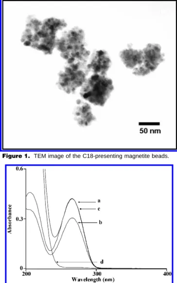
Figure 1. TEM image of the C18-presenting magnetite beads.
Figure 2. The absorption band labeled with letter a was obtained from a sample containing d(T)24(250µL, 10-6M), while the absorption
band b was obtained from the remaining solution after using the C18-presenting magnetite beads (2.5 mg) to trap their target species from the same sample under microwave-heating (60 s) followed by magnetic separation. The absorption band labeled with letter c was obtained from a sample containing d(T)24(250µL, 10-6M) with the
addition of TEAA (75 mM) (band c), while the absorption band labeled with letter d was obtained from the remaining solution after using the C18-presenting magnetite beads (2.5 mg) to trap their target species under microwave-heating (60 s) followed by magnetic separation.
suspension (40 mg/mL) while stirring at 40°C. The stirring was continued for 12 h. The particles were rinsed with methanol several times to remove unreacted TEOS and then were resus-pended in toluene (40 mL). Next, dimethyloctadecylchlorosilane (C18, 95%, 347 mg) was added to the solution with sonication for 10 min, followed by further stirring for 3-4 h in the water bath at 60 °C. The C18-presenting magnetite beads were rinsed with toluene and then were resuspended in 40 mL of methanol, giving a final concentration of 20 mg/mL.
Desalting by Presenting Magnetite Beads. The C18-presenting magnetite beads isolated from 0.5 µL (20 mg/mL) of bead solution were rinsed with triethylammonium acetate (TEAA) (0.1 M, 10 µL) followed by the addition of an oligonucleotide sample (4 µL), which had been prepared in a TEAA solution (75 mM), in an Eppendoff tube. The TEAA solution was prepared by mixing equal amounts of triethylamine and acetic acid. The mixture in the Eppendoff tube was mixed several times using a pipet. The tube was placed in a domestic microwave oven and then was microwave-heated (power: 900 W) for 30 s. The cap of the tube was kept open during microwave-heating. After which, the beads were aggregated on the wall of the tube by placing an external magnet and then the solution was removed by a pipet. The remaining beads were rinsed with a TEAA solution (0.1 M, 10 µL× 3) and deionized water (10 µL × 2). The oligonucleotides trapped on the beads were eluted by MALDI matrix (0.5 µL). The MALDI matrix was freshly prepared by dissolving 2,4,6-THAP (30 mg/mL) in a mixture of acetonitrile/DHC (200 mM) (2/1, v/v). The eluent was deposited on the MALDI sample target for MALDI MS analysis.
Instrumentation. All the mass spectra were obtained in the negative ion mode using a Biflex III linear time-of-flight mass spectrometer (Bruker Daltonics, Germany). This mass spectrom-eter was equipped with a 337 nm nitrogen laser and a 1.25 m flight path length. The IS1 and IS2 voltages were adjusted when
the mass range of the oligonucleotides was varied (see Table 1). The absorption spectra were obtained using a Varian Cary 50 spectrophotometer (Melbourne, Australia). The transmission electron microscope (TEM) image was obtained using a JEOL 2000FX (Japan).
RESULTS AND DISCUSSION
Figure 1 shows the TEM image of the magnetite beads coated with C18. Apparently, the beads are composed of several mag-netite nanoparticles (see dark dots). Moreover, the size of the beads is varied, but it is generally less than 100 nm.
Figure 2 displays the UV absorption spectra of aqueous samples containing d(T)24 before (band a) and after (band b) extraction by the C18-magnetite beads under microwave heating. The absorbance at 260 nm of the remaining solution (band b) after the removal of the magnetite beads is shown to slightly decrease as compared to that of band a. It seems that the oligonucleotides have low binding affinities with the beads. This is understandable since the surface of the C18-presenting beads is hydrophobic, and the oligonucleotides are highly charged; therefore, the binding of oligonucleotides onto the hydrophobic surface of the beads is unfeasible. Consequently, we added TEAA as the counterions for d(T)24to form neutral d(T)24-TEAA ion pairs. The change in absorbance at 260 nm in the UV absorption spectra of the remaining solution from a d(T)24 sample was examined. This was with the addition of TEAA before (see band c) and after (see band d) incubating with the C18-presenting magnetite beads. The absorbance at 260 nm of the remaining solution after incubation with the beads under microwave heating, followed by magnetic separation, dramatically drops to near zero (see band d). These results indicate that the presence of the counterions in the sample solution, that is, TEAA (75 mM), can assist the binding of oligonucleotides onto the surface of the C18-presenting magnetite beads.
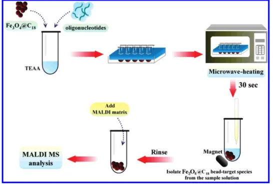
Scheme 1. Steps of Using Fe3O4@C18as the Affinity Probes to Trap Oligonucleotides from the Samples under Microwave Heating, Followed by MALDI MS Analysis (Multiple Samples Can Be Simultaneously Introduced in a Microwave Oven for Extraction)
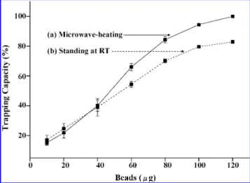
Our previous study demonstrated that the presence of mag-netite beads in aqueous solutions increased the heating rate under microwave irradiation.27Thus, we proposed that the extraction of oligonucleotides from aqueous solutions using C18-presenting magnetite beads as both the trapping agents and the microwave absorbers might be accelerated under microwave irradiation. Scheme 1 displays the steps for carrying out the extraction with the assistance of C18-presenting magnetite beads (Fe3O4@C18) under microwave heating. Multiple samples can be simultaneously introduced in a microwave oven for extraction. Thus, this technique is time and labor efficient. To investigate the effective-ness of the microwave-assisted extraction of oligonucleotides, we examined the change in absorbance at 260 nm of the remaining solution before and after incubation with different amounts of the C18-presenting magnetite beads under microwave-heating and at room temperature after magnetic separation. Figure 3 presents two plots of the trapping capacity of the C18-presenting beads for d(T)24as a function of the amount of the beads under different experimental conditions. The trapping capacity is defined as the percentage of the amount of the sample trapped on the beads divided by the original amount of the sample. Plot a shows that as the amount of beads in the solution is increased to 120 µg, d(T)24is totally trapped by the beads under microwave-heating for 60 s. That is, 1 mg of the C18-presenting beads has the capacity to trap∼625 pmol of d(T)24within 60 s under microwave-heating. Although d(T)24 can still be trapped by the beads when the extraction is carried out at room temperature, the maximum binding amount of d(T)24on the beads is only∼83% even if the amounts of the beads are increased to 120 µg (see plot b). The results indicate that the extraction of oligonucleotides using the magnetite beads as both the trapping agents and microwave-absorbers under microwave-heating is more efficient than that carried out at room temperature.
Figure 4 displays the time plots of the trapping capacity obtained by using 120 µg of the beads to trap d(T)24(3× 10⁻⁷M, 250 µL) under microwave-heating (plot a) and at room temperature (plot b) as a function of incubation time. As the extraction time is increased, the trapping capacity also increases under the two
different experimental conditions. d(T)24was totally trapped by the beads under microwave-heating for g45 s, while only ∼80% of d(T)24 was trapped on the beads at room temperature. The results demonstrate that the extraction under microwave-heating is more efficient than that carried out at room temperature. The extraction is accelerated because of the assistance of microwave heating. Furthermore, we also examined the recovery of the oligonucleotides from the beads after trapping treatment. The recovery was determined by eluting the oligonucleotides trapped on the C18-presenting magnetite beads (120 µg) with acetonitrile/ 0.1% TFA (2/1, v/v, 250 µL). The eluent was determined by UV-visible spectrophotometry. We determined that the recovery is 112 ( 15%, which is over 100%. This is understandable because the elution solvent is volatile, and it may easily be lost during analysis.
We further investigated the concentrating capacity of this approach for oligonucleotides. Because very low volumes (4∼10
µL) of samples are used for extraction, 30 s is sufficient to carry
out the extraction for oligonucleotides under microwave heating with the use of the magnetite beads. Thus, all the results shown below are obtained with the extraction time of 30 s. Figure 5a presents the direct MALDI mass spectrum of d(A)6. The depro-tonated pseudomolecular ion of d(A)6is not observed in the mass spectrum. However, when the d(A)6sample was enriched using our approach, a peak at m/z 1817.3 appears in the mass spectrum (Figure 5b). Then Figure 5c presents the direct MALDI mass spectrum of d(T)48from which it can be seen that the pseudo-molecular ion of d(T)48can barely be seen. Meanwhile, Figure 5d presents the MALDI mass spectrum of d(T)48obtained using our approach to enrich d(T)48. The deprotonated pseudomolecular ion of d(T)48 appears at m/z 14 538 with good mass resolution and intensity. Furthermore, its doubly and triply charged molec-ular ions at m/z values 7268 and 4845 are revealed in the same spectrum, respectively. These results indicate that the enrichment approach for oligonucleotides is quite effective. Furthermore, the detection limit for d(A)6is as low as∼2.8 fmol.
It is of interest to investigate the trapping capability of our approach in the selective enrichment of oligonucleotides from a Figure 3. Plots of the trapping capability (%) of the C18-presenting
magnetite beads for d(T)24(250µL, 3×10⁻⁷M) prepared in TEAA
(75 mM) as a function of the amounts of the C18-presenting magnetite beads under microwave-heating for 60 s (plot a) and standing at room temperature for 60 s (plot b).
Figure 4. Plots of the trapping capability (%) of the C18-presenting magnetite beads for d(T)24(250µL, 3×10⁻⁷M) prepared in TEAA
(75 mM) as a function of the extraction time using the C18-presenting magnetite beads as the affinity probes under microwave-heating for 60 s (plot a) and standing at room temperature for 60 s (plot b).
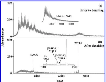
sample containing a high concentration of salts. Figure 6a presents the direct MALDI mass spectrum of a mixed-base oligonucleotide (AAAAAAAAAAAAAATGACCGCACT) containing saturated NaCl (∼6 M). A broad peak appearing at m/z ∼7400 corresponds to the singly charged ion. The broadness of the peak is contributed by the presence of a series of sodium adductions of the
oligonucleotide. Serial peaks appearing at m/z < 4000 are derived from the background of 2,4,6-THAP containing NaCl. The inset is the MALDI mass spectrum of 2,4,6-THAP containing saturated NaCl without the addition of oligonucleotides. The peak profile at m/z < 4000 looks similar to that shown in Figure 6a. Thus, we concluded that those peaks are derived from the matrix, but they are not a contribution of the mixed-base oligonucleotide. Figure 6b shows the MALDI mass spectrum of the same oligonucleotide sample containing saturated NaCl (∼6 M) after desalting by the C18-presenting magnetite beads under microwave-heating for 30 s. Two sharp peaks appearing at m/z values 3685.5 and 7371.9 correspond to the doubly and singly charged ions of the mixed-base oligonucleotide by losing two and one protons, respectively. Furthermore, the peaks at m/z < 4000 as those which appear in Figure 6a disappear. The inset shows the mass range between 7000 and 7500. There are several fragmentations derived from the mixed-base oligonucleotide. The peaks at m/z values 7058.3, 7237.5, and 7261.4 correspond to y23, [M-A-H+], and [M-C-H+]-, respectively. Overall, the results demonstrate that our desalting approach is also suitable for removing a very high concentration of salts from a mixed-base oligonucleotide sample. Additionally, we have examined the possibility by simultaneously introducing 10 samples (d(T)24) containing saturated NaCl into a microwave oven for desalting using this current approach. The desalting was done within 30 s and confirmed by MALDI MS. The deprotonated pseudomolecular ions of d(T)24with very good resolution showed up in the MALDI mass spectra after microwave-extraction (results not shown). The sodiated oligonucleotides were totally suppressed. The results indicate the feasibility of this current in the analysis of multiple samples because of the features of labor-saving and time-effectiveness.
Figure 7a shows the direct MALDI mass spectrum of d(T)100 containing 500 mM NaCl. Two broad peaks appear at m/z values ∼31 000 and ∼15 500 which correspond to the singly charged and doubly charged ions of d(T)100, respectively. The broad peaks Figure 5. (a) Direct MALDI mass spectrum of d(A)6(2.7 nM) and (b) MALDI mass spectrum of d(A)6(675 pM) obtained using the
C18-presenting magnetite beads (10µg) to trap their target species from the sample (4µL) under microwave-heating for 30 s. (c) Direct MALDI mass spectrum of d(T)48(170 nM) and (d) MALDI mass spectrum of d(T)48(42.5 nM) obtained using the C18-presenting magnetite beads (10
µg) to trap their target species from the sample (4µL) under microwave heating for 30 s. The MALDI matrix is 2,4,6-THAP (30 mg/mL) prepared in acetonitrile/aqueous diammonium hydrogen citrate (200 mM) (2/1, v/v).
Figure 6. (a) Direct MALDI mass spectrum of a mixed base oligonucleotide (AAAAAAAAAAAAAATGACCGCACT, 1.35µM) con-taining saturated NaCl (∼6 M). The inset is the MALDI mass spectrum of the MALDI matrix mixed with equal volume of saturated NaCl. (b) MALDI mass spectrum of the same mixed-base oligonucleotide (135 nM) prepared in TEAA (75 mM) containing saturated NaCl (∼6 M) obtained using the C18-presenting magnetite beads (10µg) to trap their target species from the sample (10µL) under microwave-heating for 30 s. The inset displays the same mass spectrum withm/zrange from 7000 to 7500. The MALDI matrix is 2,4,6-THAP (30 mg/mL) prepared in acetonitrile/aqueous diammonium hydrogen citrate (200 mM) (2/1, v/v). “A” stands for adenine, while “C” stands for cytosine in the inset of Figure 6b.
result from the presence of sodium adduct ions of d(T)100. Figure 7b shows the MALDI mass spectrum of d(T)100 containing 500 mM NaCl after desalting by the C18-presenting magnetite beads under microwave-heating for 30 s. The singly charged, doubly charged, triply charged, and quadruply charged ion peaks at m/z values 30 356, 15 177, 10 118, and 7589, respectively, with high intensities look much sharper than those observed in Figure 7a. Once more, the results demonstrate that our desalting approach is suitable for removing salts from the sample containing large oligonucleotides and with a high concentration of salts. CONCLUSIONS
Magnetite beads possess several unique features, including the capability of absorbing microwave radiation, ease of modifica-tion, and magnetic property, which make the beads very suitable for use as adsorbents for trapping specific analytes. However, most
previous studies on magnetite beads dealt with the use of the two latter features in affinity chromatography. Therefore, the present study is to the best of our knowledge the first time to employ the microwave radiation absorbing feature of magnetite beads in microwave-assisted extraction. With the use of this unique feature in the affinity-based method, the effectiveness of the analysis method can be readily improved. Thus, we have demonstrated in this study that microwave-assisted extraction, combined with the use of functionalized magnetite beads as the adsorbents, is ideal for effectively trapping specific analytes from complex samples. As evidence of this principle, we employed C18-presenting magnetite beads as the trapping agents for oligonucleotides in the presence of TEAA under microwave heating. It is found that a trace of oligonucleotides can be readily concentrated on the beads within 30 s under microwave-heating. Furthermore, this approach is also an effective desalting method because aqueous oligonucleotide samples containing saturated NaCl can be toler-ated without deteriorating their MALDI mass spectra after desalting using this approach. Particularly, this approach is just the beginning of utilizing the microwave radiation absorbing capability of magnetic beads in microwave-assisted extraction. Because the speed of microwave-assisted extraction is accelerated by the magnetite beads, more applications based on this approach can therefore be expected. Additionally, the time required for handling multiple samples is the same as that for one sample because it is possible to simultaneously put many samples in a microwave oven. Two major advantages of using this approach are time-effectiveness and labor-saving. Thus, this approach may be potentially employed for high-throughput analysis.
ACKNOWLEDGMENT
We thank the National Science Council (NSC), Taiwan, for financially supporting this research. We also thank Ms. Shu-Jen Weng at National Central University (Taiwan) for her technical assistance in obtaining the TEM image.
Received for review May 9, 2007. Accepted August 16, 2007.
AC0709450 Figure 7. (a) Direct MALDI mass spectrum of d(T)100(330 nM)
containing 500 mM NaCl, (b) MALDI mass spectrum of d(T)24(66
nM) prepared in TEAA (75 mM) containing 500 mM NaCl obtained using the C18-presenting magnetite beads (10µg) to trap their target species from the sample (5µL) under microwave-heating for 30 s. The MALDI matrix is 2,4,6-THAP (30 mg/mL) prepared in acetonitrile/ aqueous diammonium hydrogen citrate (200 mM) (2/1, v/v).