The purpose of this study was to reevaluate the distribution and the number of intracardiac ganglion cells in the rat. Hearts were excised from adult male Wistar rats. The distribution of the ganglia was mapped using thick, frozen sections of rat heart histochemically stained for acetylcholinesterase. The ganglion cells were divided into several groups with the largest group located dorsal to the inferior interatrial septum. The disector method was used to estimate the number of neurons in seven micron thick paraffin sections. The num-ber of neurons in the rat heart was 3629±337 (mean ±S.E., n=3).
Key words: disector method, heart, postganglionic neuron, stereology
INTRODUCTION
Heart function is tightly regulated by the sympathetic and parasympathetic nervous sys-tems. These sympathetic and parasympathetic nerves control the heart directly via axon ter-minals originating in neurons in the extracar-diac ganglia and indirectly by influencing neu-rons in the intracardiac ganglia. The intracar-diac ganglia are especially important because they are capable of maintaining normal heart function even after being completely denervat-ed, as in the case of heart transplant patients (Ardell, 1994).
The distribution of the intracardiac ganglia in the heart has been studied extensively in sev-eral mammalian species, including the rat (King & Coakley, 1958; Calaresu & St. Louis, 1967; Burkholder et al., 1992). There has been only one study that examined the number of neurons in the intracardiac ganglia of the rat (Pardini et al., 1987) and that study used the traditional Abercrombie method (Abercrombie, 1946). Recently, a stereological method, the disector method, has been devised. Because counting particles with the
disector method is not biased by particle size and shape, a total reevaluation of neuron num-bers estimated by the Abercrombie method seems to be necessary (Coggeshall, 1992). Therefore, the objective of this study was to estimate the number of neurons in the intra-cardiac ganglia of the rat using the disector method.
MATERIALS AND METHODS
Two series of experiments were performed to examine the intracardiac ganglia of adult male Wistar rats, which weighed 200-330 g. In the first series, thick sections of frozen heart tissue were processed for acetylcholinesterase histochemistry to examine the distribution of the intracardiac ganglia in the heart. In the second series, thin paraffin sections were made of the heart tissue to count cardiac ganglion neurons with the disector method.
Acetylcholinesterase staining
Seven rats were anesthetized with sodium pentobarbital (50 mg/Kg i.p.). Their hearts were perfused through a cannula inserted into the descending aorta. The perfusate consisted
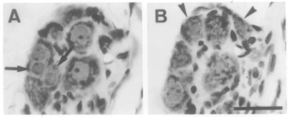
Figure 1. Photomicrograph of adjacent sections of a small intracardiac ganglion of a rat, H & E stained. Note the two neurons with round nuclei in A(arrows) are missing in B. Conversely, the nuclei of the two neurons marked with arrowheads in B are missing in A. Section A was the disector, section B the look-up section. Thus, the two neurons marked with arrows in A are the “tops”.
of heparinized saline followed by a fixative containing 1% paraformaldehyde and 1.25% glutaraldehyde in 0.1 M phosphate buffer (pH = 7.3). The heart and the great vessels and attached trachea and esophagus were excised and postfixed in the same fixatives for 1 to 2 days. These tissues were embedded in gelatin and immersed en bloc in the same fixative, with 20 % sucrose added, for an additional 3 to 5 days. Serial, 100-µm thick, frozen sections were cut on a sliding microtome. They were processed for acetylcholinestease histochem-istr y by the method of Woolf & Butcher (1981). These sections were then mounted on subbed slides. After dehydration, clearing and coverslipped, the sections were inspected under a compound microscope and photographed. Paraffin sections
Sodium pentobarbital anesthesia was administered intraperitoneally to 6 additional rats which were then perfused through the descending aorta. The perfusate consisted of phosphate buffered saline containing 0.8% sodium chloride and 0.02% potassium chlo-ride in 0.1M phosphate buffer (pH=7.3), fol-lowed by a fixative of 4% paraformaldehyde and 1.25% glutaraldehyde in the same buffer. The heart and its surrounding tissue were excised. After dehydration in an ascending
series of ethanol, the block was embedded in paraffin (mp. 60-64°C). Serial sections, 7 µm thick, were cut coronally. The sections were stained with Mayer’s Hematoxylin & Eosin (H & E). Particular attention was made to obtain sections of the rostral portion of the heart, because it is in this portion that most of the intracardiac ganglia lie. A typical series from one block was comprised of about 500 sec-tions.
A Wild M32 stereomicroscope with a cam-era lucida was used to draw the contours of sections. A Vickers M41 photoplan light microscope, equipped with CCD camera and video graphic printer (Sony UP-870MD) was used to observe and record the details of the sections. A 40× objective was used and the final magnification as calibrated against a stage micrometer was 316× on the print paper. The disector method
The disector method was used to estimate the number of the ganglion cells (Pakkenberg & Gunderson, 1989; Coggeshall, 1992; Pover et al., 1993). Equispaced sections, 3 to 9 sections apart, were chosen as the disectors. The inter-sample distances were chosen so that between 100 to 200 “tops” would be counted in a large ganglion. The sections immediately adjacent to the disectors were used as the look-up
tions. For each disector and its look-up sec-tions, all neurons were examined under a com-pound microscope and on the video printout. Only those neurons whose nuclei appeared in a disector section but not in the look-up section were counted as “tops” (figure 1). The
gan-glion area (adis) in each section was measured on a digitizer (Sigmascan, Jandel). The thick-ness of the sections (h) averaged 7 µm. The volume (Vdis) of the intracardiac ganglia within the disector was calculated as:
Vdis=adis×h
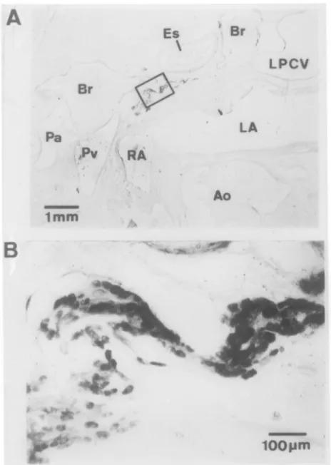
Figure 2. A. Photomicrograph of a portion of a coronal section of the rostral portion of the heart of a rat stained for acetylcholinesterase. B. Enlarged view of the boxed region in A. Note dark ganglion cells against a clear background.
Abbreviations: Ao: aorta; Br: bronchi; Es: esophagus; LA: left atrium; LV: left ventricle; LPCV:
left precaval vein; Pa: pulmonary artery; Pv: pulmonary vein; RA: right atrium; RV: right ven-tricle; SCV: superior vena cava.
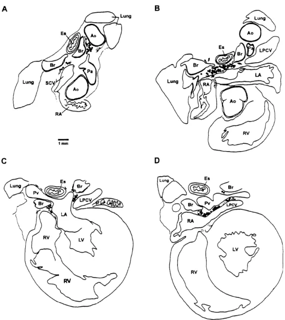
Figure 3. Distribution of ganglion cells in the heart and adjacent tissues of a rat. A, B, C and D are representative sections of the heart from rostral to caudal levels. For abbreviations see leg-end for Figure 2.
The number of neurons per mm3(N
v) is:
where Q - is the number of the tops in each area counted. The number of neurons in the
N Q V N Q a v dis v dis = × = − −
∑
∑
∑
∑
10 10 7 9 9ganglion was obtained by multiplying Nv by
total volume.
The number of neurons in small ganglia was counted manually. The total number of intracardiac ganglion cells in an individual rat was obtained by summing the number of neu-rons in all intracardiac ganglia.
interposed between the ganglion cells (Figure 1).
There were many ganglia in the heart tissue and their sizes varied greatly. Most of them were found in the fat tissues between the great vessels. Three large groups of ganglia were consistently found closely associated with the left atrium. The largest of the three groups (group 1) was located rostrally outside the junction of the left and right atria (Figure 3B). Slightly caudal, at the level where the pul-monary veins joined the left atrium, there were two groups of ganglia, one on each side of the juncture of the veins and the atrium (groups 2 and 3, Figure 3C). More rostrally, there were ganglia located between the ascending aorta and the root of the pulmonary artery, between the vena cava, pulmonary artery and bronchi, and on the left side of the pulmonary artery between the bronchi and the descending aorta (Figure 3A).
In some rats, all these ganglia were separate entities, such that there were one or two chains of ganglia extending from the great vessels to the atrial-ventricular junction. No ganglion was found in ventricular tissue. In other rats, groups of ganglia were joined here and there. For example, in some animals, group 1 joined rostrally with the rostral groups of ganglia and caudally with either group 2 or group 3 to form a large ganglion. In this case, the chain of ganglion might be Y - shaped.
Neuron size and number
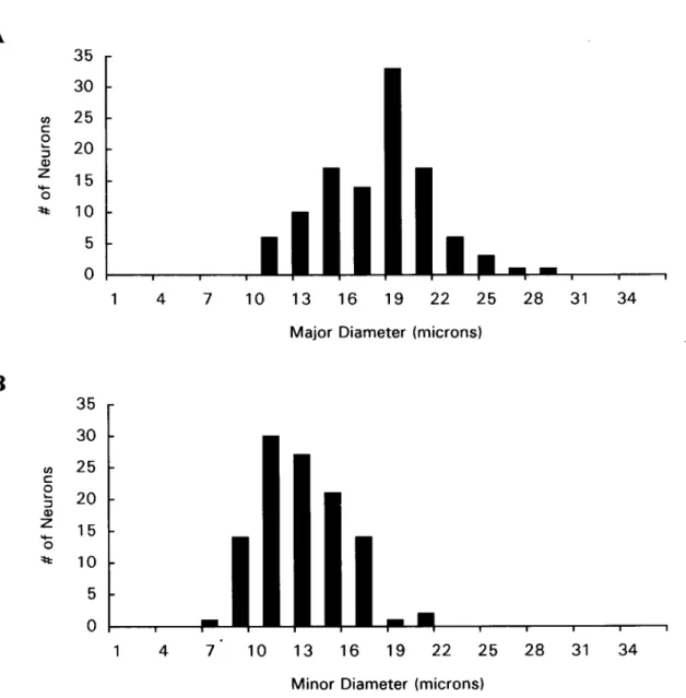
The size of 110 ganglion cells were mea-sured. The major and minor diameters were
distribution of ganglion cells in the mam-malian heart. King & Coakley (1958) summa-rized the distribution of intracardiac ganglion cells in 19 species, including the rat. More recently, this distribution has been studied by Pardini et al. (1987) and Burkholder et al. (1992). Seven clusters were found by King & Coakley (1958), 4 by Pardini et al. (1987) and Burkholder et al. (1992) found another 4. Our data agrees well with the results by Pardini et al. (1987). Our group 1 corresponds to their group in the region of the superior interatrial septum. Our group 2 corresponds to their group posterior to the inferior interatrial sep-tum and right atrium. Our group 3 corre-sponds to their group posterior to the left atri-um. Their smallest group, the one between the superior vena cava and aorta, can be also seen in our Figure 3A. Essentially, the data in all papers agree well for the larger groups of gan-glia but with differences in minor details. The variations described in the present paper may account for these minor differences. The rat has comparative smaller and less extensive intracardiac ganglia than most other mammals (King & Coakley 1958).
Our numerical data, of 3629±337 intracar-diac neurons per rat, agrees very well with the result of 3980±444 obtained by Pardini et al. (1987). Taking individual variation into account, this difference is not statistically sig-nificant. Therefore, there appears to be no dif-ference in the results obtained by the Abercrombie and disector methods in estimat-ing the number of rat intracardiac ganglion cells. It is interesting to note that the number of intracardiac ganglion cells in the cat has
Figure 4. Histograms of the size of 110 intracardiac ganglion cells measured across the major axis (A) and the minor axis (B).
REFERENCES
Abercrombie, M. (1946) Estimation of nuclear population from microtome sections. Anat. Rec. 94: 239-247.
Ardell, J. L. (1994) Structure and function of mammalian intrinsic cardiac neurons. In Neurocardiology. J. A. Armour and J. L. Ardell, eds. Oxford University Press, New York, pp. 95-114.
Burkholder, T., M. Chambers, K. Hotmire, R. D. Wurster, S. Moody and W. C. Randall (1992) Gross and microscopic anatomy of the vagal innervation of the rat heart. Anat. Rec. 232: 444-452.
Calaresu, F. R. and A. J. St. Louis (1967) Topography and numerical distribution of intracardiac ganglion cells in the cat. J. Comp. Neurol. 131: 55-66.
Acta Pathol. Microbiol. Immunol. Scand. 97: 677-681.
Pardini, B. J., K. P. Patel, P. G. Schmid and D. D. Lund (1987) Location, distribution and projections of intracardiac ganglion cells in the rat. J. Auton. Nerv. Syst. 20: 91-101. Pover, C. M., M. H. Orr Jr. and R. E.
Coggeshall (1993) A method for producing unbiased histograms of neuronal profile sizes. J. Neurosci. Meth. 49: 123-131. Woolf, N. J. and L. L. Butcher (1981)
Cholinergic neurons in the caudate-puta-men complex proper are intrinsically orga-nized: a combined Evans blue and acetyl-cholinesterase analysis. Brain Res. Bull. 7: 487-507.