Inducible Nitric Oxide Synthase and Cyclooxygenase-2 Participate in
Anti-inflammatory Activity of Imperatorin from
Glehnia littoralis
Guan-Jhong Huang,
†,+Jeng-Shyan Deng,
‡,+Jung-Chun Liao,
§Wen-Chi Hou,
∥Sheng-Yang Wang,
⊥Ping-Jyun Sung,
#,∇and Yueh-Hsiung Kuo*
,○,◆†
School of Chinese Pharmaceutical Sciences and Chinese Medicine Resources, College of Pharmacy, China Medical University,
Taichung, Taiwan
‡
Department of Health and Nutrition Biotechnology, Asia University, Taichung 413, Taiwan
§School of Pharmacy, College of Pharmacy, China Medical University, Taichung 404, Taiwan
∥Graduate Institute of Pharmacognosy, Taipei Medical University, Taipei 250, Taiwan
⊥Department of Forestry, National Chung-Hsing University, Taichung 402, Taiwan
#National Museum of Marine Biology and Aquarium, Pingtung 944, Taiwan
∇
Graduate Institute of Marine Biotechnology, National Dong Hwa University, Pingtung 944, Taiwan
○Agricultural Biotechnology Research Center, Academic Sinica, Taipei, Taiwan, Republic of China
◆
Tsuzuki Institute for Traditional Medicine, China Medical University, Taichung, Taiwan, Republic of China
ABSTRACT:
In this study, we have investigated the anti-inflammatory effects of imperatorin, a compound isolated from the
roots of Glehnia littoralis, using a lipopolysaccharide (LPS)-stimulated mouse macrophage (RAW264.7) in vitro and a
carrageenan (Carr)-induced mouse paw edema model in vivo. When RAW264.7 macrophages were treated with imperatorin
together with LPS, a significant concentration-dependent inhibition of NO production was detected. Western blotting revealed
that imperatorin blocked the protein expression of iNOS and cyclooxygenase-2 (COX-2) in LPS-stimulated RAW264.7
macrophages significantly. In the anti-inflammatory test, imperatorin decreased the paw edema at 4 and 5 h after Carr
administration and increased the activities of catalase, superoxide dismutase, and glutathione peroxidase in paw edema. We also
demonstrated that imperatorin significantly attenuated the malondialdehyde level in the edema paw at the fifth hour after Carr
injection. Imperatorin decreased the NO and tumor necrosis factor and prostaglandin E2 levels on serum at 5 h after Carr
injection. Western blotting revealed that imperatorin decreased Carr-induced iNOS and COX-2 expressions at 5 h in edema paw.
An intraperitoneal injection treatment with imperatorin also diminished neutrophil infiltration into sites of inflammation as did
indomethacin. The results suggested that imperatorin had anti-inflammatory effects in LPS-stimulated RAW 264.7 cells and
Carr-injected mice, respectively. In addition, inhibition of elevated iNOS and COX-2 protein expression as well as neutrophil
infiltration of Carr-injected paws may be involved in the beneficial effects of imperatorin.
KEYWORDS:
Chinese herb, imperatorin, anti-inflammation, NO, TNF-
α
■
INTRODUCTION
The dried roots and rhizomes of Glehnia littoralis
(Umbelli-ferae) have been used in traditional oriental medicine as
diaphoretic, antipyretic, and analgesic agents in the Taiwan.
Many researcher studies reported that G. littoralis has
anti-oxidant, antitumor, antiamnesic, blood circulation-promoting,
immunomodulatory, and antimicrobial activities.
1Quercetin,
isoquercetin, rutin, chlorogenic acid, and caffeic acid have
been isolated as the major antioxidative constituents in the
underground parts of G. littoralis.
2Imperatorin is the main
coumarin in the root of G. littoralis. Imperatorin is one of
the furanocoumarins. Furanocoumarins have biological
func-tions including antidiabetic, anticonvulsant, and vascular
vasodilation functions, increased cell differentiation in
osteo-blasts, and reduction in liver steatosis.
3Imperatorin has many
medical effects such as anticonvulsant, inflammatory,
anti-tumor, antibacterial, and anticoagulant activities.
4,5However,
little information is available on the anti-inflammatory effects of
imperatorin.
Many studies on plant-derived anti-inflammatory compounds
have investigated the potential inhibitory effects of natural
products in an in vitro system, lipopolysaccharide (LPS)-stimulated
macrophage. Using this system, bacterial LPS has become one of
the best-characterized stimuli used to induce the up-regulation
of proinflammatory proteins such as cyclooxygenase-2 (COX-2)
and inducible nitric oxide synthase (iNOS). Inducible COX-2
could be responsible for the high prostaglandins observed in
much inflammatory pathology.
6Papers also have reported that
an inflammatory effect induced by carrageenan (Carr) could be
associated with free radical formation.
7Free radical,
prosta-glandin, and nitric oxide (NO) will be released when
administer-ing with Carr for 1−5 h. The edema effect was raised to the
maximum at 3 h, and its malondialdehyde (MDA) production was
Received: October 26, 2011Revised: December 19, 2011
Accepted: December 20, 2011
Published: December 20, 2011
due to free radical attack plasma membrane.
8Thus, the
inflam-matory effect would result in the accumulation of MDA.
Therefore, in this paper, we examined the anti-inflammatory
effects of imperatorin on LPS-induced RAW264.7 cells and
Carr-induced paw edema in mice, and we detected the levels of
iNOS and COX-2 in either RAW264.7 cell or paw edema. Also,
the activities of catalase (CAT), superoxide dismutase (SOD),
and glutathione peroxidase (GPx) in the paw edema at 5 h after
Carr injection were investigated to understand the relationship
between the anti-inflammatory mechanism of the imperatorin
and the antioxidant enzymes.
■
MATERIALS AND METHODS
Chemicals. LPS (endotoxin from Escherichia coli, serotype 0127:B8), Carr (type IV), indomethacin, MTT (3-[4,5-dimethylth-iazol-2-yl]-2,5-diphenyltetrazolium bromide), and other chemicals were purchased from Sigma Chemical Co. (St. Louis, MO). Tumor necrosis factor (TNF-α) and prostaglandin E2 (PGE2) were purchased from Biosource International Inc. (Camarillo, CA). Anti-iNOS, anti-COX-2, and anti-β-actin antibody (Santa Cruz, United States) and a protein assay kit (Bio-Rad Laboratories Ltd., Watford, Herts, United Kingdom) were obtained as indicated. Poly-(vinylidene fluoride) membrane (Immobilon-P) was obtained from Millipore Corp. (Bedford, MA).
Plant Material. The root of G. littoralis was collected in Tai Chung, Taiwan. The plant material was identified by Muh-Tsuen Gun, formerly a technician of the Department of Botany, National Taiwan University. A voucher specimen has been deposited at the Herbarium of the Department of Botany, National Taiwan University, Taipei, Taiwan.
Isolation and Determination of the Active Compound. Air-dried pieces of G. littoralis root (12 kg) were extracted three times with acetone (12 L) at room temperature (7 days each time). The acetone extract was evaporated in vacuo to leave a black residue, which was suspended in H2O and then partitioned (three times) with 11 L of ethyl acetate (EtOAc). The EtOAc fraction (360 g) was chromato-graphed on silica gel using n-hexane and EtOAc of increasing polarity as an eluent and further purified by high-performance liquid chromato-graphy eluting with n-hexane:EtOAc (25:75). Imperatorin (Figure 1A) was eluted with 10% EtOAc in hexane and recrystallized with EtOH.9 Imperatorin: C16H1404, mp 97.5−99 °C. [M] + m/z 270.0897.1H NMR (CDCl3, 500 MHz): 1.69, 1.71 (each 3H, s), 4.98 (2H, d, J = 7.2 Hz), 5.59 (1H, t, J = 7.2 Hz), 6.33 (1H, d, J = 9.5 Hz, H-3), 6.79 (1H, d, J = 2.1 Hz, H-3′), 7.33 (1H, s, H-5), 7.67 (1H, d, J = 2.1 Hz, H-2′), 7.73 (1H, d, J = 9.5 Hz, H-4).13C NMR (CDC1 3, 125 MHz): 18.0 (q), 25.7 (q), 70.1 (t), 106.6 (d), 113.1 (d), 114.6 (d), 116.4 (s), 119.7 (d), 125.8 (s), 131.6 (s), 139.6 (s), 143.8 (s), 144.3 (d), 146.6 (d), 148.6 (s), 160.6 (s). UVλmax(ε) nm: 218 (22800), 249 (20600), 301 (10600). IR (KBr) cm−1: 1722, 1708 1589, 1150, 838.
Animals. Imprinting control region (ICR; 6−8 weeks, male) mice were obtained from the BioLASCO Taiwan Co., Ltd. The animals were kept in plexiglass cages at a constant temperature of 22± 1 °C and relative humidity of 55± 5% with 12 h dark−light cycle for at least 2 weeks before the experiment. They were given food and water ad libitum. All experimental procedures were performed according to the National Institutes of Health (NIH) Guide for the Care and Use of Laboratory Animals. In addition, all tests were conducted under the guidelines of the International Association for the Study of Pain.10
After a 2 week adaptation period, male ICR mice (18−25 g) were randomly assigned to four groups (n = 6) of the animals in the study. The control group received normal saline (intraperitoneal; ip). The other three groups include a Carr-treated, a positive control (Carr + Indo), and imperatorin-administered groups (Carr + imperatorin).
Cell Culture. A murine macrophage cell line RAW264.7 (BCRC no. 60001) was purchased from the Bioresources Collection and Research Center (BCRC) of the Food Industry Research and Development Institute (Hsinchu, Taiwan). Cells were cultured in plastic dishes containing Dulbecco's modified Eagle's medium (DMEM, Sigma)
supplemented with 10% fetal bovine serum (FBS, Sigma) in a CO2 incubator (5% CO2in air) at 37°C and subcultured every 3 days at a dilution of 1:5 using 0.05% trypsin−0.02% EDTA in Ca2+-, Mg2+-free phosphate-buffered saline (DPBS).
Cell Viability. Cells (2 × 10⁵) were cultured in a 96-well plate containing DMEM supplemented with 10% FBS for 1 day to become nearly confluent. Then, cells were cultured with imperatorin in the presence of 100 ng/mL LPS for 24 h. After that, the cells were washed twice with DPBS and incubated with 100μL of 0.5 mg/mL MTT for 2 h at 37°C testing for cell viability. The medium was then discarded, and 100μL of dimethyl sulfoxide (DMSO) was added. After 30 min of incubation, the absorbance at 570 nm was read using a microplate reader (Molecular Devices, Sunnyvale, CA).
Measurement of NO/Nitrite. NO production was indirectly assessed by measuring the nitrite levels in the cultured media, and serum was determined by a colorimetric method based on the Griess reaction.11 The cells were incubated with imperatorin (0, 1, 5, and 10μg/mL) in the presence of LPS (100 ng/mL) at 37 °C for 24 h. Then, cells were dispensed into 96-well plates, and 100μL of each supernatant was mixed with the same volume of Griess reagent (1% sulfanilamide, 0.1% naphthyl ethylenediamine dihydrochloride, and 5% phosphoric acid) and incubated at room temperature for 10 min, and the absorbance was measured at 540 nm with a Micro-Reader (Molecular Devices). Serum samples were diluted four times with distilled water and deproteinized by adding 1/20 volume of zinc sulfate (300 g/L) to a final concentration of 15 g/L. After centrifugation at 10000g for 5 min at room temperature, 100μL of supernatant was applied to a microtiter plate well, followed by 100 μL of Griess reagent. After 10 min of color development at room temperature, the absorbance was measured at 540 nm with a Micro-Reader. By using sodium nitrite to generate a standard curve, the concentration of nitrite was measured for absorbance at 540 nm.
Carr-Induced Edema. The Carr-induced hind paw edema model was used for determination of anti-inflammatory activity.12 Animals were ip treated with imperatorin (1, 5, and 10 mg/kg) (dissolved in 0.5% carboxymethylcellulose), Indo (10 mg/kg), or normal saline, 30 min prior to injection of 1% Carr (50μL) in the plantar side of right hind paws of the mice. The paw volume was measured after Carr injection and at 1, 2, 3, 4, and 5 h intervals after the administration of the edematogenic agent using a plethysmometer (model 7159, Ugo Basile, Varese, Italy). The degree of swelling induced was evaluated by the ratio a/b, where a was the volume of the right hind paw after Carr treatment, and b was the volume of the right hind paw before Carr treatment. Indo was used as a positive control. After 5 h, the animals were sacrificed, and the Carr-induced edema feet were dissected and stored at−80 °C. Also, blood was withdrawn and kept at −80 °C.
The right hind paw tissue was taken at 5 h. The right hind paw tissue was rinsed in ice-cold normal saline and immediately placed in cold normal saline four times their volume and homogenized at 4°C. Then, the homogenate was centrifuged at 12000g for 5 min. The supernatant was obtained and stored at−20 °C for MDA assays. The paw tissue was rinsed in ice-cold normal saline and immediately placed in cold normal saline one time their volume and homogenized at 4°C. Then, the homogenate was centrifuged at 12000g for 5 min. The supernatant was obtained and stored at −20 °C for the antioxidant enzyme (CAT, SOD, and GPx) activity assays. The protein concen-tration of the sample was determined by the Bradford dye-binding assay (Bio-Rad, Hercules, CA).
MDA Assay. MDA from Carr-induced edema foot was evaluated by the thiobarbituric acid reacting substances (TBARS) method.13 Briefly, MDA was reacted with thiobarbituric acid in the acidic medium at a high temperature and formed a red complex TBARS. The absorbance of TBARS was determined at 532 nm.
Measurement of TNF-α and PGE2 by an Enzyme-Linked Immunosorbent Assay (ELISA). The levels of TNF-α and PGE2 were determined using a commercially available ELISA kit (Biosource International Inc.) according to the manufacturer's instruction. TNF-α and PGE2were determined from a standard curve.
Determination of Antioxidant Enzyme Activity in Paw Tissue. The following biochemical parameters were analyzed to check
the protective activity of imperatorin in the paw tissues by the methods given below. The total SOD activity was determined by the inhibition of
cytochrome c reduction.14The reduction of cytochrome c was mediated by superoxide anions generated by the xanthine/xanthine oxidase system Figure 1.Chemical structure of imperatorin (A) and the effects of imperatorin on LPS-induced cell viability (B), NO production (C), TNF-α (D), and PGE2(E) in LPS-stimulated RAW264.7 cells. Cells were incubated for 24 h with 100 ng/mL of LPS in the absence or presence of imperatorin (0, 1, 5, and 10μg/mL). Imperatorin was added 1 h before incubation with LPS. The cell viability assay was performed using MTT assay. The nitrite concentration in the medium was determined using Griess reagent. TNF-α and PGE2concentrations in the medium were determined using an ELISA kit. The data were presented as means± SDs for three different experiments performed in triplicate.###p < 0.001 control group as compared to LPS-treated group.*p < 0.05, **p < 0.01, and ***p < 0.001 were compared with the LPS-alone group.
and monitored at 550 nm. One unit of SOD was defined as the amount of enzyme required to inhibit the rate of cytochrome c reduction by 50%. The total CAT activity was based on that of Aebi.15In brief, the reduction of 10 mM H2O2in 20 mM phosphate buffer (pH 7) was monitored by measuring the absorbance at 240 nm. The activity was calculated using a molar absorption coefficient, and the enzyme activities were defined as nanomoles of dissipating hydrogen peroxide per milligram protein per minute. Total GPx activity in cytosol was determined according to Paglia and Valentine's method.16The enzyme solution was added to a mixture containing hydrogen peroxide and glutathione in 0.1 mM Tris buffer (pH 7.2), and the absorbance at 340 nm was measured. The activity was calculated by using a calibration curve of GPx established from bovine whole blood. A linear relationship between the activity (unit/mL) of GPx and the reduction of NADPH absorbance at 340 nm was found, and the enzyme activities were defined as nanomoles of NADPH oxidized per milligram protein per minute.
Protein Lysate Preparation and Western Blot Analysis of iNOS and COX-2. The stimulated murine macrophage cell line RAW264.7 cells were washed with PBS and lysed in an ice-cold lysis buffer [10% glycerol, 1% Triton X-100, 1 mM Na3VO4, 1 mM EGTA, 10 mM NaF, 1 mM Na4P2O7, 20 mM Tris buffer (pH 7.9), 100 mM β-glycerophosphate, 137 mM NaCl, 5 mM EDTA, and one protease inhibitor cocktail tablet (Roche, Indianapolis, IN)] on ice for 1 h, followed by centrifugation at 12000g for 30 min at 4°C. Soft tissues were removed from individual mice paws and homogenized in a solution containing 10 mM CHAPS [3-(3-cholamidopropyl)dimethyl-ammonio]-1- propanesulfonate), 1 mM phenylmethylsulfonyl fluoride (PMSF), 5μg/mL, aprotinin, 1 μM pepstatin, and 10 μM leupeptin. The homogenates were centrifuged at 12000g for 20 min, and 30μg of protein from the supernatants was then separated on 10% sodium dodecylsulphate−polyacrylamide gel (SDS-PAGE) and transferred to polyvinylidene difluoride membranes. After transfer, the membrane was blocked for 2 h at room temperature with 5% skim milk in Tris-buffered saline-Tween (TBST; 20 mM Tris, 500 mM NaCl, pH 7.5, and 0.1% Tween 20). The membranes were then incubated with mouse monoclonal iNOS or COX-2 (1:1000 dilution) anti-body in 5% skim milk in TBST for 2 h at room temperature. The membranes were washed three times with TBST at room temperature and then incubated with a 1:2000 dilution of antimouse IgG secondary antibody conjugated to horseradish peroxidase (Sigma) in 2.5% skim milk in TBST for 1 h at room temperature. The membranes were washed three times, and the immunoreactive proteins were detected by enhanced chemiluminescence (ECL) using hyperfilm and ECL reagent (Amersham International plc., Buckinghamshire, United Kingdom). The results of Western blot analysis were quantified by measuring the relative intensity compared to the control using Kodak Molecular Imaging Software (Version 4.0.5, Eastman Kodak Company, Rochester, NY) and represented in the relative intensities.
Histological Examination. For histological examination, biopsies of paws were taken 5 h following the interplanetary injection of Carr. The tissue slices were fixed in a solution (1.85% formaldehyde, 1% acetic acid) for 1 week at room temperature, dehydrated by graded ethanol, and embedded in paraffin (Sherwood Medical). Sections (thickness 5μm) were deparaffinized with xylene and stained with hematoxylin and eosin (H&E) stain. All samples were observed and photographed with BH-2 Olympus microscopy. Every 3−5 tissue slices were randomly chosen from Carr, Indo, and imperatorin-treated (10 mg/kg) groups. Histological examination of these tissue slices revealed an excessive inflammatory response with massive infiltration of neutrophils [ploymorphonuclear leukocytes (PMNs)] by microscopy. The numbers of neutrophils were counted in each scope (400×), and thereafter, we obtained their average count from five scopes of every tissue slice.6
Statistical Analysis. Experimental results were presented as the mean± standard deviation (SD) of three parallel measurements. IC50 values were estimated using a nonlinear regression algorithm (SigmaPlot 8.0; SPSS Inc. Chicago, IL). Data obtained from animal experiments were expressed as mean standard error (±SEM). Statistical evaluation was carried out by one-way analysis of variance (ANOVA followed by Scheffe's multiple range tests). Statistical significance is expressed as*p < 0.05, **p < 0.01, and ***p < 0.001.
■
RESULTS
Cell Viability and Effect of Imperatorin on
LPS-Induced NO Production in Macrophages. The effect of
imperatorin on RAW264.7 cell viability was determined by a
MTT assay. Cells cultured with imperatorin at the
concen-trations (0, 1, 5, and 10
μg/mL) used in the presence of 100
ng/mL LPS for 24 h did not change cell viability (Figure 1B).
The RAW264.7 cell line is a mouse macrophage cell line used
to model macrophage-mediated inflammatory events in vitro,
and RAW264.7 cells were used to assess the effect of
impe-ratorin on NO synthesis. High levels of NO produced by iNOS
have been defined as a cytotoxic molecule in inflammation.
17Imperatorin did not interfere with the reaction between nitrite
and Griess reagents at 10
μg/mL (data not shown).
Unsti-mulated macrophages, after 24 h of incubation in culture
medium, produced background levels of nitrite. When
RAW264.7 macrophages were treated with different
concen-trations of imperatorin (1, 5, and 10
μg/mL) together with LPS
(100 ng/mL) for 24 h, a significant concentration-dependent
inhibition of nitrite production was detected. There was either a
significant decrease in the nitrite production of group treated
with 1
μg/mL imperatorin (p < 0.05) or a highly significant
decrease of groups treated, respectively, with 5 and 10
μg/mL of
imperatorin when compared with the LPS-alone group (p < 0.01
or p < 0.001). The IC
50value for inhibition of nitrite production of
imperatorin was about 9.59
± 0.23 μg/mL (Figure 1C).
TNF-
α mediates the production of many other cytokines during
inflammation, in particular the production of interleukin-1
β (IL-1β)
and interleukin-6 (IL-6).
18We examined the effect of
imperatorin on LPS induced up-regulation of TNF-
α. A very
low amount of TNF-
α protein was detected by a specific ELISA
for TNF-
α in controls (Figure 1D). When RAW264.7
macro-phages were treated with different concentrations of
impe-ratorin (1, 5, and 10
μg/mL) together with LPS (100 ng/mL)
for 24 h, a significant concentration-dependent inhibition of
TNF-
α production was detected. There was either a significant
decrease in the TNF-
α production of group treated with 1 μg/mL
imperatorin (p < 0.05) or a highly significant decrease of groups
treated, respectively, with 5 and 10
μg/mL of imperatorin when
compared with the LPS-alone group (p < 0.01 or p < 0.001). The
IC
50value for inhibition of TNF-
α production of imperatorin was
about 5.32
± 0.12 μg/mL (Figure 1D).
PGE
2represents the most important inflammatory product
of COX-2 activity, and it was quantified in cell-free culture
supernatant.
18As shown in Figure 1E, cells were stimulated
with LPS alone raise significant amount of PGE
2in RAW264.7
macrophages. When RAW264.7 macrophages were treated with
different concentrations of imperatorin (1, 5, and 10
μg/mL)
together with LPS (100 ng/mL) for 24 h, a significant
concentration-dependent inhibition of PGE
2production was
detected. The IC
50value for inhibition of PGE
2production of
imperatorin was about 9.46
± 0.37 μg/mL.
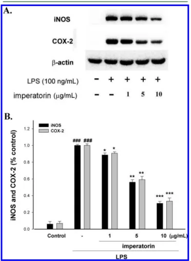
Inhibition of LPS-Induced iNOS and COX-2 Protein by
Imperatorin. To investigate whether the inhibition of NO
production was due to a decreased iNOS and COX-2 protein
level, the effect of imperatorin on iNOS and COX-2 protein
expression was studied by immunoblot. The results showed
that incubation with imperatorin (0, 1, 5, and 10
μg/mL) in the
presence of LPS (100 ng/mL) for 24 h inhibited iNOS and
COX-2 proteins expression in mouse macrophage RAW264.7
cells in a dose-dependent manner (Figure 2A). The detection of
β-actin was also performed in the same blot as an internal control.
The intensity of protein bands was analyzed using Kodak Quantity
software in three independent experiments and showed an average
of 69.2 and 66.7% down-regulation of iNOS and COX-2 proteins,
respectively, after treatment with imperatorin at 10
μg/mL as
compared with the LPS-alone (Figure 2B).
Effects of Imperatorin on Carr-Induced Mice Paw
Edema. Because imperatorin effectively inhibited iNOS and
COX-2 inductions in macrophages, studies were extended to
determine whether imperatorin affected acute phase
inflamma-tion in mice models. In this study, we used Carr-induced edema
because this model is widely employed for screening the effects
of anti-inflammatory drugs. Carr-induced paw edema is shown in
Figure 3A. Imperatorin (10 mg/kg) inhibited (p < 0.001) the
development of paw edema induced by Carr after 4 and 5 h
of treatment, significantly. Indo (10 mg/kg) significantly
decreased the Carr-induced paw edema after 4 and 5 h of
treatment (p < 0.001). On the contrary, imperatorin showed
a good dose
−response activity. Both imperatorin and Indo
show a good reduction of mice paw edema 54.4
± 0.8 and
47.9
± 0.5 inhibition percentage of volume mice paw at the
concentration of 10 mg/kg at 5 h as compared to the control,
respectively.
Effects of Imperatorin on the MDA Level in the Paw
Edema. The MDA level increased significantly in the edema
paw at 5 h after Carr injection (p < 0.001). However, the MDA
level was decreased significantly by treatment with imperatorin
(10 mg/kg) (p < 0.001) as well as 10 mg/kg Indo (Figure 3B).
Effects of Imperatorin on the NO, TNF-
α, and PGE
2Levels. In Figure 3C, the NO level increased significantly in
the edema serum at 5 h post-Carr injection (p < 0.001).
Imperatorin (10 mg/kg) significantly decreased the serum NO
level (p < 0.001). The inhibitory potency was similar to that of
Indo (10 mg/kg) at 5 h after induction. TNF-
α and PGE
2levels increased significantly in serum at 5 h post-Carr injection
(p < 0.001). However, imperatorin (5 or 10 mg/kg) decreased
the TNF-α and PGE
2levels in serum at 5 h after Carr injection
(p < 0.01 or p < 0.001) as well as 10 mg/kg Indo (Figure 3D,E).
Effects of Imperatorin on Activities of Antioxidant
Enzymes. At 5 h after the intrapaw injection of Carr, paw
tissues were also analyzed for the biochemical parameters such
as CAT, SOD, and GPx activities. CAT, SOD, and GPx activities
in paw tissue were decreased significantly by Carr administration.
CAT, SOD, and GPx activities were increased significantly
after treated with 10 mg/kg imperatorin and 10 mg/kg Indo
(P < 0.001) (Table 1).
Effects of Imperatorin on Carr-Induced iNOS and
COX-2 Protein Expressions in Mice Paw Edema. To
investigate whether the inhibition of NO production was due to
a decreased iNOS and COX-2 protein level, the effect of
imper-atorin on iNOS and COX-2 proteins expression was studied by
Western blot. The results showed that injection of imperatorin
(10 mg/kg) on Carr-induced for 5 h inhibited iNOS and COX-2
proteins expression in mouse paw edema (Figure 4A). The
intensity of protein bands was analyzed using Kodak Quantity
software in three independent experiments and showed an
average of 79.8 and 70.3% down-regulation of iNOS and COX-2
protein, respectively, after treatment with imperatorin at 10 mg/kg
as compared with the Carr-induced alone (Figure 4B). In
addition, the protein expression showed an average of 62.1 and
63.2% down-regulation of iNOS and COX-2 protein after
treatment with Indo at 10.0 mg/kg as compared with the
Carr-induced alone. The down-regulation of iNOS and COX-2
activity of the imperatorin (10 mg/kg) was better than Indo
(10.0 mg/kg).
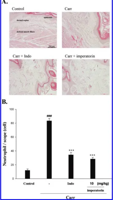
Histological Examination. Paw biopsies of Carr model
animals showed marked cellular infiltration in the connective
tissue. The infiltrates accumulated between collagen fibers and
into intercellular spaces. Paw biopsies of animals treated with
imperatorin (10 mg/kg) showed a reduction in Carr-induced
inflammatory response. Actually, inflammatory cells were
reduced in numbers and were confined to near the vascular
areas. Intercellular spaces did not show any cellular infiltrations.
Collagen fibers were regular in shape and showed a reduction
of intercellular spaces. Moreover, the hypodermal connective
tissue was not damaged (Figure 5A). Neutrophil levels were
significantly increased with Carr treatment (P < 0.01). Indo
(10.0 mg/kg) and imperatorin (10 mg/kg) could decrease
the neutrophils numbers as compared to the Carr-treated group
(P < 0.001) (Figure 5B), significantly.
Figure 2. Inhibition of iNOS and COX-2 protein expression by imperatorin in LPS-stimulated RAW264.7 cells. Cells were incubated for 24 h with 100 ng/mL of LPS in the absence or the presence of imperatorin (0, 1, 5, and 10 μg/mL). Imperatorin was added 1 h before incubation with LPS. Lysed cells were then prepared and subjected to Western blotting using an antibody specific for iNOS and COX-2. β-Actin was used as an internal control. A representative Western blot from two separated experiments is shown. Relative iNOS and COX-2 protein levels were calculated with reference to a LPS-stimulated culture. The data were presented as means± SDs for three different experiments performed in triplicate. ###p < 0.001 control group as compared to the LPS-treated group.*p < 0.05, **p < 0.01, and***p < 0.001 were compared with the LPS-alone group.
■
DISCUSSION
In the present study, we demonstrated anti-inflammatory
activities of imperatorin in in vitro and in vivo experimental
systems, using LPS-stimulated RAW264.7 macrophages and a
mouse model of topical inflammation, respectively. The
inhibitory activities against iNOS and COX-2 as shown in in
vitro assays appear to confer on imperatorin, a potent in vivo
efficacy in mouse, suggesting its potential therapeutic usage as
Figure 3.Effects of imperatorin and Indo on hind paw edema induced by Carr in mice (A), the tissue MDA concentration of foot in mice (B), Carr-induced NO (C), TNF-α (D), and PGE2(E) concentrations of serum at 5 h in mice. Each value represents a mean± SEM.###p < 0.001 as compared with the control group.*p < 0.05, **p < 0.01, and ***p < 0.001 as compared with the Carr group (one-way ANOVA followed by Scheffe's multiple range test).
an anti-inflammatory source of health food. LPS is a
prototypical endotoxin derived from Gram-negative bacterial
membrane and is the initial stimulus leading to induction of
septic shock syndrome. LPS can directly activate macrophages,
endothelial cells, and the complement-triggering production of
inflammatory mediators, such as NO, TNF-
α, interleukins, and
leukotrienes.
19However, few reports have been issued on the
anti-inflammatory effect of imperatorin and the mode of action
involved. Thus, this study aimed to evaluate the
anti-inflammatory effect of imperatorin by screening the effects of
imperatorin on LPS-induced pro-inflammatory molecules in
vitro and on acute phase inflammation in vivo.
All furanocoumarins, including byakangelicol, oxypeucedanin, and
isoimperatorin, had no effects on the inhibition of NO production.
20Table 1. Effects of Imperatorin and Indomethacin (Indo) on
Changes in CAT, SOD, and GPx Activities Were Studied on
Carr-Induced Mice Paw Edema (5 h)
agroups CAT (U/mg protein) SOD (U/mg protein) GPx (U/mg protein) control 5.28± 0.22 24.38± 0.31 23.42± 0.13 Carr 3.58± 0.31 15.64± 0.28 15.86± 0.19 Carr + Indo 4.73± 0.23** 22.53± 0.58** 21.52± 0.21** Carr + imperatorin (1 mg/kg) 3.83± 0.28* 17.23± 0.45* 16.98± 0.26 Carr + imperatorin (5 mg/kg) 4.37± 0.24** 19.82± 0.35** 18.57± 0.39* Carr + imperatorin (10 mg/kg) 4.98± 0.48*** 22.93± 0.63** 21.83± 0.24**
aEach value represents a mean± SEM. *p < 0.05, **p < 0.01, and
***p < 0.001 as compared with the Carr group (one-way ANOVA followed by Scheffe's multiple range test).
Figure 4.Inhibition of iNOS and COX-2 (A) protein expression by imperatorin induced by Carr in the foot at 5 h in mice. Suspended tissue were then prepared and subjected to Western blotting using an antibody specific for iNOS and COX-2.β-Actin was used as an internal control. A representative Western blot from two separated experi-ments is shown. Relative iNOS and COX-2 (B) protein levels were calculated with reference to a Carr-injected mouse. The data were presented as means± SDs for three different experiments performed in triplicate. ###p < 0.001 as compared with the control group. *p < 0.05, **p < 0.01, and ***p < 0.001 as compared with the Carr group (one-way ANOVA followed by Scheffe's multiple range test).
Figure 5. Representative light micrographs of mouse hind footpad H&E stained to reveal hemorrhage, edema, and inflammatory cell infiltration in (A) control mice, (B) Carr-treated mice demonstrat-ing hemorrhage with moderately extravascular red blood cells and a large amount of inflammatory leukocyte mainly neutrophils infiltration in the subdermis interstitial tissue of mice, and (C) mice given Indo (10 mg/kg) before Carr. Imperatorin (10 mg/kg) significantly shows (D) morphological alterations (100×) and (E) the numbers of neu-trophils in each scope (400×) as compared to subcutaneous injection of Carr only.###p < 0.001 control group as compared to the Carr group. ***p < 0.001 as compared with the Carr group. Scale bar = 200 μm.
Our study was performed using equally diluted fractions in
these conditions; the results indicate that imperatorin had
effects on the inhibition of NO production, and imperatorin
and phellopterin rather than byakangelicol, oxypeucedanin,
and isoimperatorin were the two major compounds responsible
for the anti-inflammatory activity in the Angelica dahurica.
21The anti-inflammatory properties of methylene chloride
fraction from G. littoralis extract (MCF-GLE) may result
from the inhibition of pro-inflammatory mediators, such as
NO, PGE
2, TNF-α, and IL-1β, via suppression of nuclear
factor-
κB (NF-κB) and mitogen-activated protein
kinase-dependent pathways.
22Excessive production of NO plays a critical role in the
aggravation of circulatory shock and chronic inflammatory
diseases, such as septic shock, inflammatory hepatic
dysfunc-tions, inflammatory lung disease, and colitis.
23As many of these
conditions exhibit rapid onset and development, often resulting
in the failure of conventional anti-inflammatory therapies and
extremely high mortality rates, a simultaneous suppression of
NO production pathways, as shown by imperatorin, may satisfy
the so far unmet need for control of the rapid progression of
the inflammatory process. In vitro models such as macrophage
cells or other cell lines are useful materials with a steady
high-level production of NO. The mechanisms by which imperatorin
inhibits macrophage functions have not been elucidated.
Results in vitro showed that imperatorin suppressed
LPS-induced production of NO (Figure 1C), the expression of
inflam-matory protein products such as iNOS and COX-2 (Figure 2A).
Examination of the cytotoxicity of imperatorin in RAW264.7
macrophages using MTT assay has indicated that imperatorin
even at 10
μg/mL did not affect the viability of RAW264.7 cells.
Therefore, inhibition of LPS-induced nitrite production by
imperatorin was not the result of a possible cytotoxic effect on
these cells.
Excess amounts of NO and PGE
2play a critical role in the
aggravation of chronic inflammatory diseases, such as hepatic
dysfunction and pulmonary disease. Recently, mounting
ev-idence that in vitro and in vivo have indicated existing cross-talk
between the release of NO and prostaglandins (PGs) in the
modulation of molecular mechanisms that regulate PGs
generat-ing pathway.
24Scientific papers observed that while the
produc-tion of both nitrite and PGE
2was blocked by the NOS inhibitors
in mouse macrophages RAW264.7 cells, these inhibitory effects
were reversed by coincubation with the precursor of NO
syn-thesis,
L-arginine. Furthermore, inhibition of iNOS activity by
nonselective NOS inhibitors attenuated the release of NO and
PGs simultaneously in LPS-activated macrophages, suggesting
that endogenously released NO from macrophages exerted a
stimulatory action on enhancing the PGs production.
Con-versely, it has been shown that COX activation in turn modulates
the
L-arginine-NO pathway, whereas COX inhibition decreases
NOS activity in human platelets.
25These results are indicative of
the cross-talk between NO and PGs pathways.
Carr-induced paw edema is a well-established model of
edema formation, which is commonly used for the screening of
anti-inflammatory drugs. The intraplantar injection of Carr
induces inflammatory responses, including increases in paw
volume (edema) and neutrophil infiltration.
26Recent studies have
shown that Carr induced peripheral release of NO as well as that
of PGE
2.
25NO plays a major role in edema formation in
inflam-matory responses and tissue injury, and Carr induced the release
of TNF-
α, which subsequently promotes IL-1 and IL-6 production
in the tissue.
27The degree of swelling of the Carr-injected paws
was maximal 3 h after injection. Statistical analysis revealed that
imperatorin and Indo significantly inhibited the development of
edema 5 h after treatment (p < 0.001) (Figure 3A). The third
phase of the edema-induced by Carr, where the edema reaches
its highest volume, is characterized by the presence of
prosta-glandins and other compounds of slow reaction.
28It was found
that the injection of Carr into the mouse paw induces the
liberation of bradykinin, which later induces the biosynthesis of
prostaglandin and other autacoids, which are responsible for the
formation of the inflammatory exudates.
29Our Carr-induced
rat paw edema model enabled us to demonstrate the ability of
imperatorin to inhibit edema induced by acute inflammation.
These results in conjunction with the marked inhibition of
LPS-induced NO and TNF-
α productions by imperatorin in
macrophages imply that the antiedema effects of imperatorin
might result from its inhibition of NO and TNF-
α syntheses in
the peripheral tissues (Figure 3C,D).
The proinflammatory cytokines such as TNF-
α and IL-1β are
small secreted proteins, which mediate and regulate immunity
and inflammation. The production of TNF-
α is crucial for the
synergistic induction of NO synthesis in interferon-γ (IFN-γ)
and/or LPS-stimulated macrophages. TNF-
α induces a number
of physiological effects including septic shock, inflammation,
and cytotoxicity.
30Also, TNF-
α is a mediator of Carr-induced
inflammatory incapacitation and is able to induce the further
release of kinins and leukotrienes, which is suggested to have an
important role in the maintenance of long-lasting nociceptive
response.
31In this study, we found that imperatorin decreased
the TNF-
α level in LPS-stimulated macrophages or after Carr
injection.
The Carr-induced inflammatory response has been linked to
neutrophils infiltration and the production of
neutrophils-derived free radicals, such as hydrogen peroxide, superoxide,
and hydroxyl radicals, as well as the release of other
neutrophils-derived mediators.
32Researchers demonstrated that the
inflam-matory effect induced by Carr is associated with free radical. Free
radical, prostaglandin, and NO will be released when
adminis-trating with Carr for 1
−6 h. MDA production is due to free
radical attack plasma membrane. Thus, the inflammatory effect
would result in the accumulation of MDA.
11Glutathione (GSH)
acts as a scavenger by scavenging NO and other oxidants. The
increased GSH level may favor reduction in MDA production.
GSH plays an important role against Carr-induced local
inflammation.
32In this study, there was significantly increased
in CAT, SOD, and GPx activities with imperatorin treatment
(Table 1). Furthermore, there were significant decreases in the
MDA level with imperatorin treatment (Figure 3B). We assume
that the suppression of MDA production is probably due to the
increases of CAT, SOD, and GPx activities.
In conclusion, these results suggested that imperatorin
pos-sessed anti-inflammatory effects. The anti-inflammatory
mech-anism of imperatorin may be related to iNOS and COX-2, and
it is associated with the increase in the activities of antioxidant
enzymes (CAT, SOD, and GPx). Imperatorin may be used as a
pharmacological agent in the prevention or treatment of disease
in which free radical formation is a pathogenic factor.
■
AUTHOR INFORMATION
Corresponding Author
*Tel: +886 4 2207-1693. Fax: +886 4 2207-1693. E-mail: kuoyh@
mail.cmu.edu.tw.
Author Contributions
+
These authors equally contributed to this work.
Funding
We are thankful for the financial support from the National
Science Council (NSC100-2313-B-039-004- and NSC
100-2320-B-039-033-), China Medical University (CMU) (CMU99-S-29,
CCM-P99-RD-042, and CMU99-COL-10), and Taiwan
Depart-ment of Heath Clinical Trial and Research Center of Excellence
(DOH101-TD-B-111-004).
■
ACKNOWLEDGMENTS
We thank Dr. Jeffrey Conrad for critically reading the
manuscript.
■
REFERENCES
(1) Ng, T. B.; Liu, F.; Wang, H. X. The antioxidant effects of aqueous and organic extracts of Panax quinquefolium, Panax notoginseng, Codonopsis pilosula, Pseudostellaria heterophylla, and Glehnia littoralis.
J. Ethnopharmacol. 2004, 93, 285−288.
(2) Yuan, Z.; Tezuka, Y.; Fan, W.; Kadota, S.; Li, X. Constituents of the underground parts of Glehnia littoralis. Chem. Pharm. Bull. 2002, 50, 73−77.
(3) Matsuura, H.; Saxena, G.; Farmer, S. W.; Hancock, R. E.; Towers, G. H. Antibacterial and antifungal polyene compounds from Glehnia littoralis ssp. leiocarpa. Planta Med. 1996, 62, 256−259.
(4) Luszczki, J. J.; Wojda, E.; Andres-Mach, M.; Cisowski, W.; Glensk, M.; Glowniak, K.; Czuczwar, S. J. Anticonvulsant and acute neurotoxic effects of imperatorin, osthole and valproate in the maximal electroshock seizure and chimney tests in mice: A comparative study. Epilepsy Res. 2009, 85, 293−299.
(5) Kwon, Y. S.; Kobayashi, A.; Kajiyama, S. I.; Kawazu, K.; Kanzaki, H.; Kim, C. M. Antimicrobial constituents of Angelica dahurica roots. Phytochemistry 1997, 44, 887−889.
(6) Huang, G. J.; Huang, S. S.; Lin, S. S.; Shao, Y. Y.; Chen, C. C.; Hou, W. C.; Kuo, Y. H. Analgesic effects and the mechanisms of anti-inflammation of ergostatrien-3β-ol from Antrodia camphorata sub-merged whole broth in mice. J. Agric. Food Chem. 2010, 58, 7445− 7452.
(7) Chang, C. T.; Huang, S. S.; Lin, S. S.; Amagaya, S.; Ho, H. Y.; Hou, W. C.; Shie, P. H.; Wu, J. B.; Huang, G. J. Anti-inflammatory activities of tormentic acid from suspension cells of Eriobotrya Japonica ex vivo and in vivo. Food Chem. 2011, 127, 1131−1137.
(8) Pferschy-Wenzig, E. M.; Kunert, O.; Presser, A.; Bauer, R. In vitro anti-inflammatory activity of larch (Larix decidua L.) sawdust. J. Agric. Food Chem. 2008, 56, 11688−11693.
(9) Takahiro, M.; Mitsuo, T.; Masaki, A. Psoralen and other linear furanocoumarins as phytoalexins in Glehnia littoralis. Phytochemistry 1998, 47, 13−16.
(10) Zimmermann, M. Ethical guidelines for investigations of experimental pain in conscious animals. Pain 1983, 16, 109−110.
(11) Chang, H. Y.; Sheu, M. J.; Yang, C. H.; Leu, Z. C.; Chang, Y. S.; Peng, W. H.; Huang, S. S.; Huang, G. J. Analgesic effects and the mechanisms of anti-inflammation of hispolon in mice. Evidence-Based Complementary Altern. Med. 2009, DOI: 10.1093/ecam/nep027.
(12) Huang, M. H.; Wang, B. S.; Chiu, C. S.; Amagaya, S.; Hsieh, W. T.; Huang, S. S.; Shie, P. H.; Huang, G. J. Antioxidant, antinociceptive, and anti-inflammatory activities of Xanthii fructus extract. J. Ethnopharmacol. 2011, 135, 545−552.
(13) Wen, C. L.; Chang, C. C.; Huang, S. S.; Kuo, C. L.; Hsu, S. L.; Deng, J. S.; Huang, G. J. Anti-inflammatory effects of methanol extract of Antrodia cinnamomea mycelia both in vitro and in vivo.
J. Ethnopharmacol. 2011, 137, 575−584.
(14) Flohe, L.; Otting, F. Superoxide dismutase assays. Methods Enzymol. 1984, 105, 93−104.
(15) Aebi, H. Catalase in vitro. Methods Enzymol. 1984, 105, 121−126.
(16) Paglia, E. D.; Valentine, W. N. Studies on the quantitative and qualitative characterization of erythrocytes glutathione peroxidase. J. Lab. Clin. Med. 1967, 70, 158−169.
(17) Pan, M. H.; Lai, C. S.; Dushenkov, S.; Ho, C. T. Modulation of inflammatory genes by natural dietary bioactive compounds. J. Agric. Food Chem. 2009, 57, 4467−4477.
(18) Lai, C. S.; Lee, J. H.; Ho, C. T.; Liu, C. B.; Wang, J. M.; Wang, Y. J.; Pan, M. H. Rosmanol potently inhibits lipopolysaccharide-Induced iNOS and COX-2 expression through downregulating MAPK, NF-κB, STAT3 and C/EBP signaling pathways. J. Agric. Food Chem. 2009, 57, 10990−10998.
(19) Hayashi, S.; Sumi, Y.; Ueno, N.; Murase, A.; Takada, J. Discovery of a novel COX-2 inhibitor as an orally potent anti-pyretic and anti-inflammatory drug: Design, synthesis, and structure-activity relationship. Biochem. Pharmacol. 2011, 82, 755−768.
(20) Kang, S. W.; Kim, C. Y.; Song, D. G.; Pan, C. H.; Cha, K. H.; Lee, D. U.; Um, B. H. Rapid identification of furanocoumarins in Angelica dahurica using the online LC-MMR-MS and their nitric oxide inhibitory activity in RAW 264.7 cells. Phytochem. Anal. 2010, 21, 322−327.
(21) Ban, H. S.; Lim, S. S.; Suzuki, K.; Jung, S. H.; Lee, S.; Lee, Y. S.; Shin, K. H.; Ohuchi, K. Inhibitory effects of furanocoumarins isolated from the roots of Angelica dahurica on prostaglandin E2 production. Planta Med. 2003, 69, 408−412.
(22) Yoon, T.; Cheon, M. S.; Lee, A. Y.; Lee, do. Y.; Moon, B. C.; Chun, J. M.; Choo, B. K.; Kim, H. K. Anti-inflammatory activity of methylene chloride fraction from Glehnia littoralis extract via suppression of NF-kappa B and mitogen-activated protein kinase activity. J. Pharmacol. Sci. 2010, 112, 46−55.
(23) Pan, M. H.; Lai, C. S.; Dushenkov, S.; Ho, C. T. Modulation of inflammatory genes by natural dietary bioactive compounds. J. Agric. Food Chem. 2009, 57, 4467−4477.
(24) Hsieh, I. N.; Chang, A. S.; Teng, C. M.; Chen, C. C.; Yang, C. R. Aciculatin inhibits lipopolysaccharide-mediated inducible nitric oxide synthase and cyclooxygenase-2 expression via suppressing NF-κB and JNK/p38 MAPK activation pathways. J. Biomed. Sci. 2011, 18, 28.
(25) Handy, R. L.; Moore, P. K. A comparison of the effects of L-NAME, 7-NI and L-NIL on carrageenan-induced hindpaw oedema and NOS activity. Br. J. Pharmacol. 1998, 123, 1119−1126.
(26) Omote, K.; Hazama, K.; Kawamata, T.; Kawamata, M.; Nakayaka, Y.; Toriyabe, M.; Namiki, A. Peripheral nitric oxide in carrageenan-induced inflammation. Brain Res. 2001, 912, 171−175.
(27) Cunha, F. Q.; Tamashiro, W. M. Tumour necrosis factor-alpha and interleukin-8 inhibit neutrophil migration in vitro and in vivo. Mediators Inflammation 1992, 1, 397−401.
(28) Tohda, C.; Nakayama, N.; Hatanaka, F.; Komatsu, K. Comparison of anti-inflammatory activities of six Curcuma rhizomes: A possible curcuminoid-independent pathway mediated by Curcuma phaeocaulis extract. Evidence-Based Complementary Altern. Med. 2006, 3, 255−260.
(29) Salvemini, D.; Wang, Z.; Bourdon, D. M.; Stern, M. K.; Curne, M. G.; Manning, P. T. Evidence of peroxynitrite involvement in the carrageenan induced rat paw edema. Eur. J. Clin. Pharmacol. 1996, 303, 217−220.
(30) Yun, K. J.; Koh, D. J.; Kim, S. H.; Park, S. J.; Ryu, J. H.; Kim, D. G.; Lee, J. Y.; Lee, K. T. Anti-inflammatory effects of sinapic acid through the suppression of inducible nitric oxide synthase, cyclooxygase-2, and proinflammatory cytokines expressions via nuclear factor-κB inactivation. J. Agric. Food Chem. 2008, 56, 10265−10272.
(31) Dawson, J.; Sedgwick, A. D.; Edwards, J. C.; Lees, P. A comparative study of the cellular, exudative and histological responses to carrageenan, dextran and zymosan in the mouse. Int. J. Tissue React. 1991, 13, 171− 185.
(32) Dawson, J.; Sedgwick, A. D.; Edwards, J. C.; Lees, P. A comparative study of the cellular, exudative and histological responses to carrageenan, dextran and zymosan in the mouse. Int. J. Tissue React. 1991, 13, 171− 185.