Article
New and Cytotoxic Components from Antrodia camphorata
Tzong-Huei Lee 1, Chien-Chih Chen 2,3, Jih-Jung Chen 4, Hui-Fen Liao 5,†, Hsun-Shuo Chang 6,7,
Ping-Jyun Sung 8,9,10, Mei-Hwei Tseng 11, Sheng-Yang Wang 12, Horng-Huey Ko 13,† and
Yueh-Hsiung Kuo 14,15,*
1 Institute of Fisheries Science, National Taiwan University, Taipei 106, Taiwan;
E-Mail: [email protected]
2 Department of Nursing, Hungkuang University, Taichung 443, Taiwan;
E-Mail: [email protected]
3 Department of Biotechnology, Hungkuang University, Taichung 443, Taiwan 4 Department of Pharmacy, Tajen University, Pingtung 907, Taiwan;
E-Mail: [email protected]
5 Department of Biochemical Science and Technology, National Chiayi University, Chiayi 600,
Taiwan; E-Mail: [email protected]
6 School of Pharmacy, College of Pharmacy, Kaohsiung Medical University, Kaohsiung 807,
Taiwan; E-Mail: [email protected]
7 Graduate Institute of Natural Products, College of Pharmacy, Kaohsiung Medical University,
Kaohsiung 807, Taiwan
8 Graduate Institute of Marine Biotechnology, National Dong Hwa University, Pingtung 944,
Taiwan; E-Mail: [email protected]
9 Department of Life Science and Institute of Biotechnology, National Dong Hwa University,
Pingtung 944, Taiwan
10 National Museum of Marine Biology and Aquarium, Pingtung 944, Taiwan
11 Department of Applied Physics and Chemistry, Taipei Municipal University of Education,
Taipei 100, Taiwan; E-Mail: [email protected]
12 Department of Forestry, National Chung-Hsing University, Taichung 402, Taiwan;
E-Mail: [email protected]
13 Department of Fragrance and Cosmetic Science, College of Pharmacy,
Kaohsiung Medical University, Kaohsiung 807, Taiwan; E-Mail: [email protected]
14 Department of Chinese Pharmaceutical Sciences and Chinese Medicine Resources,
China Medical University, Taichung 404, Taiwan
15 Department of Biotechnology, Asia University, Taichung 413, Taiwan † These authors contributed equally to this work.
* Author to whom correspondence should be addressed; E-Mail: [email protected];
Tel.: +886-4-2205-3366 (ext. 5701); Fax: +886-4-2207-1693.
Abstract: The solid-state cultured products of Antrodia camphorata as health foods has
been blooming for the past few decades in Taiwan. In continuing our studies on the chemical constituents of the solid-state cultured products of this fungus, 6-methoxy-4-methyl-2,3-(methylenedioxy)phenol (1) and 4,4'-(ethane-1,2-diyl)bis(2,3,6-trimethoxyphenol)(2) together with 2,3,6-trimethoxy-4-methylphenol (3), 1(10→6)abeo-ergosta-5,7,9,22-tetraen-3α-ol (4), citreoanthrasteroid B (5) and dankasterones A (6) and B (7) were purified by a series of column chromatography. Their structures were elucidated by spectral data analysis. For bioactivity assay, compounds 4–7 showed significant cytotoxicity toward murine colorectal CT26 and human leukemia K562 cancer cell lines with IC50 values ranging from 6.7 to
15.3 µM and from 12.5 to 23.1 µM, respectively.
Keywords: Antrodia camphorata; phenylmethanoid; dihydrostilbene; abeo-ergostane;
cytotoxicity
1. Introduction
Antrodia camphorata Wu, Ryvarden & Chang (Polyporaceae), a fungus indigenous to Taiwan, was
used by the aborigine as a hepatinica and anti-alcoholic agent initially and has been gradually used as a folk remedy for the treatment of liver cancer and various cardiovascular diseases in the past few
decades [1]. The fermentation and development of this fungus have already become one of the major components of the biotechnology industry in Taiwan recently, and some of the chemical entities isolated from the fermented products, e.g., antroquinonol [2] and ergosta-7,9,22E-trien-3β-ol [3], were selected as leads for new drug development. Although over one hundred compounds have been identified from this fungus so far [4–6], new chemical entities are still reported continually by virtue of varied culturing conditions for this fungus. In continuing our investigations on the chemical constituents of the mycelium of Antrodia camphorata, one new phenylmethanoid, 6-methoxy-4-methyl-2,3-(methylenedioxy)phenol (1), and one new stilbene, 4,4'-(ethane-1,2-diyl)bis(2,3,6-trimethoxyphenol)(2), together with
2,3,6-trimethoxy-4-methylphenol (3), 1(10→6)abeo-ergosta-5,7,9,22-tetraen-3α-ol (4),
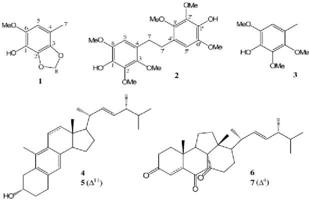
citreoanthrasteroid B (5) and dankasterones A (6) and B (7) (Figure 1) were isolated and characterized on the basis of the spectral analysis. The paper describes the isolation and identification of compounds
1–7 from A. camphorata along with their anticancer effects in murine colorectal cancer CT26 cells and
Figure 1. Chemical structures of 1–7 isolated in this study.
2. Results and Discussion
From the methanolic extracts of the solid-state cultured, fermented dried powder of A. camphorata, seven major compounds, including one new phenylmethanoid, 1, and one new stilbene, 2, along with a known phenylmethanoid, 3, and four steroids, 4–7, were isolated by sequential separation on Si-gravity column and normal-phase HPLC. Compound 3 was obtained as an amorphous white solid whose
1 H, 13C-NMR, IR, optical rotation and MS were consistent with those of synthetic
2,3,6-trimethoxy-4-methylphenol [7], and this is the first time Compound 3 has been isolated from a natural resource. Compounds 4 and 5, two rare 1(10→6)abeo-ergostane-type steroids, were identified as 1(10→6)abeo-ergosta-5,7,9,22-tetraen-3α-ol (4), obtained previously from the stromata of
Epichloe typhina [8], and citreoanthrasteroid B (5), isolated from a hybrid bacterial strain, KO 0231 [9].
Compounds 6 and 7, two uncommon 13(14→8)abeo-ergostane-type steroids, were characterized as respective dankasterones A and B, which were isolated previously from a Halichondria sponge-derived fungus, Gymnascella dankaliensis [10].
Compound 1 was afforded as a colorless amorphous solid with the molecular formula of C9H10O4 as
established through the analysis of its 13C-NMR and HRESIMS data. The IR absorption peaks of 1 at
3277, 1626 and 1527 cm−1 indicated that 1 contained a benzene moiety bearing a hydroxy functionality
as reflected in its 1H- and 13C-NMR data. The 1H-NMR of 1 exhibited a phenyl proton at δH 6.27 (s, 1H),
a benzene-borne methyl at δH 2.16 (s, 3H), a benzene-borne methoxyl at δH 3.82 (s, 3H) and
a methylenedioxy group at δH 5.91 (s, 2H), which were further confirmed by their corresponding six phenyl
resonances at δC 109.0 (d), 118.9 (s), 131.9 (s), 133.6 (s), 135.0 (s) and 136.8 (s), one methyl resonance
at δC 15.8 (q), one methoxyl resonance at δC 57.2 (q) and one dioxygenated methylene resonance at
δC 101.5 (t) in the 13C-NMR of 1. Further analysis of the 2D NMR data of 1, key cross-peaks of
δH 5.91 (H2-8)/δC 131.9 (C-3) and 135.0 (C-2), δH 6.27 (H-5)/δC 133.6 (C-1) in the HMBC spectrum and
mutual-correlated cross peaks of δH 6.27 (H-5)/δH 3.82 (OMe-6) and δH 2.16 (H3-7) in the NOESY
spectrum (Figure 2) established the locations of all of the functional groups attached to the benzene ring. Thus, 1 was deduced as the shown phenylmethanoid and was named
6-methoxy-4-methyl-2,3-(methylenedioxy)phenol.
Figure 2. Key HMBC and NOESY of 1.
Compound 2, obtained as a colorless amorphous solid, was a symmetrical chemical entity as judged from its molecular formula, C20H26O8, deduced from HRESIMS and only ten resonances in the
13 C-NMR spectrum. The IR spectrum of 2 confirmed the presence of a hydroxyl group (3410 cm−1) and
a benzene ring (1610 and 1503 cm−1). The 13C-NMR along with the DEPT of 2 displayed only ten signals
(indeed, twenty signals due to symmetry), including one methylene carbon at δC 31.4, three benzene-borne
methoxyl carbons at δC 56.4, 60.7 and 61.0 and six phenyl carbons at δC 107.2, 125.2, 137.3, 140.4,
143.2 and 145.2. The 1H-NMR spectrum showed signals for the methylene group at δH 2.78 (s, 2H),
three methoxyl groups at δH 3.79 (s, 3H), 3.80 (s, 3H) and 3.91 (s, 3H), one hydroxyl group at δH 5.45
(brs, 1H) and one phenyl proton at δH 6.38 (s, 1H). The above assignments indicated that 2 had a stilbene
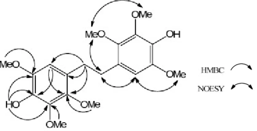
skeleton with three methoxyl and one hydroxyl groups on each benzene ring. The locations of three methoxy, one hydroxy and one phenyl proton were further corroborated by key HMBC interpretations, including δH 2.78 (H-7, -7')/δC 107.2 (C-5, -5'), 125.2 (C-4, -4') and 145.2 (C-3, -3'), δH 3.79
(OMe-3, -3')/δC 145.2 (C-3, -3'), δH 3.80 (OMe-6, -6')/δC 143.2 (C-6, -6'), δH 3.91 (OMe-2, -2')/δC 140.4
(C-2, -2'), δH 5.45 (OH-1, -1')/δC 137.3 (C-1, -1'), 140.3 (C-2, -2'), 143.2 (C-6, -6') and δH 6.38
(H-5, -5')/δC 137.3 (C-1, -1') and 145.2 (C-3, -3'), as well as key NOESY correlations, including δH 6.38
(H-5, -5')/δH 2.78 (H-7, -7') and 3.80 (OMe-6, -6') and δH 3.79 (OMe-3, -3')/δH 2.78 (H-7, -7') and 3.91
(OMe-2, -2') (Figure 3). Accordingly, 1 was determined as the shown dihydrostilbene and was named 4,4′-(ethane-1,2-diyl)bis(2,3,6-trimethoxyphenol). Compound 2 was speculated from the oxidative coupling between two molecules of Compound 3.
For the anticancer activity, Table 1 shows the IC50 values of compounds 1‒7 against murine colorectal
cancer CT26 cells and human leukemia K562 cells. Compounds 1‒3 exhibited no obvious effect toward CT26 and K562 cells with their IC50 values higher than 20 μM, and 4–7 showed significant cytotoxicity
toward murine colorectal CT26 and human leukemia K562 cancer cell lines with IC50 values ranging
from 6.7 to 18.2 μM and from 12.5 to 23.1 μM, respectively. At the same condition, the IC50 value of
Figure 3. Key HMBC and NOESY of 2.
Table 1. IC50 values of compounds 1–7 against colorectal cancer CT26 and leukemia
K562 cells. Compounds IC50 (μM) CT26 K562 1 >20 >20 2 >20 >20 3 >20 >20 4 15.3 19.9 5 18.2 12.5 6 6.7 >20 7 8.4 23.1
The IC50 value of staurosporine, the positive control, against K562 cells was 16.7 nM.
3. Experimental
3.1. General
Optical rotations were measured on a JASCO DIP-1000 polarimeter (Tokyo, Japan). 1H and 13C-NMR
were acquired on a Bruker DMX-500 (Ettlingen, Germany). Low resolution and high resolution mass spectra were obtained using an API4000 triple quadrupole mass spectrometer (Applied Biosystems, Foster City, CA, USA) and a Synapt High Definition Mass Spectrometry system with an ESI interface and a TOF analyzer (Waters Corp., Manchester, UK), respectively. IR spectra were recorded on a JASCO FT/IR 4100 spectrometer (Tokyo, Japan). TLC was performed using silica gel 60 F254 plates
(200 µm, Merck, Taipei, Taiwan).
3.2. Fungal Material
Freeze-dried powder of Antrodia camphorata was provided by Kang Jian Biotech Corp. Ltd, Nantan, Taiwan R.O.C.
3.3. Extraction and Isolation
The dried powder (2.83 kg) of solid-state cultured Antrodia camphorata was extracted with 12 L methanol for three times, and the concentrated residues (323.8 g) were suspended in H2O and further
partitioned three times with equal volumes of ethyl acetate, then concentrated in vacuum to dryness (131.5 g). The ethyl acetate extract was applied onto an open column with silica gel. The column was eluted with mixtures of n-hexane, ethyl acetate and methanol, and each 1 L was collected as one fraction. Fractions 16–33 were combined and evaporated to dryness (1.4 g), which were further purified by HPLC on a semi-preparative Phenomenex Luna Si column (5 µm, 10 × 250 mm) with n-hexane–ethyl acetate (90:10, v/v) as the eluent, 2 mL/min, obtaining 1 (4.6 mg), 2 (6.4 mg) and 3 (8.9 mg). Fractions 92–124 were combined and evaporated to dryness (33.1 g), which were further purified by HPLC on the same column with n-hexane–ethyl acetate (50:50, v/v) as the eluent, 2 mL/min, obtaining 4 (5.6 mg), 5 (7.4 mg), 6 (10.3) and 7 (9.2 mg).
6-Methoxy-4-methyl-2,3-(methylenedioxy)phenol (1): Colorless amorphous solid; IR (neat): νmax 3277,
3015, 1626, 1527, 1440, 1341, 1208, 1129, 1043, 925 cm−1. 1H-NMR (CDCl3, 400 MHz): δH 2.16 (3H,
s, H-7), 3.82 (3H, s, OMe-6), 5.91 (2H, s, H-8), 6.27 (1H, s, H-6). 13C-NMR (CDCl3, 100 MHz): δC 15.8
(C-7), 57.2 (Ome-6), 101.5 (C-8), 109.0 (C-5), 118.9 (C-4), 131.9 (C-3), 133.6 (C-1), 135.0 (C-2), 136.8 (C-6). ESIMS: m/z = 183 [M+H]+. HRESIMS: m/z = 183.0653 [M+H]+ (calcd. for C9H11O4, 183.0657).
4,4'-(Ethane-1,2-diyl)bis(2,3,6-trimethoxyphenol) (2): Colorless amorphous solid; IR (neat): νmax 3410,
3012, 2956, 2860, 1610, 1503, 1420, 1325, 1248, 1198, 1125, 1076, 970, 880. 1H-NMR (CDCl3, 400 MHz):
δH 2.78 (4H, s, H-7, -7'), 3.79 (6H, s, OMe-3, -3'), 3.80 (6H, s, OMe-6, -6'), 3.91 (6H, s, OMe-2, -2'),
5.45 (2H, brs, OH-1, -1'), 6.38 (2H, s, H-5, -5'). 13C-NMR (CDCl3, 100 MHz): δC 31.4 (C-7, -7'), 56.4
(OMe-3, -3'), 60.7 (OMe-6, -6'), 61.0 (OMe-2, -2'), 107.2 (C-5, -5'), 125.2 (C-4, -4'), 137.3 (C-1, -1'), 140.4 (C-2, -2'), 143.2 (C-6, -6'), 145.2 (C-3, -3'). ESIMS: m/z = 395 [M+H]+. HRESIMS: m/z = 395.1710
[M+H]+ (calcd. for C20H27O8, 395.1706).
3.4. Cells and Viability Assay
Murine colorectal cancer CT26 cells were cultured in RPMI1640 medium (Gibco, Grand Island, NY, USA) supplemented with 10% heat-inactivated fetal bovine serum (FBS; Hyclone, Logan, UT) and 2 mM L-glutamine at 37 °C in a humidified 5% CO2 incubator. The viability of CT26 cells was
determined by using the 3-(4,5-dimethylthiazol-2-yl)-2,5-diphenyltetrazolium bromide (MTT, Sigma) colorimetric assay. Human chronic myeloid leukemia K562 cells were cultured in RPMI 1640 medium, 10% FBS and 2 mM L-glutamine at 37 °C in an incubator. The viability of K562 cells with compound treatments for one day were measured using the Trypan blue dye exclusion test. The IC50 values of
compounds in CT26 and K562 cells were determined by using SigmaStat software (Jandel Scientific, San Rafael, CA, USA).
4. Conclusions
In this study, three C6-C1 derivatives, as well as four phytosteroids with rearranged skeletons were
isolated from the industrial fermented products of Antrodia camphorata. Of the compounds isolated, four phytosteroids exhibited significant cytotoxicity against cancer cell lines, which may, to some extent, account for the traditional uses of this fungal product as a health food to treat cancer. More experiments should be performed to deduce the action modes of these compounds.
Acknowledgments
The work was supported by the grant from CMU under the Aim for Top University Plan of the Ministry of Education, Taiwan, and Taiwan Ministry of Health and Welfare Clinical Trial and Research Center of Excellence (MOHW103-TDU-B-212-113002).
Author Contributions
Hui-Fen Liao, Yueh-Hsiung Kuo, and Horng-Huey Ko designed research; Tzong-Huei Lee, Chien-Chih Chen, and Hsun-Shuo Chang performed research; Jih-Jung Chen, Ping-Jyun Sung,
Mei-Hwei Tseng, and Sheng-Yang Wang contributed new analytical tools and reagents; Tzong-Huei Lee and Yueh-Hsiung Kuo wrote the paper.
Conflicts of Interest
The authors declare no conflict of interest.
References
1. Yang, S.S.; Wang, G.J.; Wang, S.Y.; Lin, Y.Y.; Kuo, Y.H.; Lee, T.H. New constituents with iNOS inhibitory activity from mycelium of Antrodia camphorata. Planta Med. 2009, 75, 512–516. 2. Lee, T.H.; Lee, C.K.; Tsou, W.L.; Liu, S.Y.; Kuo, M.T.; Wen, W.C. A new cytotoxic agent from
solid-state fermented mycelium of Antrodia camphorata. Planta Med. 2007, 73, 1412–1415.
3. Huang, G.J.; Huang, S.S.; Lin, S.S.; Shao, Y.Y.; Chen, C.C.; Hou, W.C.; Kuo, Y.H. Analgesic effects and the mechanisms of anti-inflammation of ergostatrien-3beta-ol from Antrodia camphorata submerged whole broth in mice. J. Agric. Food Chem. 2010, 58, 7445–7452.
4. Huang, Y.; Lin, X.; Qiao, X.; Ji, S.; Liu, K.; Yeh, C.; Tzeng, Y.; Guo, D.; Ye, M. Antcamphin A–L, ergostanoids from Antrodia camphorata. J. Nat. Prod. 2014, 77, 118–124.
5. Lu, M.C.; El-Shazly, M.; Wu, T.Y.; Du, Y.C.; Chang, T.T.; Chen, C.F.; Hsu, Y.M.; Lai, K.H.; Chiu, C.P.; Chang, F.R.; et al. Recent research and development of Antrodia cinnamomea.
Pharmacol. Ther. 2013, 139, 124–156.
6. Geethangili, M.; Tzeng, Y.M. Review of pharmacological effects of Antrodia camphorata and its bioactive compounds. Evid. Based Complement. Altern. Med. 2011, 2011, 212641:1–212641:17. 7. Foti, M.C.; Daquino, C.; Mackie, I.D.; DiLabio, G.A.; Ingold, K.U. Reactions of phenols with the
2,2-diphenyl-1-picrylhydrazyl radical. Kinetics and DFT calculations applied to determine ArO-H bond dissociation enthalpies and reaction methanism. J. Org. Chem. 2008, 73, 9270–9282.
8. Koshino, H.; Yoshihara, T.; Sakamura, S.; Shimanuki, T.; Sato, T.; Tajimi, A. A ring B aromatic sterol from stromata of Epichloe typhina. Phytochemistry 1989, 28, 771–772.
9. Nakda, T.; Yamamura, S. Three new metabolites of hybrid strain KO 0231, derived from
10. Amagata, T.; Tanaka, M.; Yamada, T.; Doi, M.; Minoura, K.; Ohishi, H.; Yamori, T.; Numata, A. Variation in cytostatic constituents of a sponge-derived Gymnascella dankaliensis by Manipulating the carbon source. J. Nat. Prod. 2007, 70, 1731–1740.