Six Genera of Physaraceae (Myxomycetes) in Taiwan
Chin-Hui Liu (1*) and Jong-How Chang(1)
1. Institute of Plant Science, National Taiwan University, No. 1, Sec. 4, Roosevelt Road, Taipei 106, Taiwan. * Corresponding author. Email: huil4951@ntu.edu.tw
(Manuscript received 12 March 2011; accepted 9 April 2012)
ABSTRACT: Species of six genera Badhamia, Craterium, Fuligo, Leocarpus, Physarella, and Willkommlangea (Physaraceae) reported from Taiwan are critically revised. Two new records, Craterium concinnum and Leocarpus fragilis and an unknown species of Craterium are described and illustrated in this paper. Keys to the species of Badhamia, Craterium, and Fuligo, and a key to the genera of Physaraceae from Taiwan are also provided.
KEY WORDS: Myxomycetes, Physaraceae, Taiwan, taxonomy.
INTRODUCTION
The fruiting bodies of all members in Physaraceae are often limy, with non-crystalline lime granules and dark-colored spore mass. Their capillitia are typically composed of calcareous nodes connected by slender and hyaline threads (physaroid), or of calcareous tubes and thickened nodes (badhamioid). In Taiwan, 7 genera out of the 10 world records are known. The distinct characteristics separating the genera from each other are shown in the key to the genera provided. In this paper we compile data of six genera: Badhamia, Craterium,
Fuligo, Leocarpus, Physarella, and Willkommlangea,
leaving the largest genus Physarum to a separate paper. Characteristic examination for the fruiting bodies of these specimens were made by light and scanning electron microscopy as described previously (Liu et al., 2002a).
TAXONOMIC TREATMENTS
Key to genera of Physaraceae in Taiwan1. Capillitium of two morphologically distinct systems ..…..…….… 2 1. Capillitium essentially homogenous ………...….……...… 4 2. Fruiting body plasmodiocarpous, peridium opaque, encrusted with
red spots and white lime granules on the surface ………....………... Willkommlangea 2. Fruiting body sporangiate, if plasmodiocarpous, then usually
accompanied by sporangia …………...…… 3 3. Sporangia ovate; outer peridium yellowish brown, cartilaginous,
smooth, shinning; capillitium a limy network, connected with and interpenetrating a limeless net of flatted tubules ……… Leocarpus 3. Sporangia cylindrical, deeply perforated from above, appearing as a
hollow cup, rarely plasmodiocarpous; peridium rough; capillitium composed of stout calcareous spines and a net work of slender threads bearing a few calcareous nodes …...…………... Physarella 4. Capillitium a network of calcareous tubes of nearly uniform
diameter; limeless connecting tubules few or none ………...………..… Badhamia
4. Capillitium a network of limeless tubules with connected calcareous nodes at many or all the junctions ………. 5 5. Fruiting body an aethalium; pseudocapillitium present …… Fuligo 5. Fruiting body sporangiate or plasmodiocarpous, rarely approaching
aethalioid; pseudocapillitium lacking …...…... 6 6. Fruiting body plasmodiocarpous, cylindrical, pendent, often
anastomosing to form a 3-dimensional network ……….. ..……… Fuligo aurea (= Erionema) 6. Fruiting body sporangiate or plasmodiocarpous, rarely pendent;
never forming a 3-dimensional network in the plasmodiocarpous fruiting body ...… 7 7. Fruiting body sporangiate; dehiscence often circumscissile or by a
preformed lid, the lower portion always persisting as a deep cup ………..………. Craterium 7. Sporangiate or plasmodiocarpous, rarely somewhat aethalioid;
dehiscence irregular or lobate, never circumscissile; the lower portion of peridium persisting as at most as a shallow or irregular cup ……….………..……… Physarum
Key to species of Badhamia inTaiwan
1. Spores in clusters; peridium double; sporangia usually yellow, greenish yellow, or dull yellow, rarely iridescent ……..… B. nitens 1. Spores free; peridium single; sporangia white, grayish or pale
gray ………..2 2. Usually stalked ………..……….….……...….….3 2. Usually sessile or shortly stipitate ………..………...……. 4 3. Spores angular in profile, with large reticulum, 1~6 in a hemisphere
on the surface; stalk pale straw-colored, weak ……...…. B. gracilis 3. Spores not angular in profile, without large reticulum on the
surface; stalk white, limy throughout ………... B. formosana 4. Spores minutely punctate, usually ovoid; stalk, if present,
red ……….... B. panicea 4. Spores densely spinulose or warted, usually globose; stalk, when
present, yellowish brown or nearly black ….………..…… 5 5. Capillitium radiating from the base to the periphery of sporangia;
stalk, when present, dark, nearly black ………..… B. affinis 5. Capillitium reticulate; stalk, when present, yellowish or brown,
only dark at the base ………... B. macrocarpa
Badhamia affinis Rostaf., Sluzowce monogr. 143. 1874.
It was reported as a new record without any description and illustration (Wang et al., 1981).
Taiwania Vol. 57, No. 3
As pointed out in the reference (Nennenga-Bremekamp, 1991), it is difficult to distinguish this species from B. macrocarpa. The capillitium is radiated from the base of the sporangium in
B. affinis, not reticulate as that in B. macrocarpa, and is
rarely branched and not or hardly interconnected.
Badhamia formosana C.H. Liu and Y.F. Chen,
Taiwania 47: 291. 2002.
Description and illustration: Liu et al. (2002b).
Badhamia gracilis (T. Macbr.) T. Macbr., in T. Macbr.
& G. W. Martin, The Myxomycetes (New York): 35. 1934.
Description and illustration: Liu (1990).
Badhamia macrocarpa (Ces.) Rostaf., Sluzowce
monogr. 143. 1874.
It was reported in a list by Nakazawa (1929), but no specimen was deposited in Taiwan.
Badhamia nitens Berk., Trans. Linn. Soc. London 21:
153. 1853.
It was reported in a list by Nakazawa (1929), but no specimen was deposited in Taiwan.
Badhamia panicea (Fr.) Rostaf., in Fuckel, Jahrb.
Nassauischen Vereins Naturk. 27-28: 71. 1873 It was reported as a new record without any description and illustration by Wang et al. (1981). A species very close to B. macrocarpa, but it has smoother and usually ovoid spores (Nennenga-Bremekamp, 1991).
Key to species of Craterium in Taiwan
1. Fructification sessile or rarely short-stalked ………...… 2 1. Fructification stalked ………... 3 2. Sporangia reddish brown ……….. C. reticulatum 2. Sporangia white, or whitish ...…… C. leucocephaleum var. sessile 3. Sporangia not bright yellow (if globose, then white or grayish white), obconical or deep cup-like, with a preformed lid of dehiscence ………4 3. Sporangia bright yellow ……….. 6 4. Sporangia turbinate or funnel-shaped, brownish; peridium double, thick ……… 5 4. Sporangia deep cup-like or globose, white, grayish; peridium single ……….…….C. leucocephaleum 5. Lime nodes small, ochraceous or brownish ….……. C. concinnum 5. Lime nodes large, white ……….…….…. C. minutum 6. Sporangia oboviod or turbinate, lime node yellow, dehiscence often irregular from the top ………...…….. C. aureum 6. Sporangia globose or prolate, lime node white, dehiscence petaloid at the upper part ……….… Craterium sp.
Craterium aureum (Schumach.) Rostaf., Sluzowce
monogr. 124. 1874.
Specimens examined: TAIWAN, Taipei City: Yungmin Lake, on fallen leaves, CHL B1414, Mar. 27, 1997; CHL B1415, Apr. 1, 1998; Peitou, Yangmingshan National Park, on fallen leaves of
Liquidambar formosana, CHL B2309, Aug. 24, 2001.
It was reported as a new record without any description and illustration (Wang et al., 1981). There are some differences in our specimens from the typical
C. aureum. The pseudocolumella is not observed and our
spores are smaller (7.0-8.5 μm), which are 8-10 μm in diameter in the references (Martin and Alexopoulos, 1969; Nannenga-Bremekamp, 1991).
Craterium concinnum Rex, Proc. Acad. Nat. Sci. Phila.
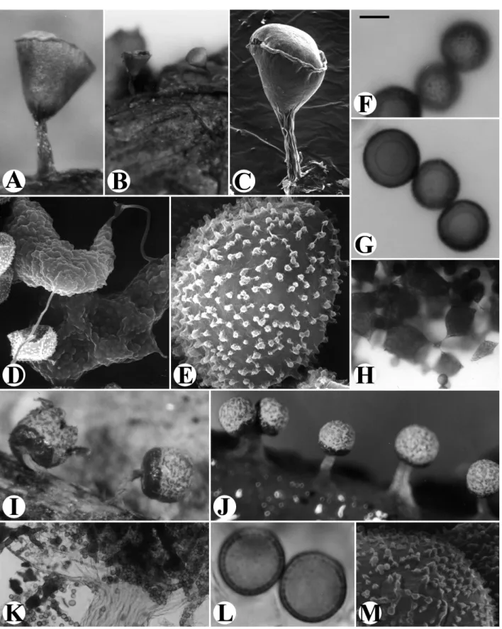
45: 370. 1893. Figs. 1A-H Fructification sporangiate, loosely gregarious, (0.15-) 0.38-0.68 mm in total height. Sporangia obovate or goblet-shaped, pale reddish brown to brown, stipitate, 0.12-0.38 mm in diameter, with a paler and convex operculum. Peridium double, the outer layer cartilaginous, thick, closely attached to the membranous inner layer; dehiscent along the rim of the lid as a whole. Stalk about half the total height or often a little shorter, brownish, furrowed. Capillitium abundant, consisted of small, rounded or angular, brownish lime nodes connected by short and transparent threads. Spores nearly black in mass, dusky brown by transmitted light, globose, 8.5-10.5 μm in diameter, minutely and densely warted.
Specimen examined: TAIWAN, Taipei City: Peitou, Yangmingshan National Park, on decayed twigs and leaves, Y.F.
Chen504, Aug. 7, 1995.
Distinguished by its small fruiting bodies, the pale and convex operculum, and the small and brownish lime nodes, which separate it from C. minutum. In C. minutum the operculum is often sunken, the lime nodes are white and larger.
Craterium leucocephalum (Pers. ex J.F. Gmel.) Ditmar,
in Sturm, Deutschl. Fl. Pilze 1: 21. 1813.
Craterium leucocephaleum var. scyphoides (Cooke & Balf.) G.
Lister, in List, Mycet. ed. 2. 97. 1911.
Description and illustration: Shi (1981).
Specimens examined: TAIWAN, Taipei City: main campus of National Taiwan Univ., on straw, CHL B357, Apr. 3, 1984; Peitou, Yangmingshan National Park, on twigs, CHL B2246, Nov. 30, 2000.
This is a common, distinctive species and easy to be recognized in the field by a hand lens. The distinctive character is the stalked, cylindrical, white sporangium with an operculum-like apex.
Fig. 1. A-H. Craterium concinnum. A-B: Fruiting bodies. C: One fruiting body, by SEM. D: Capillitial threads and lime nodes, by SEM. E: Surface markings of spore, by SEM. F: Surface view of spores. G: Marginal view of spores. H: Capillitial threads and lime nodes. I-M. Craterium sp. I-J: Fruiting bodies. K: Limeless basal part of peridium. L: Marginal view of spores. M: Surface markings of spore, by SEM. Scale bar: A = 130 μm; B, J = 320 μm; C = 91 μm; D = 3.8 μm; E = 0.95 μm; F, G, L = 5 μm; H = 25 μm; I = 210 μm; K = 70 μm; M = 0.84 μm.
Taiwania Vol. 57, No. 3 Craterium leucocephalum var. sessile C.H. Liu, I.G
Huang & J.H. Chang, Taiwania 46: 326. 2001. Description and illustration: Liu et al. (2001).
Craterium minutum (Leers) Fr., Syst. Mycol. 3: 151.
1829.
Description and illustration: Chung and Liu (1997b).
Specimens examined: TAIWAN, Taipei City: Peitou, Yangmingshan National Park, on fallen leaves of Liquidambar
formosana, CHL B2284, Oct. 22, 2000. Taichung: Hoping, Mt. Tao, on
decayed twigs and leaves, CHL B1155, Feb. 2, 1994. Nantou County: Sinyi, Guangao, on fallen twigs, CHL B777, Jan. 7, 1988.
This species is characterized by the deep, brownish cup, the cartilaginous outer peridium, and the lid which is often sunken or at least depressed at the margins. Specimen CHL B777 is a collection with larger and stalked sporangia (0.5-0.9 mm in diameter, 1.0-1.27 mm in total height) containing large spores of 10-11 μm in diameter.
Craterium reticulatum Nann.-Bremek. & Y. Yamam.,
Proc. K. Ned. Akad. Wetensch. C. 90: 314. 1987.
Craterium leucocephalum var. rufum G. Lister, A monograph
of the Mycetozoa 3 ed. 78. 1925.
Description and illustration: Chung and Liu (1997b).
Specimen examined: TAIWAN, Hualien County: Kuanyuan Forest Recreation Area, on plant debris, C.-H. Chung
M1401, Apr. 19, 1996.
It is similar to C. leucocephalum var. sessile in the shape of sporangia (sessile, long and conical) but different by the lid which in var. sessile is white (due to the dense white lime granules) and dehiscent as a whole piece along a circumscissile, fine line, while in
C. reticulatum the lid is brown and fragmented at
dehiscence.
Craterium sp. Figs. 1I-M
Fructification sporangiate, stipitate, gregarious, erect, or nodding in some, 0.9-1.0 mm in total height. Sporangium globose or prolate, 0.4-0.5 mm in diameter. Peridium membranous, the upper two-third deposited with bright yellow lime granules, the remaining part thickened, translucent, smooth, limeless, brown to pale brownish yellow under transmitted light, dehiscent lobately or irregularly from the top to about one third of the sporangium, below persistent as a deep cup. Stalk erect or curved in few, pale brownish yellow, wrinkled, limeless, about 1/2-2/3 times of totalheight in length. Hypothallus membranous, pale brownish yellow.
Columella lacking. Capillitium dense, netted, lime nodes white, angular, abundant, capillitial threads hyaline, tubular. Spores dark brown in mass, brown under transmitted light, minutely warted, globose or subglobose, 8.5-11.5 (-13.5) μm in diameter. Plasmodium yellow.
Specimen examined: TAIWAN, Pingtung County: Nanjen Lake, on dead log, Y.F. Chen186, plasmodium collected on Jan. 31, 1996, fruiting bodies harvested on Feb. 12, 1996.
The distinct characters are the prolate or globose sporangia, the membranous peridium covered by bright yellow lime granules on the upper two-third portion of the sporangium, and the petaloid dehiscence at the upper part, the remaining persistent as a deep cup which is a distinct feature of the genus Craterium. The above combined characters are not identical with any known species of Craterium.
Key to species of Fuligo in Taiwan
1. Fructification plasmodiocarpous, cylindrical, pendent, often anastomosing to form a 3-dimensional net ……….……… F.aurea
1. Fructification aethalioid …..………...….. 2 2. Aethalium usually yellow, sometimes violet or white; spores 6-9 μm
in diameter, lime nodes small, fusiform ..………... F. sepitica 2. Spores usually exceeding 10 μm in diameter; lime nodes large,
angular or irregular in shape …………...……….…….. 3 3. Cortex thick, lime nodes connected by hyaline threads; spores
spinulose, spinules often connected by narrow ridges into a broken reticulum ……….…….…. F. cinerea 3. Cortex thin, sometimes lacking; capillitium badhamioid, with a few
hyaline threads; spores minutely warted …….……… F. intermidia
Fuligo aurea (Penzig) Y. Yamam., Myxomycete Biota
Japan 390. 1998. Figs. 2A-G
Erionema aureum Penz., Myxomyc. Fl. Buitenzorg 37. 1898.
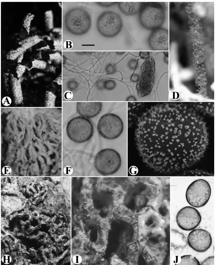
Fructification plasmodiocarpous, pendent, on the surface of substrate, composed of many tubular plasmodiocarps, each tube with one end adhering to the hypothallus, intertwined to form a 3-dimensional net, up to more than 5 cm in total length, more than 1.5 cm in total width. Plasmodiocarps bright yellow or greenish yellow, tubular, 0.22-0.32 mm in diameter, sometimes dichotomously branched. Peridium membranous, covered with a thick layer of yellow lime granules, usually forming a long crusted tail at the free end of the plasmodiocarp. Hypothallus crust-like, yellowish or bright yellow. Capillitium netted, strongly elastic, the threads colorless, with few small, yellow, fusiform lime nodes. Spores dark brown in mass, brown under transmitted light, globose, subglobose, 6.5-8 (-10.5) μm in diameter, minutely warted, with clustered warts.
Specimens examined: TAIWAN, Taipei City: Mt. Samoa, on dead wood, Y.F. Chen11, Aug. 6, 1995, Y.F. Chen507, Aug. 7, 1995. Pingtung County: Nanjen Lake, inside a tree hole of a dead wood, Y.F. Chen255, Jan. 31, 1996.
Fig. 2. A-G. Fuligo aurea. A & E: Fruiting bodies. B: Surface view of spores. C: Capillitial threads and lime node. D: Capillitium. F: Marginal view of spores. G: Surface markings of a spore, by SEM. H-J. Willkommlangea reticulata. H: Fruiting bodies. I: Dehiscent fruiting body. J: Spores. Scale bar: A = 320 μm; B, F = 4 μm; C = 10 μm; D = 210 μm; E = 0.77 mm; G = 1 μm; H = 1 mm; I = 350 μm; J = 5 μm.
Taiwania Vol. 57, No. 3
The pendent tubular plasmodiocarps and the strongly elastic capillitium are the characteristics separating this species from other Fulilgo species.
It was named under the genus Erionema as E.
aureum. The three dimensional network of fruiting
bodies resembles the small aethalia of Fulligo septica without a cortex, and it was then transferred to the genus
Fuligo (Yamamoto, 1998).
Fuligo cinerea (Schwein.) Morgan, Journ. Cincinnati
Soc. Nat. Hist. 19: 33. 1896.
Description and illustration: Chung and Liu (1997a).
Fuligo intermidia T. Macbr., N. Amer. Slime-Moulds
ed. 2. 30. 1922.
It was reported in a list by Nakazawa (1929), but no specimen was deposited in Taiwan.
Fuligo septica (L.) F. H. Wigg., Prim. Fl. Holsat. 112.
1780.
Fuligo septica var. flava Pers., Roemers Neues Mag. Bot. I: 88.
1794.
Fuligo candida Pers., Obs. Myc. I: 92. 1796.
Fuligo septica var. rosea Nann.-Bremek., Proc. K. Ned. Akad.
Wet. Ser. C. 76: 485. 1973.
Description and illustration: Liu (1980).
Specimens examined: TAIWAN, Taipei City: main campus of National Taiwan Univ., on bark, CHL B1478, Apr. 30, 1998. Taipei County: Pinglin, on decayed hard wood, CHL B2066, May 21, 2000. Nantou County: Yuchih, on decayed hard wood, CHL B45, Nov. 26, 1979. Tainan City: on decayed wood, CHL B608, Aug. 4, 1986. Pingtung County: Nanjen Lake, on dead logs, Y.F. Chen256, Aug. 2, 1996.
Fuligo candida, F. septica var. flava, and F. septica
var. rosea, previously reported from Taiwan (Chen et al., 2005) are distinguished from F. septica by colors of aethalia or lime nodes, but those varieties do not have other characteristics to separate them from the type var.
septica. According to Martin and Alexopoulos (1969),
the three should be confined in the synonyms under F.
septica.
Leocarpus fragilis (Dickson) Rostaf., Sluzowce
monogr. 132. 1874. Figs. 3A-H Fructification sporangiate, crowded in clusters, 1.2-1.6 mm in total height. Sporangia shortly stipitate or sessile, ovoid to subglobose, 0.8-1.2 mm in diameter, yellowish brown. Peridium triple, the outer cartilaginous, smooth, shining, the middle thick and calcareous, the inner membranous, hyaline. Stalk weak, whitish or pale ochraceous. Hypothallus prominent, pale
yellow, wrinkled. Capillitium reticulate, duplex, composed of a network of rigid, calcareous nodes (badhamioid), particularly toward the outside, connected with a network of slender, colorless tubules. Spores blackish brown in mass, brown by transmitted light,minutely warted, 10-13 (-15) μm in diameter. Plasmodium not observed.
Specimens examined: TAIWAN, Hualien County: Kuanyuan Forest Recreation Area, on fallen twig, CHL B398, CHL
B399, Nov. 14, 1994.
It is a very distinctive species. The crowded and often clustered sporangia, the fragile, smooth and shining outer peridium, and the rigid, limy capillitium are the distinctive characters of this species.
Physarella oblonga (Berk. & M.A. Curtis.) Morgan, J.
Cincinnati Soc. Nat. Hist. 19: 7. 1896. Figs. 3I-K Description and illustration: Liu (1980).
Willkommlangea reticulata (Alb. & Schwein.) Kuntze,
Revis. Gen. Pl. 2: 875. 1891. Figs. 2H-J
Cienkowskia reticulata (Alb. & Schwein.) Rostaf., Sluzowce
monogr. 91. 1874.
Description and illustration: Liu (1982) and Chung and Liu (1997b).
The duplex capillitium is very conspicuous after the capillitium ruptured. This species is distinctive on the net-like plasmodiocarp, the limy peridium, and the scattered red spots on the peridium. It was first recorded (as Cienkowskia reticulata) from Taiwan in a list by Nakazawa (1929), and the species description here is based on the examination of the specimen CHL M336 (Liu, 1982).
LITERATURE CITIED
Chen, Y.-F., P.-A. Yea, J.-H. Chang and C.-H. Liu. 2005.
Myxomycetes in Hsien-Chi-Yen, Taipei City. Coll. and Res. 18: 15-23.
Chung, C.-H. and C.-H. Liu. 1997a. Noets on some
Myxomycetes from Kenting National Park. Taiwania 42: 28-33.
Chung, C.-H. and C.-H. Liu. 1997b. Myxomycetes of Taiwan
VIII. Taiwania 42: 274-288.
Liu, C.-H. 1980. Myxomycetes of Taiwan I. Taiwania 25:
141-151.
Liu, C.-H. 1982. Myxomycetes of Taiwan III. Taiwania 27:
64-85.
Liu, C.-H. 1990. Myxomycetes of Taiwan VI. Badhamia gracilis. Taiwania 35: 57-63.
Liu, C.-H., J.-H. Chang and I.-G. Huang. 2001.
Myxomycetes of Taiwan XIII. One new record and one new variety. Taiwania 46: 325-331.
Fig. 3. A-H. Leocarpus fragilis. A-B: Fruiting bodies. C: Capillitium. D: Badhamioid network of capillitium, by SEM. E: Spores. F: Peridium of 3-layered, with spores and capillitium on the inside, by SEM. G: Broken sporangium, by SEM. H: Surface markings of one spore, by SEM. I-K. Physarella oblonga. I-J: Fruiting bodies. K: Surface markings of spores, by SEM. Scale bar: A = 0.53 mm; B = 0.77 mm; C = 40 μm; D = 29 μm; E = 9 μm; F = 19 μm; G = 265 μm; H = 1.4 μm; I = 1.8 mm; J = 0.9 mm; K = 1.1 μm.
Taiwania Vol. 57, No. 3
Myxomycetes of Taiwan XIV. Three new records of Trichiales. Taiwania 47: 97-105.
Liu, C.-H., Y.-F. Chen, J.-H. Chang and F.-H. Yang. 2002b.
Myxomycetes of Taiwan XVI. One new species and one new record of Physaraceae. Taiwania 47: 290-297.Martin,
G. W. and C. J. Alexopoulos. 1969. The Myxomycetes.
Univ. Iowa Press; Iowa City, IA. 477 pp.
Nakazawa, R. 1929. A list of Formosan Mycetozoa. Trans.
Nat. Hist. Soc. Formosa 19: 16-30.
Nannenga-Bremekamp, N. E. 1991. A Guide to Temperate
Myxomycetes. Biopress; Bristol, UK. 409 pp.
Shi, H. 1981. Myxomycetes in Yangmingshan area, I. Bull.
Hsinchu Teacher’s Coll. 7: 392-410.
Wang, S.-M., Y.-W. Wang and S. Huang. 1981. The revised
checklist of Myxomycetes in Taiwan. Biol. Bull. Natl. Taiwan Normal Univ. 16: 1-12.
Yamamoto, Y. 1998. The Myxomycetes Biota of Japan. Toyo
Shorin Publishing; Tokyo, Japan. 700 pp. (in Japanese)