With Chitosan-Coated Superparamagnetic Iron Oxide Nanoparticles
J.-H. Juang, J.-J. Wang, C.-R. Shen, C.-H. Kuo, Y.-W. Chien, H.-Y. Kuo, Z.-T. Tsai, and T.-C. Yen
ABSTRACT
Although only 10% of islet recipients maintain insulin independence, 80% of them are
C-peptide positive at 5 years after transplantation. To better understand the fate of
transplanted islets, a magnetic resonance imaging (MRI) technique has been used to
detect Feridex-labeled islet grafts in rodents. In this study, we used a novel MRI contrast
agent, chitosan-coated superparamagnetic iron oxide (CSPIO) nanoparticles, to monitor
mouse islet grafts. Male inbred C57BL/6 mice were used as donors and recipients of islet
transplantation. The islet cytotoxicity was evaluated by fluorescein diacetate and
pro-pidium iodide staining for RAW cells incubated with CSPIO. After being incubated
overnight with and without CSPIO (10 mg/mL), 300 islets were transplanted under the left
kidney capsule of each mouse. After transplantation, 3.0-Tesla MRI of the recipients was
performed biweekly until 19 weeks. At the end of study, the islet graft was removed for
insulin and Prussian blue staining. The cell death rates in RAW cells did not increase with
increasing CSPIO concentrations or incubation time. The grafts of CSPIO-labeled islets
were visualized on MRI scans as distinct hypointense spots homogeneously located at
the upper pole of left kidney. Their MRI signal was 30%–50% that of control islets and
was maintained throughout the follow-up period. At 18 weeks, the histology of
CSPIO-labeled islet graft revealed the insulin- and iron-stained areas to be almost
identical. Our results indicate that isolated mouse islets labeled with CSPIO
nanopar-ticles can be effectively and safely imaged by using MRI as long as 18 weeks after
transplantation.
R
ecently, the Edmonton Protocol has markedly im-proved the success rate of human islet transplanta-tion.1 However, ⱖ2 pancreases are usually required toachieve normoglycemia. Moreover, long-term function of the transplanted islets has been disappointing.2,3Allograft
failure may be due to nonimmunologic (eg, insufficient -cell mass and islet engraftment problems) as well as immunologic (eg, immune rejection, toxicity of immuno-suppressants and autoimmune recurrence) factors.4
Al-though only 10% of recipients maintain insulin indepen-dence, 80% of them are C-peptide positive at 5 years after islet transplantation.2To better understand the fate
of transplanted islets, a magnetic resonance imaging (MRI) technique has been used to detect Feridex (dextran-coated superparamagnetic iron oxide [SPIO])–labeled rat, porcine, and human islets transplanted into rodents.5–7In the
present study, we used a novel MRI contrast agent, chitosan-coated SPIO (CSPIO) nanoparticles,8to monitor mouse islet
grafts.
From the Division of Endocrinology and Metabolism, Depart-ment of Internal Medicine, Chang Gung University and Chang Gung Memorial Hospital (J.-H.J., Y.-W.C.), Department of Med-ical Imaging and Radiologic Sciences (J.-J.W.), and Department and Graduate Institute of Medical Biotechnology and Laboratory Science (C.-R.S., H.-Y.K.), Chang Gung University, Taoyuan, the Molecular Imaging Center, Chang Gung Memorial Hospital, Taoyuan (C.-R.S., Z.-T.T., T.-C.Y.), and the Department of Bio-logic Science and Technology, National Chiao Tung University, Hsinchu (C.-H.K.), Taiwan.
J.-H. Juang and J.-J. Wang contributed equally to this work. Supported by grants from Chang Gung Memorial Hospital (CMRPG370871, CMRPG370872, CMRPG370361, and CMRPG370362), Taoyuan, Taiwan.
Address reprint requests to Dr Jyuhn-Huarng Juang, Division of Endocrinology and Metabolism, Chang Gung Memorial Hos-pital, 5 Fu-Shin Street, Kweishan, Taoyuan, Taiwan. E-mail: [email protected]
0041-1345/10/$–see front matter © 2010 by Elsevier Inc. All rights reserved. doi:10.1016/j.transproceed.2010.05.103 360 Park Avenue South, New York, NY 10010-1710
MATERIALS AND METHODS Animals
Male inbred C57BL/6 mice, aged 8 –12 weeks, were used as donors and recipients of islet transplantation.9The animal experiments
were approved by the Animal Ethics Committee of Chang Gung Memorial Hospital.
Islet Isolation
Under anesthesia with sodium amobarbital, pancreases were dis-tended with 2.5 mL RPMI-1640 medium (Gibco, Grand Island,
NY, USA) containing 1.5 mg/mL collagenase (collagenase from Clostridium histolyticum, type XI; Sigma Immunochemicals, St Louis, Mo), excised and incubated in a water bath at 37°C.9The
islets were separated by a density gradient (Histopaque-1077; Sigma Immunochemicals), and purified islets were then hand-picked under a dissecting microscope.
Islet Labeling
Isolated islets were overnight incubated with CSPIO (10 mg/mL) in culture medium.8After incubation, islets were washed with culture
medium and used for in vitro studies or islet transplantation.
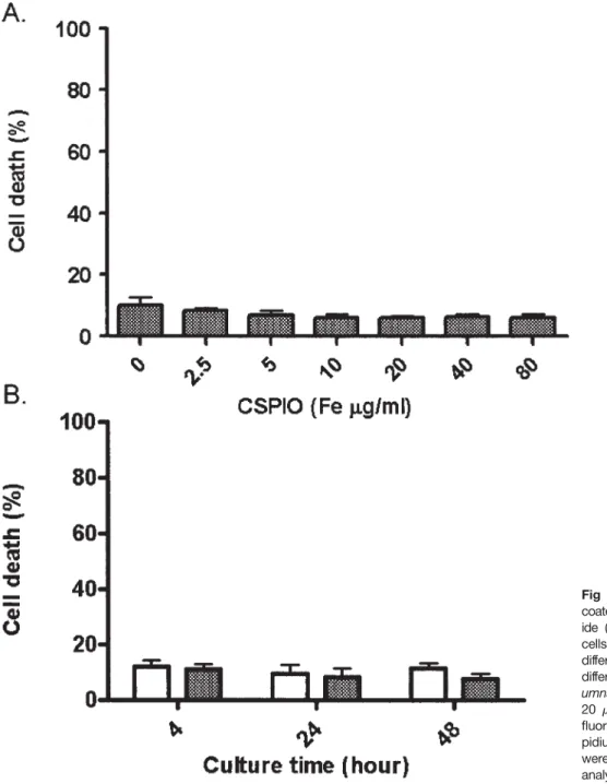
Fig 1. Cytotoxicity of chitosan-coated superparamagnetic iron ox-ide (CSPIO) on RAW cells. RAW cells were incubated with CSPIO of different concentrations (A) and different time periods (B; open
col-umns: control; shaded colcol-umns: Fe
20g/mL) and then stained with fluorescein diacetate and pro-pidium iodide. Cell death rates were assayed by flow cytometry analysis.
Islet Cytotoxicity
RAW cells were incubated with CSPIO and then stained with fluorescein diacetate and propidium iodide. Cell viability and death rate were assayed by flow cytometry analysis.10
Islet Transplantation
Three hundred islets cultured with and without CSPIO were syngeneically transplanted under the left kidney capsule of each mouse.9To accomplish this, the islets were centrifugated in PE-50
tubing (Clay Adams, Parsippany, NJ) connected to a 200-L pipette tip. With the mouse under amobarbital anesthesia, the left kidney was exposed through a lumbar incision. A capsulotomy was performed in the lower pole of the kidney and the tip of the tubing advanced under the capsule of the upper pole, the site of final injection. The capsulotomy was left unsutured.
In Vivo MRI of Transplanted Islets
After transplantation, serial MRI of the recipients (2 with control islets and 2 with CSPIO-labeled islets) were performed biweekly. One control animal and 1 CSPIO-labeled animal died at 9 and 16 weeks, respectively. The other control animal and 1 CSPIO-labeled animal were followed until 19 and 18 weeks, respectively. Images were acquired on a 3.0-Tesla MRI scanner (Magnetom Trio with TIM; Siemens, Erlangen, Germany) using a homemade surface coil. A T2*-weighted gradient-recalled echo sequence was acquired for all subjects. The MR signal intensity of the islet graft was quantified using the contralateral kidney as a reference.
Removal of the Islet Graft
Eighteen weeks after transplantation, animals intended for graft removal were anesthetized with amobarbital. An abdominal inci-sion was made and the kidney was exposed. Under dissecting
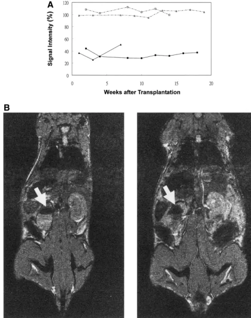
Fig 2. A. Evolution of the mag-netic resonance (MR) signal inten-sity of islet isografts in controls (open squares and triangles) and an-imals with chitosan-coated super-paramagnetic iron oxide (CSPIO)– labeled islets (solid squares and
tri-angles). The MR signal intensity of
the contralateral kidney was used as a reference. B. The 2-week (left) and 18-week (right) MR image of the CSPIO-labeled islet graft showing a distinct hypointense spot homogeneously located at the upper pole of left kidney (arrows).
microscope, the kidney capsule surrounding the graft was excised and removed with the adherent graft.11
Immunohistochemistry of the Islet Graft
The removed grafts were fixed in formalin solution and processed for paraffin embedding and sectioning. Sections of grafts were stained for iron with Prussian blue and for the endocrine-cells with a guinea pig anti-swine insulin antibody (Dako Co., Glostrup, Denmark).11
RESULTS
After incubation with CSPIO, the cell death rates in RAW cells did not increase with increasing CSPIO concentrations or incubation time (Fig 1). In contrast to the control, the grafts of CSPIO-labeled islets were visualized on MRI scans as distinct hypointense spots homogeneously located at the upper pole of the left kidney. There was a 30%–50% signal loss throughout the follow-up period (Fig 2). At 18 weeks, the CSPIO-labeled islet graft was positive for insulin and iron staining and the insulin- and iron-stained areas were almost identical (Fig 3).
DISCUSSION
An MRI technique has been used to detect Feridex-labeled rat, porcine, and human islets transplanted into rodents.5–7
Dextran-coated Feridex is approved by the US Food and Drug Administration for human use as a liver imaging contrast agent. Unfortunately, in November 2008 the com-pany ceased to manufacture Feridex. Recently, Tsai et al. developed a novel MRI contrast agent, CSPIO, by coating SPIO with chitosan to increase the content of magnetite.8
In the present study, we demonstrated that isolated mouse islets labeled with CSPIO nanoparticles could be visualized by MRI after transplantation.
Chitosan has been applied in numerous biomedical ap-plications owing to its nontoxicity, biocompatibility, and biodegradability.12 Here, we showed that the cell death
rates in RAW cells did not increase with increasing CSPIO concentrations or incubation time. This indicates that CSPIO can be safely used to label cells for MRI tracking.
On MRI scans, the iron oxide leads to an extensive signal loss owing to magnetic field inhomogeneity. Therefore, the grafts of Feridex-labeled5–7 and CSPIO-labeled islets
showed distinct hypointense spots homogeneously located at the transplantation sites. In the present study, we have for the first time quantified the graft MR signal intensity by using the contralateral kidney as a reference. The MR signal intensity of CSPIO-labeled islet grafts was 30%–50% that of control grafts. Moreover, this signal loss was main-tained for 18 weeks, indicating the persistent existence of CSPIO at the transplantation site. To our knowledge, this is the longest MRI follow-up for SPIO-labeled islet grafts. At 18 weeks, the graft histology revealed that the insulin- and iron-stained areas were almost identical. This further sup-ports the notion that the uptake of CSPIO by isolated islets leads islet grafts to be visualized by MRI after
transplanta-tion. In conclusion, isolated mouse islets labeled with CSPIO nanoparticles can be effectively and safely imaged by using MRI as long as 18 weeks after transplantation.
REFERENCES
1. Shapiro AMJ, Lakey JRT, Ryan EA, et al: Islet transplanta-tion in seven patients with type 1 diabetes mellitus using a glucocorticoid-free immunosuppressive regimen. N Engl J Med 343:230, 2000
2. Ryan EA, Paty BW, Senior PA, et al: Five-year follow-up after clinical islet transplantation. Diabetes 54:2060, 2005
3. Shapiro AMJ, Ricordi C, Hering BJ, et al: International trial of the Edmonton Protocol for islet transplantation. N Engl J Med 355:1318, 2006
4. Juang JH: Islet transplantation: an update. Chang Gung Med J 27:1, 2004
5. Evgenov NV, Medarova Z, Pratt J, et al: In vivo imaging of immune rejection in transplanted pancreatic islets. Diabetes 55: 2419, 2006
6. Tai JH, Foster P, Rosales A, et al: Imaging islets labeled with magnetic nanoparticles at 1.5 Tesla. Diabetes 55:2931, 2006 Fig 3. The chitosan-coated superparamagnetic iron oxide– labeled islet graft stained with insulin (top, brown color) and Prussian blue (bottom, blue color) at 18 weeks after islet transplantation.
7. Evgenov NV, Pratt J, Pantazopoulos P, et al: Effects of glucose toxicity and islet purity on in vivo magnetic resonance imaging of transplanted pancreatic islets. Transplantation 85:1091, 2008
8. Tsai ZT, Wang JF, Kuo HY et al: In situ preparation of high relaxivity iron oxide nanoparticles by coating with chitosan. J Magn Magn Mater 322:208, 2010
9. Juang JH, Hsu RS, Kuo CH, et al: Normoglycemic environ-ment is important for the growth and function of islet isograft. Transplant Proc 30:565, 1998
10. Ross DD, Joneckis CC, Ordóñez JV, et al: Estimation of cell survival by flow cytometric quantification of fluorescein diacetate/ propidium iodide viable cell number. Cancer Res 49:3776 – 82, 1989 11. Juang J-H, Kuo C-H, Wu C-H, et al: Exendin-4 treatment expands graft -cell mass in diabetic mice transplanted with a marginal number of fresh islets. Cell Transplant 17:641, 2008
12. Kumar MN, Muzzarelli RA, Muzzarelli C, et al: Chitosan chemistry and pharmaceutical perspectives. Chem Rev 104:6017, 2004