行政院國家科學委員會專題研究計畫成果報告
單發性結節的自動化檢測方法
An Automated Inspection Method for Solitary Pulmonary Nodule
計畫編號:NSC 89-2212-E-110-030
執行期限:90 年 8 月 1 日至 91 年 7 月 31 日
主持人:嚴成文 教授 國立中山大學機械與機電工程學系
共同主持人:吳銘庭 主治醫師 高雄榮民總醫院放射線部
潘慧本 主治醫師 高雄榮民總醫院放射線部
一、中文摘要 醫療自動化是自動化學門的推動重點之一,而 本計畫的重要動機之一是因為發我們發現許多工 業上所用的自動檢測及電腦視覺技術,若經適當的 發展與整合,應可非常有效的應用於許多病症研判 的醫療問題上,使醫療成本能因病症的早期發現及 早期治療而大幅降低。然而在此之前自動化技術與 病症研判的問題卻甚少結合,由於癌症是我國國人 十大死因的首位,故本計畫選擇其中為害最重的肺 癌做為研究對像。 透過自動檢測及電腦視覺技術的使用,本研究 的主要目的在於發展一套智慧型的型態鑑別系統 來判斷單發性結節的良惡性,以降低對入侵性的診 斷方法的依賴性。 為加強本計畫的實用性,本計畫所發展的技術 將儘量降低將所需的人為參與。此外本研究也已得 到高雄榮民總醫院的全力支持,在電腦斷層攝影設 備的使用及病例的取得上,都已完全得到妥善的安 排。我們並預計將本計畫的結果整合於高雄榮總的 無軟片電腦影像傳輸系統內,成為整體醫療資訊系 統中重要的診斷工具之一。 關鍵詞:肺癌;電腦斷層攝影;自動檢測;電腦視 覺;類神經網路;型態鑑別;單發性結節 AbstractMedical automation is one of the important research goals of the automation division of the NSF. For this project, one of motivations is that we have discovered that, via appropriate development and integration, many of the industry based automated inspection and computer vision methods can be applied to a lot of medical diagnosis problems. With early treatment that resulted from early detection, costs for medical and social expenses can be reduced significantly. However, automation techniques and medical diagnosis problems have seldom met. Since cancer is the number one death cause in Taiwan, we choose lung cancer, which causes the most cancer-related deaths in Taiwan, as the target of this project.
Via the application of automatic inspection and computer vision techniques, the goal of this project is develop an intelligent pattern recognition system to diagnose solitary pulmonary nodules (SPNs).
To increase the practicability of the system, this study will try to minimize the requirement for human intervention. This research has also have the full support of the Veterans General Hospital-Kaohsiung in the use of CT facilities. The acquisition of lung CT cases has also been properly arranged. We are also planning on integrating the proposed method with the hospital’s filmless computer image transmission system and thus make the proposed technique
becomes an integrated part of the medical information system.
Keywords: Lung cancer; computed tomography;
automatic inspection; computer vision; pattern recognition; solitary pulmonary nodule
二、緣由與目的 近數十年來歐美國家及臺灣肺癌病人有顯著的 增加。根據衛生署的統計資料顯示,過去數年來國 人男性癌症死亡原因中,肺癌僅次於肝癌,為第二 位的癌症死亡原因;在女性,肺癌則均高居癌症死 亡原因的第一位。但在 1997 年,雖然肺癌在男、 女死亡原因的排名沒有改變,但是肺癌總死亡數已 經超越肝癌,躍居國人癌症死亡原因之首位。近年 來儘管醫學已有相當進展,但是肺癌的治療成果還 是非常令人失望,整體而言,在台灣肺癌的5 年存 活率僅約10%左右 [國家衛生研究所, 1998]。然而 過去的研究結果也明確的顯示,愈能早期發現肺 癌,其存活率即愈高;舉例而言,單發性結節在10 mm 以下即被檢測出的病人的 5 年存活率高達約 80% [Lillington, 1997]。然而肺癌在初期,與一般良 性腫瘤之間的差異不大,相當難以判斷,目前臨床 上的做法是進一步追蹤其是否成長、及其輪廓、鈣 化狀況是否變得較為容易分辨。然而最後的確定方 法,也唯有將其切除並做分析。本研究的目的是希 望在早期發現時,能有效的協助醫師進行診斷,以
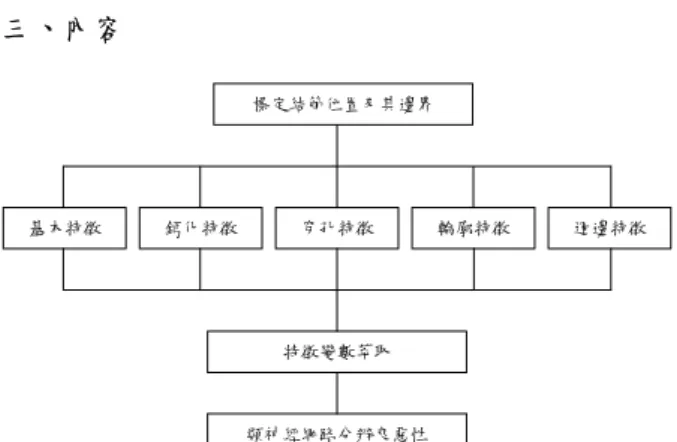
期提高早期良惡性檢測的準確率。 三、內容 標定結節位置及其邊界 基本特徵 鈣化特徵 穿孔特徵 輪廓特徵 週邊特徵 特徵變數萃取 類神經網路分辨良惡性 圖1. 單發性結節良惡性檢測系統架構圖 本論文的系統架構如圖1 所示,我們依序說明 如下︰ 1. 標定結節位置及其邊界 由電腦自行偵測我們要處理的結節位置,以 目前來說是不可能的。但是若要求由人為的 方式,將結節的邊界畫出來,又很容易受到 人為的不確定性所影響,因此我們選擇由人 為方式框選結節的大略位置,再透過影像處 理的技術,將結節的邊界描繪出來。 2. 計算特徵變數 本論文使用了多組特徵變數,分別敍述如 下︰ A. 基本特徵 : 年齡、性別、結節尺寸、吸煙病史等是以往 研究中常使用的特徵,故將其列為基本特 徵。目前已使用上的有結節尺寸。 B. 鈣化特徵 : 描述結節內部鈣化型態的特徵變數。在結節 內部的鈣化會形成區塊群,在臨床上常藉由 鈣化區塊分部狀況,來做為判斷結節良惡性 的依據之一。由於鈣化與非鈣化的CT 值難 以掌握,因此設定 CT 值較高的前 10% 的 像素點為組成區塊群,並採用由Dhawan 於 1996 年提出的方法[17],基於一些統計學常 用 的 方 法 來 量 化 區 塊 的 分 佈 情 形 及 其 強 度,以其能描繪鈣化型態。此方法可以得到 10 個特徵變數,其說明如下︰ 1) 區塊的數目 2) 區塊面積的平均值 3) 區塊面積的標準差 4) 所有區塊內像素點 CT 值的平均值 5) 所有區塊內像素點 CT 值的標準差 6) 區塊兩兩距離的平均值 7) 區塊兩兩距離的標準差 8) 所有區塊與結節質心距離的平均值 9) 所有區塊與結節質心距離的標準差 10) 系統位能︰平均灰均值×所有區域面積 /區塊數 除此之外,針對結節內部及其邊界處的 灰階及梯度值,亦使用統計學方法,嘗試找 出良惡性之間的不同點,因此定義了以下4 個特徵變數,分別敘述如下︰ 11) 結節灰階值的平均值 12) 結節灰階值的標準差 13) 結節內部(不含邊界)的梯度平均值 14) 結節邊界上的梯度平均值 C. 穿孔特徵︰ 描述結節內部穿孔的特徵變數。在臨床上, 結節是否有穿孔的現象、及穿孔的型態也是 良惡性診斷的依據。在實作上以CT 值較低 的 10% 像素點組成區塊群,並同樣採用 Dhawan 的方法,取得 10 個特徵變數。 D. 輪廓特徵(Uniresolution Features)︰ 輪廓特徵是臨床上判定腫瘤良惡性的重要 特徵之一,我們計算了三組關於輪廓的特徵 變數,本組資料是非複利葉的特徵變數,第 4 組則是複利葉特徵變數。本組所採用的輪 廓 特 徵 , 是 傳 統 的 Uniresolution Features[15]。 此方法是以 radial distance measurements 為基礎。如圖 2 所示,radial distance 的計算方式是先計算出腫瘤的質 心 , 再 計 算 出 每 一 個 邊 界 點 與 質 心 的 Euclidean 距離。則選擇任意的起始點,以順 時 針 的 方 式 記 錄 下 這 些 邊 界 點 與 質 心 的 Euclidean 距 離 , 即 為 radial distance measurements。Uniresolution Features 一共有 6 個特徵變數,分別說明如下︰
圖2. Radial Distance Measurements 1) Tumor circularity, C C 的定義為 A P C 2 = ...(1) 其中P 為腫瘤周長,A 為腫瘤的面積。 2) Radial distance mean, davg
∑
= = N i avg d i N d 1 ) ( 1 ...(2)3) Radial distance standard deviation
∑
= − = N i avg d i d N 1 2 ) ) ( ( 1 σ ...(3) 4) Entropy if the radial distance histogram∑
= =100 1 ) log( k k k p p E ...(4) 若將radial distance 的最大到最小值之間分成100 等分,則 pk就是radial distance 在第 k 等 分的機率。 5) Area ratio, A
∑
= − = N i avg avg d i d N d A 1 ) ) ( ( 1 ...(5) 其中,當d(i)≦davg時不列入計算。 6) Roughness index, R = + − =∑
+ = L N j i d i d j R j L j i ,...., 1 where , ) 1 ( ) ( ) ( [ ]∑
= = N L j j R N L R / 1 ) ( ...(6) E. 輪廓特徵(Fourier Descriptor)︰ 這是第二組描述輪廓特徵的方法︰複利葉 描述元(Fourier Descriptor)。複利葉描述元是 利用離散複利葉轉換的方式,描述出輪廓的 頻率響應[16]。假設我們找到一組邊界點資 料(x[m],y[m]),其中 1≦m≦L,L 為資料點 數。經由複利葉轉換,可以得到複利葉係數[ ]
[ ]
( )[ ]
∑
[ ]
( )∑
= − = − = = L m m L jk L m m L jk e m y L k b e m x L k a 1 / 2 1 / 2 1 1 π π ...(7) 其中 0≦k≦L-1。因為 a[0]、b[0]僅代表影 像中心的資訊,故忽略之。接著令[ ]
[ ]
[ ]
[ ] [ ]
[ ]
1 2 2 r k r k s k b k a k r = + = ...(8) 其中1≦k≦L-1。則 s[k]即為複利葉描述元。 s[1]恆等於 1,在我們的研究中選取 k = 2 ~ 15。 F. 輪廓特徵(GLIS)︰ 這是第三組描述輪廓的特徵變數。這組特徵 與前兩組的不同點在於計算的對象不是輪 廓本身,而是輪廓內外一定範圍內帶狀區域 的 texture features。使用此組特徵的主要原 因是結節的通常會有極為尖銳但不明顯的 毛邊存在,在邊界截取上這是相當困難的處 理對象,故希望透過計算邊界附近區塊的 texture features 來偵測出這些毛邊的特性。 計 算 texture features 的 方 法 來 自 Dhawan(1996) 使 用 的 Gray-Level Image Structure(GLIS) 特徵變數。其方法是基於影 像的二階灰階長方圖(second-order histogram, H(yq,yr,d)),H(yq,yr,d)所代表的意義是兩兩相 隔d,灰階值分別為 yq、yr的像素點出現的 機率。GLIS 一共有 10 個特徵變數,分別如 下︰ 1) Entropy of H(yq,yr,d)[
] [
]
∑ ∑
= = − = t q t r y y y y y y r q r q y H y y y H 1 1 ) , , ( log ) , , ( Entropy d d ...(9) 2) Contrast of H(yq,yr,d)[
]
∑ ∑
= = ∂ = t q t r y y y y y y r q r q y H y y y 1 1 ) , , ( ) , ( Contrast d ...(10)3) Angular Second Moment of H(yq,yr,d)
[
]
∑ ∑
= = = t q t r y y y y y y r q y y H 1 1 2 ) , , ( moment order -second Angular d ...(11)4) Inverse difference moment
q r y y y y y y q r r q y y y y y y H t q t r ≠ ∂ + =
∑ ∑
= = for ) , ( 1 ) , , ( moment difference Inverse 1 1 d ...(12) 5) Correlation of H(yq,yr,d)[
]
∑
∑
∑ ∑
= = = = = = − = t q t r r m q m t q t r r m q m y y y r q r m y y y r q q m y H y H y y y y y y y H y H r q y y H y H y y H y H y y H 1 1 1 1 ) , , ( ) d , ( and ) , , ( ) d , ( where ) , , ( measure n Correlatio ) , ( ) , ( ) , ( ) , ( d d d d d d d σ σ µ µ ...(13) 6) Mean of H(yq,yr,d)∑
= = t q y y y q m qH y y 1 ) , ( Mean d ...(14) 7) Deviation of H(yq,yr,d)∑
∑
= = − = t q t p y y y q m y y y p m p q y H y H y y 1 1 ) , ( ) , ( Deviation 2 d d (15)8) Entropy of Hdiff(i,d)
[
] [
]
∑
∑
∑
= − = = = = − = t r q q t r t y i y y y y y y y r q diff i i i diff diff y y H i H i H i H 1 1 1 ) , , ( ) , ( where ) , ( log ) , ( Entropy d d d d ...(16)9) Angular second moment(ASM) of Hdiff(i,d)
[
]
∑
= = it i i diff diff i H i H 1 1 2 ) , ( ) , ( of ASM d d ...(17)∑
= = it i i diff diff i iH i H 1 1 ) , ( ) , ( of Mean d d ...(18) G. 週邊特徵︰ 結節以外的週邊區域也常常是臨床上診斷 的依據,例如︰玻璃狀組織、衛星病灶等。 由於在進行邊界截取之前,會先框選結節所 在的區域,因此結節週邊區域直接定義為框 選區域扣除結節區域。特徵計算方式同樣採 用Dhawan(1996) 的 GLIS 特徵,共 10 個。 3. 特徵變數萃取 以上使用的特徵變數有7 組共有 67 個,而目 前我們所有可用的資料筆數也不過 146 筆, 相對而言,維度實在太大,因此我們希望透 過特徵變數萃取方法以期在盡可能保留資料 特徵的情況下,降低資料的維度。這裡所嘗 試採用的特徵變數萃取有兩種方法,其一是 統計學常用的Forward Selection 方法,其二是 以主軸分析為基礎的萃取方法,稱為主軸萃 取。Forward Selection 的主要方法是︰(1)選 出有最佳分類能力的特徵。(2)與之前選出的 特徵配合下,有最佳分類能力的特徵。如此 逐一選出適合的特徵。另主軸萃取方法,是 將多個特徵的資料,透過主軸分析計算得到 多個主軸及相對的特徵值。根據特徵值的大 小順序,可以排列出各主軸的代表性,特徵 值愈高,相對的主軸愈能代表原始資料。最 後取前 n 個特徵值較大的主軸(n 小於原資料 維度),則原始資料經此 n 個主軸即可轉換成 n 個維度的新資料空間。經實證,只要 n 取得 夠多,就有機會可以使 NN 分類器的分類結 果與原維度空間相同。 4. 類神經網路分類器 綜合上述特徵變數的計算及選取的步驟,緊 接著是透過分類器來進行腫瘤良惡性的分 類。分類器的選用上,我們採用新發展的 Committee Coordinator(CC)。由於本研究的實 驗資料受限,使用傳統類神經網路或進階的 委員會機器均有資料點數不足的疑慮。為了 解決這個問題,我們引入了 Bagging 的方法 [20]。Bagging 是一種統計學的方法,透過不 斷取樣的過程,經證明可以以少數資料來模 擬出原始資料的分佈型態。在我們的實作 上,將分類問題分成兩階段︰(1)單純 MLP 組 成之委員會機器。(2)將第(1)階段訓練資料的 輸出值差之絕對值低於門檻值者,送入第(2) 階段做為訓練資料,並使用Bagging 的方法建 立100 個 MLP 組成的委員會機器,以針對特 別難分的資料來做訓練。除此之外,我們亦 採用 Averaging Committee(AC)及 Weighted Averaging Committee(WAC)兩種委員會機器 來做比較。以上委員會機器的組成均採用以 逆傳遞式方法訓練的MLP。 四、實驗結果與討論 4.1 各演算法的實驗結果 1. 標定結節位置及其邊界 目前的進度已算完成,僅需人為手動框出大概 輪廓之後,即可自動產生結節的邊界,其結果 如圖3、4。圖 3 是經人為手動框選出結節大略 位置,並將該區域影像放大後所得結果。而圖 圖3. 框選出來的影像 圖4. 結節的邊界 4 則顯示對圖 3 影像,經影像處理技術,取得 的影像邊界結果。由圖4 可以清楚的看出,我 們的方法已經可以有效的掌握結節的邊界。 2. 特徵變數萃取 使用的Forward Selection 及主軸萃取方法均已 完成並經過實例測試。實驗方法如下︰首先將 原始資料,利用特徵變數萃取的方法,依順序 紀錄特徵變數的選取順序,接著將原始資料隨 機分成訓練及測試資料各為 90%、10%,並根 據萃取結果,依序加入特徵變數,分別計算其 分類正確率。當對原始資料做分群時,使用了 ten-fold cross vali- dation 的方法將資料分成 10 組,故每加入一個特徵變數時,即計算10 組資 料的分類正確率,並予以平均。實驗資料共三 組 , 分 別 為 乳 癌(Breast Cancer) 、 糖 尿 病 (Diabetes)、信用卡(Credit Card)。其實驗結果如 圖5、6、7 所示。以目前的實驗結果來看,ForwardSelection 具有較佳的特徵變數萃取能力。 1 2 3 4 5 6 7 8 9 Feature Numbers 88 89 90 91 92 93 94 95 96 97 98 Cl ass if icati on Acc ur
acy(%) Feature Extraction
Forward Selection 1 2 3 4 5 6 7 8 9 88 89 90 91 92 93 94 95 96 97 98 圖5. 使用 Forward Selection 及主軸萃取方法 —乳癌 1 2 3 4 5 6 7 8 60 62 64 66 68 70 72 1 2 3 4 5 6 7 8 Feature Numbers 60 62 64 66 68 70 72 Cla ssif ica tion Accu rac y(%) Feature Extraction Forward Selection 圖6. 使用 Forward Selection 及主軸萃取方法 —糖尿病 0 2 4 6 8 10 12 14 78 80 82 84 86 0 1 2 3 4 5 6 7 8 9 10 11 12 13 14 Feature Numbers 78 79 80 81 82 83 84 85 86 Cla ssif ica tion Accu racy(%) Feature Extraction Forward Selection 圖7. 使用 Forward Selection 及主軸萃取方法 —信用卡 3. 類神經網路分類器 分類器的測試同樣採用乳癌(Breast Cancer)、糖 尿病(Diabetes)、信用卡(Credit Card)三組實際資 料。實驗時將資料隨機分成訓練、驗證及測試 資料三組,訓練資料佔60%、驗證資料佔 20%、 測試資料佔20%。共隨機分組 50 次,每次均建 立三種分類器,計算各分類器50 次實驗結果的 平均值及標準差。其結果如表1-1 ~ 1-3,結果 顯示以新發展的Committee Coordinator(CC)方 法,確實可以比傳統的Averaging Committee(AC) 及Weighted Averaging Committee(WAC)有較佳 的分類精度。 表1-1 分類器之測試-乳癌 分類器 精度平均值(%) 精度標準差 AC 96.6056 1.54347 WAC 96.6056 1.54347 CC (T=0.62*) 96.9296 1.35543 *T︰Committee Coordinator 的門檻值 表1-2 分類器之測試-糖尿病 分類器 精度平均值(%) 精度標準差 AC 75.8581 3.13659 WAC 75.8581 3.13659 CC (T=0.46) 76.3484 3.15958 表1-3 分類器之測試-信用卡 分類器 精度平均值(%) 精度標準差 AC 87 2.69975 WAC 87 2.69975 CC (T=0.87) 87.3857 2.6239 4.2 單發性結節良惡性檢測的實驗結果 本論文的實驗資料是肺部電腦斷層掃瞄(CT) 影像,由高雄榮民總醫院所提供,其儲存格式是醫 界常用的DICOM 格式。目前已收集到的資料已接 近 200 個,而已確定良惡性結果堪用的資料有一 半,良性48 筆、惡性 98 筆,共 146 筆資料。首先 利用邊界截取的方法,得到每個病歷上的結節邊 界,並透過特徵變數的計算方法,產生病歷的特徵 資料,目前我們計算的特徵變數維度有 67 個。由 於 特 徵 變 數 維 度 太 多 , 目 前 分 別 採 用 較 理 想 的 Forward Selection 演算法來篩選,以降低特徵變數 維度。由於肺腫瘤實驗資料的筆數比起先前的乳癌 等資料更少,故資料僅分成訓練及驗證兩組。訓練 資料佔 80%、測試資料佔 20%。同樣隨機分組 50 次,每次均建立三種不同分類器,並記錄實驗結果 於表2。結果亦顯示 Committee Coordinator 有較佳 的分類結果,而且分類精度也較另外兩種分類器要 穩定(標準差較小)。
表2 肺腫瘤良惡性檢測實驗結果 分類器 精度平均值(%) 精度標準差 AC 74.863 2.38575 WAC 75 2.30409 CC 77.274 0.614026 五、結論 目前的實驗結果的精度已從初期的 70%進步 到目前的77%以上。其結果之所以能有所進展的原 因包含︰ 1. 資料不斷的累積。資料愈多,類神經網路分類 器的可靠度及精度也會隨著提升,因此我們仍 舊持續的在收集資料。 2. 新特徵的加入。在研究過程中,我們會持續發 現有用的特徵變數,並加入到實驗中。則新的 特徵自然提升我們的分類結果。 3. 分類器的改進。類神經網路分類器的不斷改 良,尤其是針對資料點不足採用的Bagging 方 法,是有效的提升分類精度的一大功臣。 現在我們並不滿足於目前的分類結果,在我們 的研究上,而是以能夠達到80%以上分類精度為主 要的目標。目前我們朝向兩個主要方向來努力︰ 1. 尋找更有代表性的特徵變數。在目前的分類問 題上,良性的判別正確率相對比較低,因此我 們考慮從中觀察是否有未曾注意到的特徵。 2. 測試其他可能的分類方法。類神經網路分類器 的結合方法有許多,現在使用到的AC、WAC、 CC 只是其中的三種。目前我們還繼續在測試 及發展其他方法,希望能更有效的提升實驗的 分類結果。 六、參考文獻
[1] Cavouras, D., Prassopoulos, P. and Pantelidis, N., “Image analysis methods for solitary pulmonary nodule characterization by computed tomography,” European Journal of Radiology, 1992,Vol. 14, pp. 169-172.
[2] Colice, G. L., “Chest CT for known or suspected lung cancer,” Chest, 1994, Vol. 106, pp. 1538-1550.
[3] Delorme, S. Keller-Reichenbecher, M.-A., Zuna, I., Schlegel, W., and Kaick, G. V., “Quantitative assessment of high-resolution computed tomography findings by computer-assisted texture-based image analysis,” Investigative
Radiology, 1997, Vol. 32, pp. 566-574.
[4] Gurney, J. W., Lyddon, D. M. , and Mckay, J. A., “Determining the liklihood of malignancy in solitary pulmonary nodules with Bayesian analysis, Part II. Application,” Radiology, 1993, Vol. 186, pp. 415-422.
[5] Haralick, R. M., “Statistical and structural approaches to texture,” Proceedings of IEEE,
1979, Vol. 67, pp. 786-804.
[6] Kim, H. and Nam K., “Object recognition of one-DOF tools by a back-propagation neural net,” IEEE Transactions on Neural Networks, 1995, Vol. 6, pp. 484-487.
[7] Lillington, G. A., “Management of solitary pulmonary nodules,” Postgradrate Medicine, 1997, Vol. 101, pp. 145-150.
[8] Murino, V., Ottonello, C., and Pagnan, S., 1998, “Noisy texture classification: A higher-order statistics approach,” Pattern Recognition, Vol. 31, pp. 389-393.
[9] J. R. Parker, “Algorithms for Image Processing And Computer Vision”, Wiley Computer
Publishing, 1997.
[10] Siegelman, S. S., Zerhouni, E. A., Leo, F. P., Khouri, N. F. and Stitik, F. P., “CT of the solitary pulmonary nodule,” American Journal
of Roentgenology, 1980, Vol. 135, pp. 1-13.
[11] Siegelman, S. S., Khouri, N. F., Leo, F. P., Fishman, E. K., Braveman, R. M., and Zerhouri, E. A., “Solitary pulmonary nodules: CT
assessment,” Thoracic Radiology, 1986, Vol. 160, pp. 307-312.
[12] Tan, T. N., “Noise robustness of texture features,” Image and Vision Computing, 1997, Vol. 15, pp. 815-817.
[13] Webb, W. R., “Radiologic evaluation of the solitary pulmonary nodule,” American Journal
of Roentgenology, Vol. 154, pp. 701-708.
[14] Zwirewich, C. V., Vedal, S., Miller, R. R. and Muller, N. L., “Solitary pulmonary nodule: high-resolution CT and radiologic-pathologic correlation,” Thoracic Radiology, 1991, Vol. 179, pp. 469-476.
[15] Lori Mann Bruce and Reza R. Adhami, “Classifying Mammographic Mass Shapes Using the Wavelet Transform Modulus-Maxima Method,” IEEE Trans. Med. Imag., 1999, Vol.18, No.12, December.
[16] Hongbong Kim and Kwanghee Nam, “Object Recognition of One-DOF Tools by a
Back-propagation Neural Net,” IEEE
Transaction on Neural Network, 1995, Vol. 6.
No. 2. March.
[17] Atam P. Dhawan, Yateen Chitre, Christine Kaiser-Bonasso, Myron Moskowitz, “Analysis of Mammographic Microcalcifications Using Gray-Level Image Structure Features,” IEEE
Trans. Med. Imag., 1996, Vol. 15, No. 3, June,
pp.246-259.
[18] Boaz Lerner, Hugo Guterman ,Mayer Aladjem, Its’hak Dinstein, “A comparative study of neural network based feature extraction paradigms,” Pattern Recognition Letters 20(1999) 7-14.
[19] 國家衛生研究院, 1998, 肺癌診治共識,
http://www.nhri.org.tw/b5/cancer/reselu.htm
[20] Breiman, L. 1996a. Bagging predictors.