Preparation and Characterization of Gelatin/Oligomeric Proanthocyanidins
Composite Microspheres
Ching-Wen Lou
1, Jin-Jia Hu
2,b*, Chao-Chiung Huang
3,c*, Shih-Yu Huang
1, Hsiu-Hui
Yeh
4and Jia-Horng Lin
4,5,a*
1
Institute of Biomedical Engineering and Material Science, Central Taiwan University of Science and Technology, Taichung 406, Taiwan, R.O.C.
2
Department of Biomedical Engineering, National Cheng Kung University, Taiwan, R.O.C
3
Department of Textiles & Clothing, Fu Jen Catholic University, Taipei County 242, Taiwan, R.O.C.
4
Laboratory of Fiber Application and Manufacturing, Department of Fiber and Composite Materials, Feng Chia University, Taichung City 407, Taiwan, R.O.C.
5
School of Chinese Medicine, China Medical University, Taichung 40402, Taiwan, R.O.C. *corresponding email: [email protected], [email protected], [email protected].
Keywords:Gelatin, Oligomeric proanthocyanidins, Swelling, Composite microsphere
Abstract
Gelatin is a heterogeneous mixture of single or multi-stranded polymers made of amino acids. Its film formability, air permeability, biocompatibility, and hemostatic activity render it appropriate for wound dressing. In this study, we used a syringe to drop gelatin solution into oligomeric proanthocyanidins solution to prepare the composite microspheres. Physical properties of the composite microsphere, such as swelling, stability in water, contact angle, and Fourier transform infrared spectroscopy were examined. We aimed at investigating its potential use in promoting wound healing along with wound dressings. The greatest swelling (1480 %) of the gelatin/OPC composite microsphere was achieved in 10 min. The composite microspheres dissolved 90% in the first 60 min during water stability test. Therefore, the developed composite microsphere has great hydrophilic and can be used in biomedical applications.
Introduction
A functional wound dressing is one with a specific function to be used in a certain situation. For example, an absorbent dressing aiming at absorb body fluid in the wound, an anti-sticking dressing can be removed easily without introducing secondary damage to the wound, and a drug-contained dressing can gradually release drug promoting healing of the wound. In clinical practice, it is desired to have a variety of dressings that are specifically designed for wounds at different stages in order to achieve optimal wound healing. These specific functions include but not limited to abilities of absorbing a large amount of pus, inhibiting growth of bacteria and other microorganisms, preventing contraction, and promoting healing.Gelatin is a hydrolyzed product of collagen and generally extracted from skin, bone, and tendons of animals. It is a tasteless, colorless, semi-transparent, rigid, amorphous biopolymer [1]. Because gelatin is a biopolymer with great biocompatibility, it has many applications in biomedical fields [2-6]. Oligomeric proanthocyanidins (OPC) is in fact a mixture including monomers and polymers with varying degrees of polymerization. Their chemical structures are similar; they are different in molecular configurations and positions of polyphenols. Previous reports shows that OPC has great hemostatic activity and air permeability, and can promote cell proliferation and reduce scar formation; thus it can be used as one of wound dressing ingredients [7-11].
In this study, we used a syringe to drop gelatin solution into OPC solution. Gelatin/OPC composite microspheres were produced due to the hydrogen bonds formed between gelatin and OPC molecular. We performed tests of swelling, stability in water, water contact angle, Fourier
transform infrared spectroscopy (FTIR) aiming to understand the related physical and chemical properties of gelatin/OPC composite microsphere as a drug carrier.
Materials and Methods Materials
Gelatin (Type A, porcine skin) was purchased from Sigma-Aldrich. Oligomeric proanthocyanidins (95% purity) was purchased from Compson trading Co, Taiwan.
Methods
Preparation of Gelatin/OPC composite microspheres
Gelatin was dissolved in deionized water to prepare 10 wt% and 15 wt% gelatin solutions. OPC was dissolved in 95 % ethanol to prepare 10 wt% and 15 wt % OPC solutions. The gelatin solution was added to 250 ml OPC solution using a syringe to form gelatin/OPC composite microspheres. The composite microspheres were left in the OPC solution for 90 min and then removed, washed with deionized water to remove uncross-linked reagents. Finally, the composite microspheres were dried in an oven.
Swelling Measurements
A given weight of gelatin/OPC composite microspheres was placed in the deionized water at room temperature for 10, 20, 30, 40, 50, and 60 min. At certain intervals, the sample was removed from water and weighted. The swelling ratio was calculated using Equation (1).
(%) Wt Wo 100
Sw
Wo
(1)
where Wo is the dried weight of the sample, Wt is the wet weight of the swollen sample after t min, and t= 10, 20, 30, 40, 50, 60 min.
Stability in water
A given weight of Gelatin/OPC composite microspheres was placed in the deionized water at 37°C with agitation for 1, 2, 3, or 4 hour. At certain intervals, the sample was removed from water and placed in oven 37 °C at 24 hour. The water stability was calculated using Equation (2).
(%) Wo Wn 100
S
Wo
(2)
where Wo is the dried weight of the sample, Wn is the dried weight of the sample after n hour, and n= 1 hour.
Wetability
Drops of water were placed on the surface of the testing sample. Contact angles were measured and averaged.
Fourier Transform Infrared Spectroscopy (FTIR) Spectra
FTIR analyses were performed using a Fourier transform infrared spectroscopy (FTIR-8400S). The spectrum of a sample in the range of 4000–400 cm−1 was obtained, which can be analyzed to estimate functional groups of the sample.
Results and Discussion Swelling measurements
The swelling of gelatin/OPC composite microspheres made from 10 % gelatin solution and 10 % OPC solution was 1480 % within the first 10 min (Figure 1). When gelatin/OPC composite
microspheres were placed in deionized water, they absorbed a significant amount of water due to the amino groups, which is a hydrophilic functional group in gelatin. The swelling increased and reached the equilibrium where the composite microspheres cannot absorb more water. As gelatin/OPC composite microspheres started to dissolve, some microspheres with loosen structure broke down, resulting in a decrease in swelling. When 15 wt% OPC solution was used, the swelling of the gelatin/OPC composite microspheres decreased in comparison with 10 wt% OPC solution. Hydrogen bonds formed between gelatin and OPC stabilize the composite microsphere. When more concentrated OPC is used, the number of hydrogen bonds formed between gelatin and OPC increases and hence reduces the number of hydrogen bonds formed between gelatin and water, reducing the swelling.
Figure 1. The swelling of gelatin/OPC composite microspheres at different time points
Figure 2 The weight loss of gelatin/OPC composite microspheres after soaking in water for different time periods.
Stability in water
Figure 2 showed that increase in gelatin and OPC content in the composite microsphere reduces its weight loss significantly in first hour. However, after first hour, the weight loss of all groups appeared to be similar. This may be due to the amino group, which is strongly hydrophilic, in gelatin. The microspheres swelled tremendously, broken down, and then dissolved.
Wetability
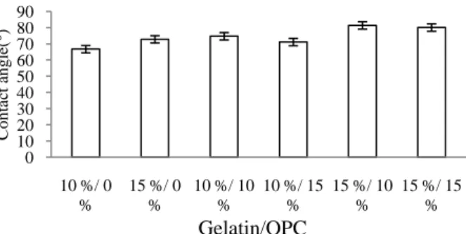
A specimen is hydrophilic if the water contact angle of the specimen is less than 90° and hydrophobic if that of the specimen is greater than 90°. Figure 3 showed that the contact angles of all gelatin/OPC composite membrane and pure gelatin membrane are less than 90°; these membranes are essentially hydrophilic. Compared with composite membranes with different gelatin contents, it appeared that the more gelatin the more stable of the membrane. Indeed, the more gelatin the more amino and hydroxyl functional groups and hence hydrogen bonds, which stabilize the structure, in the composite.
Figure 3. The contact angles of gelatin/OPC composite membrane and pure gelatin membrane.
FTIR
The membrane made of 10 wt% gelatin has peaks at 1560-1640 cm-1 and 1420 cm-1, which demonstrated the existence of NH2 and OH functional groups in gelatin. On the other hand, the membrane made of 15 wt% OPC has peaks at 3300 cm-1 and 1000-1250 cm-1, 2000-1500 cm-1, and
0 500 1000 1500 2000 2500 3000 3500 10 20 30 40 50 60 S w el li n g r at io (%) Time(min) Gelatin10﹪ OPC10﹪ Gelatin10﹪ OPC15﹪ Gelatin15﹪ OPC10﹪ Gelatin15﹪ OPC15﹪ 0 10 20 30 40 50 60 70 80 90 100 1 2 3 4 W ei g h t lo ss rat io (%) Time(hr) Gelatin10 ﹪OPC10 ﹪ Gelatin10 ﹪OPC15 ﹪ Gelatin15 ﹪OPC10 ﹪ Gelatin15 ﹪OPC15 ﹪ 0 10 20 30 40 50 60 70 80 90 10 %/ 0 % 15 %/ 0 % 10 %/ 10 % 10 %/ 15 % 15 %/ 10 % 15 %/ 15 % C o n ta ct a n g le (° ) Gelatin/OPC
1500-1300 cm-1, which demonstrated the existence of phenol group, benzene group, O-H, and C-H, respectively. As to gelatin/OPC composite microsphere, we found peaks at 1737 cm-1 and 1420 cm-1, which represent hydrogen bond and hydroxyl bond, respectively.
Figure 4 The FTIR of pure gelatin 10 wt%, pure OPC 15 wt%, and gelatin/OPC composite microspheres.
Conclusion
The fabrication process of gelatin/OPC composite microspheres was developed. Many of the physical properties of the composite microspheres were examined. We believed that based on these properties the developed gelatin/OPC composite microspheres can be used as a drug carrier to be used along with wound dressing.
Acknowledgement
This work would especially like to thank National Science Council of the Republic of China, Taiwan, for financially supporting this research under Contract NSC 94-2212-E-035-008.
Reference
[1] Y. Yumin and D. Richard: Carbohydratio Research, Vol.343 (2008), p. 2657–2666.
[2] W. J. Habraken, J.G. Wolke, A.G. Mikos and J.A. Jansen: J Biomed Mater Res B Appl Biomater. Vol. 91(2009), p.555-561.
[3] R. Cortesi, E. Esposito, M. Osti, G. Squarzoni and E. Menegatti, Eur J Pharm Biopharm Vol. 47 (1999), p.153–160.
[4] W.J.E.M. Habraken, L.T. de Jonge, J.G.C. Wolke, L. Yubao, A.G. Mikos and J.A. Jansen: J Biomed Mater Res A Vol. 3(2008), p.643-655.
[5] N.T. Annan, A.D. Borza and L.T. Hansen: Food Res Int Vol. 41(2008), p.184–193.
[6] W.J.E.M. Habraken, O.C. Boerman, J.G.C. Wolke, A.G. Mikos and J.A. Jansen: J. Biomed. Mater. Res. Part A Vol. 91A (2009), P.614-622.
[7] W. Zhai, J. Chang, K. Lin, J. Wang, Q. Zhao and X. Sun: Biomaterials Vol. 27(2006), p.3684–3690.
[8] B. Han, J. Jaurequi, B.W. Tang and M.E.Nimni: J Biomed Mater Res A Vol. 65(2003), p.118–124.
[9] S. Kim, M.E. Nimni, Z. Yang and B. Han: J Biomed Mater Res B Vol. 75(2005), p.442–450. [10] D. Bagchi, M. Bagchia, S.J. Stohsa, D.K. Dasb, S.D. Rayc, C.A. Kuszynskid, S.S. Joshid and
H.G. Prues: Toxicology Vol.148 (2000), p.187–197.
[11] B. Hughes-Formella, O. Wunderlich, R. Williams: Skin Pharmacol. Physiol. Vol. 20 (2007), p. 43–49.