Design and Development of Immunomodulatory
Antigen Delivery Systems Based on
Peptide/PEG-PLA Conjugate for Tuning Immunity
Fanny Coumes,†,∥ Chiung-Yi Huang,‡,∥ Chung-Hsiung Huang,‡ Jean Coudane,† Dominique
Domurado,†,§ Suming Li,¶ Vincent Darcos,†,* and Ming-Hsi Huang,‡,⊥,*
†Max Mousseron Institute of Biomolecules, UMR CNRS 5247, University of Montpellier,
Faculty of Pharmacy, 34093 Montpellier Cedex 5, France
‡National Institute of Infectious Diseases and Vaccinology, National Health Research Institutes,
35053 Miaoli, Taiwan
§Institut National de la Santé et de la Recherche Médicale, 34000 Montpellier, France
¶Institut Europeen des Membranes, UMR CNRS 5635, University of Montpellier, 34095
Montpellier, France.
⊥Graduate Institute of Immunology, China Medical University, 40402 Taichung, Taiwan.
KEYWORDS: cancer immunotherapy; conjugated polymers; vaccine adjuvants
ABSTRACT: Cancer vaccines are considered to be a promising tool for cancer immunotherapy. However, a well-designed cancer vaccine should combine a tumor-associated antigen (TAA) with the most effective immunomodulatory agents and/or delivery system to provoke intense immune responses against the TAA. In the present study, we introduced a new approach by conjugating the immunomodulatory molecule LD-indolicidin to the hydrophilic chain end of the polymeric emulsifier poly(ethylene glycol)-polylactide (PEG-PLA), allowing the molecule to be located close to the surface of the resulting emulsion. A peptide/polymer conjugate, named LD-indolicidin-PEG-PLA, was synthesized by conjugation of the amine end-group of LD-indolicidin to the N-hydroxysuccinimide-activated carboxyl end-group of PEG. As an adjuvant for cancer immunotherapeutic use, TAA vaccine candidate formulated with the LD-indolicidin-PEG-PLA-stabilized squalene-in-water emulsion could effectively help to elicit a T helper (Th)1-dominant antigen-specific immune response as well as anti-tumor ability, using ovalbumin (OVA) protein/EG7 cells as a TAA/tumor cell model. Taken together, these results open up a new approach to the development of immunomodulatory antigen delivery systems for vaccine adjuvants and cancer immunotherapy technologies.
INTRODUCTION
Immunotherapy is a type of treatment designed to use the patient’s own immune system to treat illness. Recent strategies for developing cancer immunotherapy have focused on the molecular identification of tumor-associated antigens (TAAs) and the induction of antigen-specific T cell-mediated immune responses that can induce tumor regression.1-4 In a typical approach,
autologous TAA-specific T cells are expanded ex vivo and then re-infused into patients.1,2 To
avoid the complications of leukapheresis and infusion schedules, another promising approach is vaccination to elicit TAA-specific T cell-mediated immune responses in vivo.3,4 However,
designing effective TAA-based therapeutic vaccine candidates against cancer is much more challenging than developing autologous cellular immunotherapies due to limited efficacy, which is caused by the fact that immune responses are suppressed in cancer patients.3 Hence, these
vaccines require the incorporation of an agent, dubbed an adjuvant (from the Latin adjuvare, meaning “to help”), to elicit efficient immune responses against tumor cells.3,4
Vaccine adjuvants are incorporated into many vaccines to enhance vaccine immunogenicity and efficacy.5,6 They can be broadly divided into two classes based on their principal
mechanisms of action: immunomodulatory adjuvants and delivery systems.6 In contrast to the
former, which are supposed to trigger immune responses, the latter mainly work as carriers to deliver biologically active agents (e.g., antigens and immunomodulatory adjuvants) to immune cells in a targeted and prolonged manner, thus efficiently promoting vaccine immunogenicity.6
Among the vaccine adjuvants evaluated in human trials, oil-in-water (O/W) emulsions have been successfully applied to increase the antigen-sparing efficacy in pandemic influenza vaccine preparedness.7 Recently, our research group investigated the use of synthetic, amphiphilic, and
development of vaccine delivery.8-12 Emulsified vaccine delivery systems can be stabilized
during the homogenization process and can be disintegrated and further resorbed in vivo.12
Immunogenicity studies in mice using ovalbumin (OVA),8 inactivated influenza virus,9 or protein
subunits10 as a model showed that such emulsions are able to induce potent antigen-specific
antibody responses. To extend these applications to a therapeutic vaccine or immunotherapy against cancers, our previous studies demonstrated that a human papillomavirus (HPV) E7 protein-derived peptide antigen formulated with an emulsion could induce greater anti-cancer immunity compared with peptide alone or peptide formulated with conventional aluminum-based mineral salts (Alum) adjuvant.11,12 To enlarge the selection of vaccine adjuvants for the design of
new-generation vaccines, we have also synthesized an analogue peptide, dubbed LD-indolicidin, that contains the regular enantiomeric sequence of the host defense peptide indolicidin (ILPWKWPWWPWRR-NH2) with alternating L- and D-amino acid substitution.13 Interestingly,
LD-indolicidin not only increases the resistance of the peptide segment to enzymatic degradation but also enhances cell-mediated immune responses better than the native form of indolicidin does when used as an adjuvant in an inactivated H5N1 influenza virus vaccine.13 Although our results
strongly suggest that LD-indolicidin may be a potential immunomodulatory adjuvant for controlling effective/harmful immune responses, its poor water solubility is an obstacle that has considerably restrained the potential of using LD-indolicidin in further applications.
Here, we report the design and development of an immunomodulatory antigen delivery system comprising an O/W emulsion-based delivery system and an immunomodulatory analogue peptide. A peptide/polymer conjugate, namely LD-indolicidin-poly(ethylene glycol)-polylactide (LD-indolicidin-PEG-PLA), was synthesized to provide a good vehicle for vaccine delivery. Conjugation of LD-indolicidin to water-soluble macromolecular carriers provides an approach to
aqueous solubilization. Moreover, attaching LD-indolicidin molecules to the hydrophilic PEG chain end of polymeric emulsifiers should allow the molecules to be located close to the surface of the emulsion. LD-indolicidin-PEG-PLA conjugate-stabilized emulsions present several advantages, including stability for long-term storage, proper size for cell uptake, and facilitation of Th1 polarization. Finally, the synergistic effect of emulsion and LD-indolicidin adjuvants lead good prognosis in tumor-bearing mice and offered the possibility of complementary enhancement to develop novel immunomodulatory antigen delivery systems for vaccine adjuvants and cancer immunotherapy technologies.
MATERIALS AND METHODS
Materials. Lactide was obtained from Purac Biochem (Goerinchem, The Netherlands). α-Hydroxy-ω-carboxyl PEG (Mn = 5000 g/mol) was purchased from RAPP Polymere GmbH
(Tuebingen, Germany). Toluene, dichloromethane (DCM) and dimethylformamide (DMF) were supplied by Sigma-Aldrich (Saint-Quentin-Fallavier, France) and were dried over calcium hydride for 24 hours at room temperature and then distilled under reduced pressure. Triethylamine was supplied by Sigma-Aldrich (Saint-Quentin-Fallavier, France) and dried over potassium hydroxide for 24 hours at room temperature and then distilled under reduced pressure. All other materials were also obtained from Sigma-Aldrich (Saint-Quentin-Fallavier, France) and were used without any further purification.
Characterization. 1H nuclear magnetic resonance (1H NMR) spectra were recorded on a
Bruker spectrometer (AMX300) operating at 300 MHz. Chemical shifts were referenced to the peak of residual non-deuterated solvents. Two-dimensional diffusion ordered spectroscopy (2D DOSY) NMR measurements were performed at 300 K on a Bruker Avance AQS600 NMR
spectrometer operating at 600 MHz and equipped with a Bruker multinuclear z-gradient inverse probe head capable of producing gradients in the z direction with a strength of 55 G.cm-1. The
DOSY spectra were acquired with the ledbpgp2s pulse program in Bruker Topspin software. All spectra were recorded with 32 K time-domain data points in t2 dimension and 32 t1 increments.
The gradient strength was logarithmically incremented in 32 steps, from 2 % up to 95 % of the maximum gradient strength. All measurements were performed with a compromise diffusion delay Δ of 200 ms to keep the relaxation contribution to the signal attenuation constant for all samples. The gradient pulse length δ was 5 ms to ensure full signal attenuation. The diffusion dimension of the 2D DOSY spectra was processed via Bruker Topspin software (version 2.1). The DOSY maps were obtained with Bruker Topspin software (version 2.1).
Size exclusion chromatography (SEC) was performed at room temperature using a Viscotek GPCmax system equipped with a Viscotek guard column (10×4.6 mm) and two Viscotek columns (LT 5000L, mixed medium, 300×7.8 mm), with a Viscotek VE 3580 refractometric detector and a Viscotek VE 3210 UV/Vis detector. Molar mass (Mn) and dispersity (Đ) were
expressed with respect to polystyrene standards from Polymer Laboratories. THF was used as the solvent, with a flow rate of 1 mL/min.
Synthesis of LD-indolicidin. LD-indolicidin was synthesized in-house by the solid-phase method using an automated peptide synthesizer (model PS-3 from Protein Technologies, Inc.) employing the fluorenyl methoxycarbonyl (Fmoc) group for α-amino group protection.13 The
resin used was derived from NovaSyn TGR resin with the modified Rink linker (Merck, Darmstadt, Germany). Tryptophan and lysine residues were protected with tert-butoxycarbonyl (tBoc), and arginine residues were protected with a 2,2,4,6,7-pentamethyldihydrobenzofuran-5-sulfonyl (Pbf) group. The final de-blocking step was carried out with a mixture of
TFA/triisopropylsilane/water (94:3:3). The crude peptides were recovered by precipitation with diethyl ether as a non-solvent and were characterized by analytical reverse-phase HPLC. Mass spectrometry analyses were performed on an Agilent 1100 Series LC/MSD high-performance ion-trap mass spectrometer to ensure that the target peptide was obtained.
Synthesis of carboxyl-PEG-block-PLA (HOOC-PEG-PLA). Ring-opening polymerization was carried out in solution using the standard Schlenk technique under an argon atmosphere. Lactide (0.88 g, 2 mmol, 20 equiv), Sn(Oct)2 (8.1 mg, 2×10⁻² mmol, 0.2 equiv), HOOC-PEG (0.5
g, 0.1 mmol, 1 equiv), and anhydrous toluene (3 mL) were placed in an oven-dried Schlenk tube. The tube was fitted with a rubber septum. The solution was further degassed by three freeze-pump-thaw cycles. The resulting mixture was stirred at 100°C for 5 hours. At the end of the reaction, the Schlenk tube was cooled to room temperature first. The reaction was stopped by addition of an excess of 1 N HCl. The reaction mixture was then poured into cold diethyl ether. The precipitated polymer was finally collected by filtration and dried in vacuo to yield 0.61 g of copolymer (yield 45 %).
Mn,SEC = 9100 g/mol; Đ = 1.08
Mn,NMR = 6300 g/mol 1H NMR (300 MHz, CDCl
3) δ (ppm) = 5.2 (m, O-CH(CH3)-C(O)), 4.3 (m, O-CH(CH3)-OH), 3.6
(m, CH2-CH2-O), 2.6 (t, 2H, C(O)-CH2-CH2-(CO2H)), 2.5 (t, 2H, C(O)-CH2-CH2-(CO2H)), 1.6
(m, O-CH(CH3)-C(O)).
Synthesis of N-hydroxysuccinimide-activated carboxyl-PEG-block-PLA (NHS-PEG-PLA). HOOC-PEG-PLA (0.60 g, 9.5×10⁻² mmol, 1 equiv) and DCM (4 mL) were placed in a
round-bottom flask. NHS (0.02 g, 1.9×10⁻¹ mmol, 2 equiv) was then added slowly at 0°C, and the
1.9×10⁻¹ mmol, 2 equiv) was dissolved in dry DCM (3 mL) and transferred into the solution
through the septum. The mixture was then stirred at room temperature for 24 hours. The solution was cooled to 0°C, and filtered to remove insoluble DCC. Finally, the polymer was collected by precipitation in diethyl ether, followed by filtration and subsequent vacuum drying in vacuo to yield 0.54 g of pure NHS-PEG-PLA (yield 85 %).
Mn,SEC = 9000 g/mol; Đ = 1.1
Mn,NMR = 6700 g/mol 1H NMR (300 MHz, CDCl
3) δ (ppm) = 5.2 (m, 1H, O-CH(CH3)-C(O)), 4.3 (m, O-CH(CH3)-OH),
3.7 (m, 4H, CH2-CH2-O), 2.9 (t, 2H, C(O)-CH2-CH2-(CO2N)), 2.8 (m, 4H, C(O)-CH2-CH2-C(O)),
2.6 (t, 2H, C(O)-CH2-CH2-(CO2N)), 1.6 (m, 3H, O-CH(CH3)-C(O)).
Synthesis of LD-indolicidin-PEG-PLA. LD-indolicidin (0.17 g, 8.9×10⁻² mmol, 1.1 equiv)
and DMF (2 mL) were placed into an oven-dried Schlenk tube. Triethylamine (TEA, 0.9 mL) was added to reach a pH between 7 and 9. A solution of NHS-PEG-PLA (0.54 g, 8×10⁻² mmol, 1
equiv) in dry DMF (5 mL) was then added dropwise into the Schlenk tube. The reaction was left stirring at room temperature for 18 hours. The solution was then precipitated in cold diethyl ether to remove the TEA and unreacted LD-indolicidin, yielding 0.593 g of LD-indolicidin-PEG-PLA (yield 76 %).
1H NMR (300 MHz, DMF-d
7) δ (ppm) = 11.5 (m, H Peptide), 7.6-6.8 (m, H Peptide), 5.2 (m,
1H, O-CH(CH3)-C(O)), 4.3 (m, O-CH(CH3)-OH), 4.2 (m, H Peptide), 3.7 (m, 4H, CH2-CH2-O),
2.9 (t, 2H, C(O)-CH2-CH2-C(O)NH), 2.6 (t, 2H,C(O)-CH2-CH2-C(O)NH), 1.6 (m, 3H,
O-CH(CH3)-C(O)), 1,1 (m, H Peptides).
Synthesis of PEG-PLA. Methoxy PEG-block-PLA copolymer (PEG-PLA) was synthesized for comparison with the LD-indolicidin-PEG-PLA conjugate. Lactide (3 g, 20.8 mmol), Sn(Oct)2
(90 mg, 22×10-2 mmol), and MeO-PEG (9 g, 1.8 mmol) were placed into a polymerization
ampoule. The mixture was further degassed and sealed under vacuum. ROP was carried out in bulk under vacuum at 140°C for 24 hours. The copolymer was dissolved in acetone and then poured into ethanol. The precipitated polymer was finally collected by filtration and was dried in vacuo to yield pure PEG-PLA.
Mn,SEC = 8500 g/mol; Đ = 1.08 1H NMR (300 MHz, CDCl
3) δ (ppm) = 5.2 (m, O-CH(CH3)-C(O)), 4.2 (m, O-CH(CH3)-OH), 3.6
(m, CH2-CH2-O), 3.3 (s, CH3-O-CH2-CH2-)), 1.6 (m, O-CH(CH3)-C(O)).
Polymer-stabilized emulsions. For the preparation of polymer-stabilized emulsions, 134 mg of PEG-PLA, 0.8 mL of phosphate-buffered saline (PBS), and 1.1 mL of squalene oil were emulsified using a Polytron® PT 3100 homogenizer (Kinematica AG, Swiss) at 6000 rpm for 5
min. A total of 4 mg of indolicidin was mixed with the emulsion to produce the LD-indolicidin/emulsion mixture. LD-indolicidin-PEG-PLA conjugate (18 mg) with an amount of the LD-indolicidin moiety equivalent to that in the preceding mixture was introduced into a PBS solution together with PEG-PLA (120 mg) to yield the conjugate-emulsion. The emulsified formulations were stored at 4°C until use. Emulsified vaccine preparation was investigated by re-dispersing 200 µL of stock emulsion in 1800 µL of aqueous solution and mixing the two in a test-tube rotator at 5 rpm for least 1 hour. Protein antigen was introduced in the aqueous solution to yield a vaccine formulation. The stability of the emulsion stocks was recorded at 4°C, 25°C and 37°C based on visual observation. The emulsion was re-dispersed in PBS, and the particle size was measured using an optical microscope (Olympus DP70, Olympus Inc., Tokyo, Japan) and the laser light scattering technique (Brookhaven 90Plus particle size analyzer, Brookhaven Instruments Limited, NY, USA).
Mice and ethics statement. Five-week-old female C57BL/6 mice were obtained from the National Laboratory Animal Center. All mice were housed at the Laboratory Animal Facility of the NHRI, Miaoli County, Taiwan. All animal studies were approved by the NHRI Institutional Animal Care and Use Committee (NHRI-IACUC-101040-AC).
Immunization and T cell immunoassays. C57BL/6 mice (3 mice/group) were injected subcutaneously (s.c.) in both hind footpads with 10 μg/mice of OVA (Grade V, Sigma, St. Louis, MO, USA) alone or formulated with LD-indolicidin (20 μg/mice), with LD-indolicidin-PEG-PLA conjugate (90 μg/mice), or with the emulsions/mixtures. Seven days after injection, spleens were collected from the immunized mice, and 5×10⁶ splenocytes were re-stimulated in triplicate
in the absence or presence of 50 μg/mL target OVA protein for 48 hours. The interferon (IFN)-γ, interleukin (IL)-2, IL-4 and IL-10 concentrations in the supernatants were measured by ELISA according to the manufacturer’s instructions (DueSet® ELISA Development kit, R&D Systems,
Inc., Minneapolis, MN).
Total RNA from the splenocytes in each group was extracted using TRI Reagent (Sigma, St. Louis, MO, USA) according to the manufacturer’s instructions. The steady-state mRNA expression of T-bet, GATA3 and β-actin was measured by reverse transcription-polymerase chain reaction (RT-PCR). All isolated RNA samples were confirmed to be free of DNA contamination, as determined by the absence of product after PCR amplification in the absence of reverse transcription. For reverse transcription, 10 μg of total RNA was reverse-transcribed into cDNA using the Maxime RT PreMix Kit (iNtRON Biotechnology) and random primers. The reverse transcription proceeded at 45°C for 60 min and then at 95°C for 5 min. Next, 2X PCR Master mix Solution (iNtRON Biotechnology, Kyungki-Do, Korea) and 10 pmol of each forward and reverse primer specific for the gene of interest were added to each cDNA sample for PCR.
The samples were heated to 94°C for 2 min and cycled 30-37 times at 94°C for 30 seconds, 55°C for 45 seconds, and 72°C for 60 seconds, followed by an additional step at 72°C for 5 min. The PCR products were electrophoresed in 2 % agarose gels and stained with 0.1 μg/mL SYBR®
Green (Thermo Fisher Scientific, Inc., CA, USA) for visualization. Quantification was performed by assessing the optical density of the DNA bands using the ImageJ image processing and analysis program (MD, USA), as described elsewhere.14 The results are expressed as the
density ratio between the gene of interest and the reference standard (β-actin).
Tumor challenge study. The EG7 cell line (American Type Culture Collection, CRL-2113), a stable transfectant of the murine OVA-expressing EL4 thymoma (H-2b), was maintained in complete RPMI 1640 medium supplemented with G418 (0.4 mg/ml; Calbiochem). A total of 2×10⁵ EG7 tumor cells per mouse were first inoculated s.c. into the mouse flank. Upon the
appearance of palpable tumors, 10 µg of OVA, which was non-formulated or formulated with candidate adjuvants, was injected s.c. at the base of the tail of mice on day 7. Tumor sizes were measured by using a caliper (Digimatic Caliper, Mitutoyo, Japan) in two vertical dimensions twice per week. Tumor volumes were calculated according to the following formula: (length x width2)/2. The mice were euthanized when the tumor volume exceeded 2000 mm3 or when they
experienced severe faintness. The median survival was calculated using the Gehan-Breslow-Wilcoxon method.
Statistical analysis. The graphs and statistical analyses were performed using GraphPad Prism version 5.02 (GraphPad Software, Inc.). Comparison of T cell immunity and tumor volume between groups was conducted by use of ANOVA followed by a Bonferroni post-test. The differences were considered significant at p < 0.05.
RESULTS AND DISCUSSION
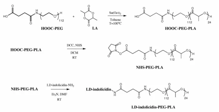
Synthesis of LD-indolicidin-PEG-PLA conjugate. LD-indolicidin-PEG-PLA conjugate was prepared by combination of ring-opening polymerization and conjugation chemistry (Figure 1). An α-carboxyl-PEG-block-PLA (HOOC-PEG-PLA) copolymer was first obtained by ring-opening polymerization of lactide using α-hydroxy-ω-carboxyl PEG as a macroinitiator. The carboxyl moiety of HOOC-PEG-PLA was then activated by reaction with NHS. Finally, LD-indolicidin was attached to the PEG-PLA copolymer by conjugation of the amine end-group of LD-indolicidin to the NHS-activated carboxyl end-group of PEG. The resulting copolymer was characterized by SEC and 1H NMR. SEC of HOOC-PEG-PLA revealed a monomodal
distribution, with an Mn,SEC and Đ of 9100 g/mol and 1.1, respectively (see Supporting
Information Figure S1). Moreover, a clear shift toward a higher molecular weight was observed in comparison with the starting PEG, thus evidencing block copolymer formation. The molecular structure of HOOC-PEG-PLA was also determined by 1H NMR (see Supporting Information
Figure S2). The peaks at 5.2 and 1.6 ppm were associated with the PLA backbone, whereas the signal at 3.6 ppm was assigned to protons from the PEG. The peaks at 2.6 and 2.5 ppm were related to the methylene protons close to the carboxyl end-group. The molar ratio between the two components was calculated from the relative intensity of the methyne protons of PLA units at 5.2 ppm and the methylene protons of PEG units at 3.6 ppm, and was found to be 4.9. Accordingly, the molar mass (Mn,NMR) was calculated from the 1H NMR signals, and was found to
be 6300 g/mol.
The HOOC-PEG-PLA was allowed to react with NHS in the presence of DCC to yield α-block-PLA (PLA). The SEC traces of HOOC-PEG-PLA and NHS-PEG-PLA showed unchanged molar masses and dispersities (see Supporting Information Figure S1b),
indicating that no chain cleavage had occurred during activation of the carboxyl group. The incorporation of the NHS moiety was evidenced by a downfield shift of the methylene protons close to the carboxyl end-group and by the appearance of NHS methylene protons at 2.8 ppm (see Supporting Information Figure S2b). NHS anchoring was found to be quantitative.
The synthesis of LD-indolicidin was performed according to a previously reported procedure.13 LD-indolicidin was attached to the PEG-PLA copolymer by conjugating the amine
end-group of LD-indolicidin to the NHS-activated carboxyl end-group of PEG, yielding peptide/copolymer conjugate. The efficiency of the peptide ligation was confirmed by 2D DOSY NMR, a powerful tool to characterize block copolymers.15 DOSY NMR is a 2D NMR technique
in which the signal decays exponentially due to the self-diffusion behavior of molecules. This phenomenon leads to two dimensions: the first dimension (F2) accounts for the conventional chemical shift, and the second one (F1) accounts for self-diffusion coefficients (D). Thus, each component in a mixture can be virtually separated based on its own diffusion coefficient on the diffusion dimension. Figure 2 shows the DOSY map of the peptide/polymer conjugate in dilute DMF-d7. The 1H NMR spectrum exhibited signals corresponding to the PLA block (δ = 5.2 and
1.6 ppm), PEG block (δ = 4.3 and 3.7 ppm), and peptide block (δ = 6.8-7.6 ppm). The 1H NMR
signals of the three different blocks exhibited the same diffusion coefficient, or D = 9×10-11 m2/s,
in agreement with efficient ligation of the peptide to the copolymer. No free peptide was observed because the diffusion coefficient of the peptide in dilute DMF-d7 was D = 1.6×10⁻¹⁰
m2/s. The diffusion coefficient of the residual protons of DMF-d
7 was found to be D = 1.26×10⁻⁹
m2/s. As peptide/polymer conjugate cannot be characterized by conventional SEC, DOSY NMR
is often considered as chromatographic NMR, providing evidence of the successful attachment of peptide.
Amphiphilic copolymers generally present a core-shell micellar architecture in aqueous solution, with a core consisting of hydrophobic blocks and a shell composed of chains.6 Dynamic
light scattering (DLS) measurements were performed to confirm the micelle sizes in the present study (see Supporting Information Figure S3). The data showed that the PEG-PLA polymeric aqueous solution contained micelles with a unimodal distribution and an average diameter of 25 ± 5 nm. It appears that coupling the LD-indolicidin moiety to the end of the PEG did not significantly alter the size or the size distribution of the polymeric micelles (23 ± 1 nm). In contrast, LD-indolicidin was found to be insoluble in PBS at room temperature.
Preparation of LD-indolicidin-PEG-PLA-stabilized emulsion. In the preliminary experiment, we found that coupling LD-indolicidin to PEG-PLA induces phenotypic and functional maturation of murine bone marrow derived dendritic cells (BMDCs) in vitro (see Supporting Information Figure S4). We next sought to investigate the adjuvant potency by using PEG-PLA or LD-indolicidin-PEG-PLA conjugate as an emulsifier component to stabilize the interfaces between the oil and the aqueous solution. PEG-PLA or LD-indolicidin-PEG-PLA was dissolved in PBS, mixed with squalene oil, and then homogenized, resulting in an isotropic emulsion (Figure 3a). Squalene was selected as the core oil because it is a natural human metabolite and the precursor of cholesterol, and therefore the precursor of vitamin D and many hormones.16 Squalene has been used as a skin moisturizer in cosmetics and as an adjuvant in
licensed human vaccines.7,16 In the current study, the dispersion type of emulsion was
investigated by the droplet test and light scattering. PEG-PLA- and LD-indolicidin-PEG-PLA-stabilized squalene droplets could stand in aqueous solution only for a few seconds and then diffused in the water, indicating the continuous phase of the water.8 This result is similar to the
findings of our previous work, which showed that the dispersion type of PEG-PLA-stabilized squalane (a saturated form of squalene) emulsions belongs to the O/W emulsion type.12
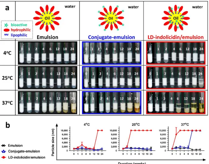
DLS showed that the PEG-PLA-emulsified particles possessed a unimodal distribution, with an average diameter of approximately 1000 nm. The emulsion remained stable for 24 weeks when stored at 4°C or 25°C. However, mixing the emulsion with LD-indolicidin significantly destroyed the squalene/water interface. The particle size increased due to the disassociation of the immiscible squalene and water beyond week 2 and week 18 at 25°C and 4°C, respectively (Figure 3b). It is interesting to note that little difference was observed between PEG-PLA- and conjugate-stabilized emulsions, indicating that PEG-PLA bearing the peptide LD-indolicidin moiety could preserve the emulsifier behavior intrinsic to PEG-PLA. In contrast, emulsion instability occurred during storage at 37°C. This feature confirms that loss of the PLA moiety of the PEG-PLA emulsifier directly affected the stability of a PEG-PLA- or conjugate-stabilized emulsion, leading to emulsion disintegration and squalene/water phase separation.12 We also
attempted to study the morphology of the prepared emulsions by electron microscopy techniques; however, the morphology could not be observed by either SEM or TEM because the cryo-fixed emulsion samples fractured under vacuum (data not shown).
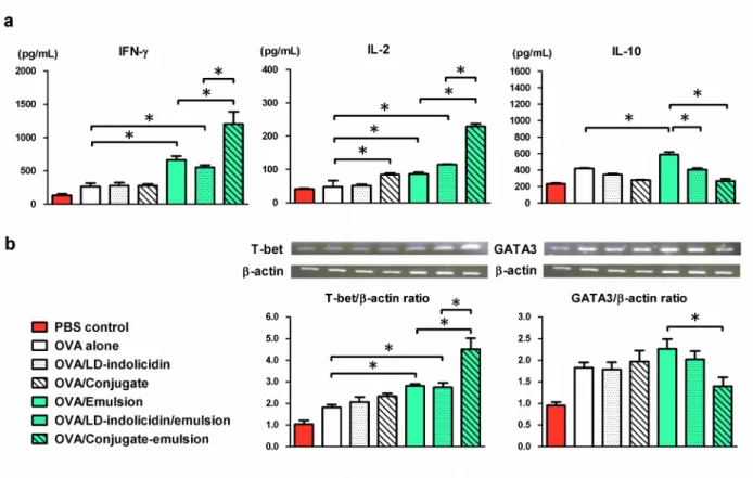
LD-indolicidin-PEG-PLA-stabilized emulsion reshapes cell-mediated immunity in vivo. To determine the impact of the candidate compounds on immunity, we immunized mice through footpad injection with OVA alone or OVA formulated with different candidate formulations. T cell cytokine responses were measured in the spleen following re-stimulation of the cells in vitro with the OVA antigen. Figure 4a showed that following vaccination, OVA alone and OVA formulated with LD-indolicidin or the conjugate did not conspicuously induce antigen-specific cytokine production, such that the IFN-γ, IL-2, and IL-10 concentrations were at the same level
as those in the control group. On the other hand, OVA formulated with squalene-based emulsion did enhance a notable cellular response, and the IFN-γ, IL-2 and IL-10 concentrations detected in the splenocyte supernatants were significantly higher than those in the non-adjuvanted group. Nevertheless, the cytokine IL-4 was found to be at an undetectable level. It should be noted that IFN-γ and IL-2 are predominant T helper (Th)1-type cytokines relevant to cytotoxic T lymphocyte (CTL) activity, whereas IL-4 and IL-10 are common Th2-type cytokines.6
Interestingly, sufficiently elevated IFN-γ and IL-2 secretion was detected in splenocyte supernatants collected from mice treated with conjugate-emulsions, whereas the Th2-type cytokine IL-10 was at the reduced level compared with the level in the no-adjuvant group (Figure 4a). We next characterized the mRNA expression of transcription factors, including T-bet (Th1) and GATA3 (Th2),14 in both T cell subsets to assess T cell differentiation.
Immunization with OVA formulated with the conjugate or the emulsion augmented T-bet and GATA3 mRNA expression compared with immunization with OVA alone (Figure 4b). Nevertheless, immunization with conjugate-emulsion increased T-bet mRNA expression but diminished GATA3 mRNA expression compared with immunization without the formulation (Figure 4b). These findings suggest that LD-indolicidin-PEG-PLA conjugate may be a potential immunomodulatory agent that shifts the Th1/Th2 immune balance toward Th1 polarization.
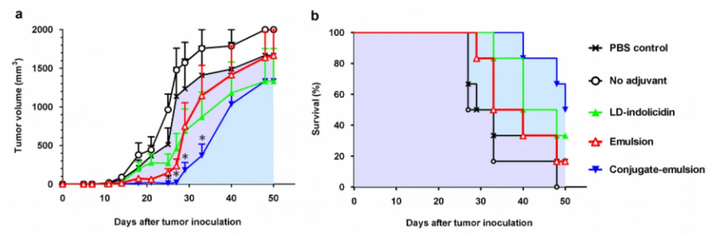
Formulating TAA with LD-indolicidin-PEG-PLA-emulsified particles enhances anti-tumor efficacy. To evaluate the adjuvanticity of LD-indolicidin-PEG-PLA conjugate-emulsified particles, we applied a cancer immunotherapy consisting of OVA protein/EG7 cells (a thymoma cell line stably transfected with OVA complementary DNA) as a TAA/tumor cell model.17 Mice
were first inoculated with 2×10⁵ EG7 tumor cells/mouse and were then immunized with a single
Figure 5. Without any treatment (PBS control group), the tumors grew progressively, and the mice started to die within 30 days (Figure 5a). No protection was observed for the mice that received OVA alone; in this case, all mice died before day 60. Vaccination with OVA plus LD-indolicidin or emulsion provided a better protective effect than no adjuvant did but did not eliminate the inoculated EG7 cells. Nevertheless, it is noteworthy that the conjugate-emulsion was able to broaden the immunotherapeutic efficacy of OVA and to prolong the median survival of EG7-bearing mice from 30 days to 50 days. Furthermore, only the mice that received the conjugate-emulsion-formulated OVA vaccine reached up to 50 % survival 50 days post-tumor implantation (Figure 5b).
Many studies aimed at developing recombinant TAA vaccines for the treatment of cancers such as melanoma and non-small-cell lung carcinoma.2,3 In this approach, the TAA vaccine
antigen is first taken up by presenting cells (APCs) following injection. The antigen-loaded APCs then fragment the antigen into antigenic peptides for presentation through the MHC class I and MHC class II pathways to CTLs and T-helper cells, respectively.5 The former cells
can in turn attack tumor cells that express cognate antigenic determinants, and T-helper cells can provide help to B cells during antibody production, leading to tumor cell death.1 However, many
studies have demonstrated that immunization with antigenic peptides or proteins alone is not sufficient to elicit MHC class I- and II- restricted responses.3 To achieve effective vaccination,
the first step is to provide sufficient danger/alarm signals through vaccine antigens and/or an adjuvant. In this regard, the development of an adjuvant with high potential to enhance cellular responses is the key to improving the prospects of TAA-based cancer vaccines. Combination adjuvants comprising a particulate delivery system and an immunomodulatory compound are promising choices, as they offer the possibility of complementary and synergistic enhancement
of immune responses against vaccine antigens. Our tumor challenge studies demonstrate that antigen alone neither inhibits tumor growth nor induces tumor regression. We also confirmed that the immunogenicity of OVA antigen is enhanced by using a single adjuvant, such as an immunomodulatory adjuvant, and an antigen delivery system (Figure 4). LD-indolicidin-PEG-PLA conjugate facilitates the immune cell activation elicited by the LD-indolicidin moiety, thus generating valuable tumor inhibition and tumor regression (Figure 5).
Concerning the size of the particles, it is also noteworthy that small solutes or nanoparticles (< 50 nm) are internalized by APCs through macropinocytosis, whereas O/W emulsions with submicron size can be internalized by APCs through phagocytosis, without specific recognition.18 In contrast, toll-like receptor ligands such as CpG oligodeoxynucleotides and host
defense peptides are internalized by DCs through receptor-mediated endocytosis.19 It should be
noted that the dimensions of conjugate-stabilized squalene-emulsion particles allow internalization by APCs, probably via both receptor-mediated endocytosis and phagocytosis, thus facilitating the induction of cell-mediated immunity (Figure 4). Therefore, LD-indolicidin-PEG-PLA conjugate-stabilized emulsions present several advantages, including stability for long-term storage, proper size for DC uptake, facilitation of Th1 polarization, and enhancement of the anti-tumor efficacy of candidate TAAs. Further investigations are underway to integrate this immunotherapy with cancer chemotherapeutic agents, such as docetaxel or paclitaxel, to prolong the survival of tumor-bearing mice. Such applications will require further investigations whether the incorporation of these bioactive molecules can influence the stability and immunomodulatory efficacy of LD-indolicidin-PEG-PLA-based formulations.
Here, we reported the first investigation of co-administration of LD-indolicidin peptide together with an emulsified vaccine delivery system. Targeting immunomodulatory molecules to the hydrophilic chain end of polymeric amphiphile provides an approach to aqueous solubilization. Moreover, LD-indolicidin-PEG-PLA conjugate could serve as a hydrophilic emulsifier to stabilize the squalene/water interfaces and yield stable oil-in-water particles with proper size for cell uptake. As an adjuvant for cancer immunotherapeutic use, TAA vaccine candidate formulated with such an emulsion could help to effectively elicit appropriate antigen-specific T cell immunity, thus inducing tumor regression. Our results are of great interest for the design of cancer immunotherapy.
Figure 1. Synthesis of LD-indolicidin-PEG-PLA conjugate. HOOC-PEG-PLA copolymer was first obtained by ring-opening polymerization of lactide monomer in solution at 100°C using α-hydroxy-ω-carboxyl PEG at 5000 g/mol as a macroinitiator and Sn(Oct)2 as a catalyst. The
HOOC-PEG-PLA was allowed to react with NHS in the presence of DCC to yield NHS-PEG-PLA in the second step. LD-indolicidin was attached to the PEG-NHS-PEG-PLA copolymer by conjugating the amine end-group of LD-indolicidin to the NHS-activated carboxyl end-group of PEG, yielding the peptide/copolymer conjugate LD-indolicidin-PEG-PLA.
Figure 2. DOSY NMR spectra of LD-indolicidin-PEG-PLA conjugate in DMF-d7. The 1H NMR
spectrum exhibited signals corresponding to the PLA block (δ = 5.2 and 1.6 ppm), PEG block (δ = 4.3 and 3.7 ppm), and peptide block (δ = 6.8-7.6 ppm). The three different blocks exhibited the same diffusion coefficient (blue), in agreement with efficient ligation of the peptide to the NHS-PEG-PLA copolymer.
Figure 3. (a) Visual aspects and (b) laser light scattering analysis of the emulsion upon storage at 4°C, 25°C, or 37°C for 24 weeks. At the very beginning, homogeneous, fine particles with a mean size of 1000 nm were observed for all three emulsions. In the cases of PEG-PLA emulsions and conjugate-emulsions, the formulations remain stable for 24 weeks when they were at 4°C or 25°C, whereas the LD-indolicidin/emulsion mixture became instable at 25°C after 24 weeks. For calculation purposes, an undetectable particle size was scored as a diameter equal to 10000 nm. The data are expressed as the mean value with standard deviation of three samples.
Figure 4. Analysis of T cell immunity. (a) Cytokine secretion responses and (b) mRNA expression levels. C57BL/6 mice (3 mice/group) were injected once s.c. in both hind footpads with 10 μg/mL OVA alone or OVA formulated with various adjuvant candidates. Seven days after the immunization, splenocyte suspensions (5×10⁶ cells/mL) were pooled and incubated in
the presence or absence of 50 μg/mL OVA protein for 72 hours. Supernatants from triplicate cultures were collected to measure the concentrations of IFN-γ, IL-2 and IL-10 by ELISA via paired antibodies. The data are presented as cytokine release in the presence of OVA minus release in the presence of medium only. In parallel, the mRNA expression of T-bet and GATA3 was measured by RT-PCR. The data are expressed as the mean value ± standard deviation. Statistical significance was determined by performing ANOVA followed by a Bonferroni post-test. *p < 0.05. The data are representative of two independent experiments.
Figure 5. Anti-tumor efficacy of OVA protein formulated with different adjuvants and administered to C57BL/6 mice bearing EG7 tumor cells. (a) Tumor volume and (b) survival rate. Mice were inoculated s.c. in the flank with EG7 tumor cells (2×10⁵ cells/mouse). Upon the
appearance of palpable tumors, six mice per group were injected s.c. at the tail base with 10 µg/dose OVA protein with or without adjuvant on day 7. Tumor sizes were assessed twice per week using calipers to determine the volume of each tumor. The tumor volumes are shown (mm3). The data are expressed as the mean value ± standard deviation. Gray-filled area: PBS
control group; blue-filled area: conjugate-emulsion group. The tumor volumes were compared on days 25, 27, 29 and 33, following the onset of tumor growth in the vaccine group. Statistical significance was determined by performing ANOVA followed by a Bonferroni post-test. *p < 0.05 compared with no adjuvant group.
ASSOCIATED CONTENT
Supporting Information
Additional information related to molecular characterization and cell culture study. This material is available free of charge via the Internet at http://pubs.acs.org.
AUTHOR INFORMATION
Corresponding Author
*Address: Vincent Darcos, Max Mousseron Institute of Biomolecules, UMR CNRS 5247, University of Montpellier, Faculty of Pharmacy, 34093 Montpellier Cedex 5, France. E-mail: [email protected]; Address: Ming-Hsi Huang, National Institute of Infectious Diseases and Vaccinology, National Health Research Institutes, No. 35 Keyan Road, Zhunan Town, Miaoli County 35053, Taiwan. E-mail: [email protected].
Author Contributions
∥Fanny Coumes and Chiung-Yi Huang contributed equally to this work.
Notes
The authors declare no competing financial interest.
ACKNOWLEDGMENTS
This work was supported by grant 104A1-IVPP26-014 from the National Health Research Institutes of Taiwan and by grant NSC-102-2320-B-400-001-MY2 from the Ministry of Science and Technology of Taiwan. The authors are grateful to the CNRS, the French Ministry of Education, and the Max Mousseron Institute of Biomolecules for the grant to Fanny Coumes.
Additionally, the authors are grateful to Mr. Sheng-Kuo Chiang and Mr. Chih-Wei Lin for their help in preparing the materials and to Sylvie Hunger for the NMR analysis.
REFERENCES
(1) Ledford, H. Nature 2014, 516, 156.
(2) Cheever, M. A.; Higano, C. S. Clin. Cancer Res. 2011, 17, 3520-3526. (3) Tefit, J. N.; Serra, V. Expert Rev. Vaccines 2011, 10, 1207-1220. (4) Brichard, V. G.; Lejeune, D. Vaccine 2007, 25S, B61-B71.
(5) Reed, S. G.; Orr, M. T.; Fox, C. B. Nat. Med. 2013, 19, 1597-1608.
(6) Huang, M. H.; Leng, C. H.; Liu, S. J.; Chen, H. W.; Sia, C.; Chong, P. In Immunogenicity; Villanueva, C. J., Ed.; Nova Science Publishers: New York, 2011; p 61-90.
(7) Vogel, F. R.; Caillet, C.; Kusters, I. C.; Haensler, J. Expert Rev. Vaccines 2009, 8, 483-492.
(8) Huang, M. H.; Chou, A. H.; Lien, S. P.; Chen, H. W.; Huang, C. Y.; Chen, W. W.; Chong, P.; Liu, S. J.; Leng, C. H. J. Biomed. Mater. Res. B Appl. Biomater. 2009, 90, 832-841.
(9) Huang, M. H.; Huang, C. Y.; Lin, S. C.; Chen, J. H.; Ku, C. C.; Chou, A. H.; Liu, S. J.; Chen, H. W.; Chong, P.; Leng, C. H. Microb. Infect. 2009, 11, 654-660.
(10) Lin, S. C.; Jan, J. T.; Dionne, B.; Butler, M.; Huang, M. H.; Wu, C. Y.; Wong, C. H.; Wu, S. C. PLoS One 2013, 8, e66719.
(11) Song, Y. C.; Cheng, H. Y.; Leng, C. H.; Chiang, S. K.; Lin, C. W.; Chong, P.; Huang, M. H.; Liu, S. J. J. Control. Release 2014, 173, 158-165.
(12) Chen, W. L.; Liu, S. J.; Leng, C. H.; Chen, H. W.; Chong, P.; Huang, M. H. Biomaterials 2014, 35, 1686-9165.
(13) Chang, C. Y.; Lin, C. W.; Chiang, C. K.; Chen, P. L.; Huang, C. Y.; Liu, S. J.; Chong, P.; Huang, M. H. ACS Med. Chem. Lett. 2013, 4, 522-526.
(14) Huang, C. H.; Liu, D. Z.; Jan, T. R. J. Nat. Prod. 2010, 73, 1033-1037.
(15) Bakkour, Y.; Darcos, V., Li, S.; Coudane, J. Polym. Chem. 2012, 3, 2006-2010. (16) Allison, A. C. Methods 1999, 19, 87-93.
(17) Ding, Q.; Chen, J., Wei, X.; Sun, W.; Mai, J.; Yang, Y.; Xu, Y. Pharm. Res. 2013, 30, 60-69.
(18) Shen, K. Y.; Song, Y. C.; Chen, I. H.; Leng, C. H.; Chen, H. W.; Li, H. J.; Chong, P.; Liu, S. J. J. Immunol. 2014, 192, 4233-4241.
(19) Kovacs-Nolan, J.; Latimer, L.; Landi, A.; Jenssen, H.; Hancock, R. E. W.; Babiuk, L. A.; van Drunen Littel-van den Hurk, S. Vaccine 2009, 27, 2055-2064.