" " " " " "
Metabolite Profiling and Comparison of Bioactivity
in Antrodia cinnamomea and Antrodia salmonea
Fruiting Bodies
Authors
Chieh-Yin Chen 1, 2, Shih-Chang Chien 3, Nai-Wen Tsao 1, Chiem-Sing Lai 1, Ya-Yun Wang 1, Wen-Wei Hsiao 2,
Fang-Hua Chu 4, Yueh-Hsiung Kuo 5, 6, Sheng-Yang Wang 1, 7
Affiliations
The affiliations are listed at the end of the article
Key words l Antrodia cinnamomea l Antrodia salmonea l Polyporaceae l metabolites profiling l anti-inflammatory activity l cytotoxicity Correspondence
Prof. Sheng-Yang Wang National Chung-Hsing University
Department of Forestry 250 Kou Kung Road Taichung 402 Taiwan Phone: + 886 422 840 345 ext. 138 Fax: + 886 422 873 628 [email protected] Abstract !
Antrodia cinnamomea is a precious edible mush-room endemic to Taiwan that has been claimed to have significant health promotion activities. trodia salmonea is a new species of the genus An-trodia. In this study, we compared the metabolites and bioactivity of A. cinnamomea and A. salmonea fruiting bodies. The volatiles of A. cinnamomea and A. salmonea were characterized and 3,4,5-trime-thoxybenzaldehyde was found to be the most abundant compound in A. cinnamomea; the other abundant compounds were δ-guaiene, isolongifo-lene, 1-octen-3-ol, 4-terpinenol, α-guaiene, and p-cymene. In A. salmonea, the main volatiles were α-cedrene, 1-octen-3-ol, D-limonene, cadinadiene, germacrene D, isolongifolene, and α-muurolene. Furthermore, five ergostane-type triterpenoids and two lanostane-type triterpenoids were se-lected as index compounds characterizing A. cin-namomea and A. salmonea extracts. The content of each compound varied between the two spe-cies. (R,S)-antcin B was the most abundant com-pound in A. cinnamomea fruiting bodies (75.18 ± 0.11 µg/mg). However, (R,S)-antcin C (184.85 ± 0.96 µg/mg) was the major triterpenoid in the A. salmonea fruiting body. Furthermore, two com-pounds, antcin M and methyl antcinate K, were only present in the A. salmonea fingerprint; therefore, antcin M and methyl antcinate K may be important for distinguishing between A.
cinna-Introduction
!
Antrodia cinnamomea (syn. Antrodia camphorata and Taiwanofungus camphorate) is a precious edi-ble mushroom that has long been the most highly valued medicinal fungus in Taiwan. Traditionally, A. cinnamomea has been used as a folk remedy for various diseases including cancer, hypertension, abdominal pains, and diarrhea [1]. The extract of
momea and A. salmonea fruiting bodies. Finally, examination of anti-inflammation activity and cy-totoxicity showed that A. salmonea had more anti-inflammatory activity than A. cinnamomea; how-ever, A. salmonea was more cytotoxic than A. cin-namomea. In conclusion, the composition and bio-activity of the fruiting bodies of A. cinnamomea and A. salmonea varies. Therefore, it is recom-mended that further toxicological evaluation and investigation of the biological activity of A. salmo-nea is carried out to ensure its safe and efficacious use as an alternative to A. cinnamomea.
Abbreviations
!
DMEM:
Dulbeccoʼs modified Eagle medium
FBS:
fetal bovine serum
GC/MS:
gas chromatography/mass spectrom-etry
HPLC: high-performance liquid chromatog-raphy
LPS: lipopolysaccharide
MTT: 3-[4,5-dimethylthiazol-2-yl]-2,5-di-phenyltetrazolium bromide NMR: nuclear magnetic resonance NO: nitric oxide
SPME: solid-phase microextraction
UPLC-MS: ultra-performance liquid chromatog-raphy-mass spectrometry
A. cinnamomea has also been used as a food intox-icant since antiquity [2]. Owing to its perceived efficacy, A. cinnamomea dry fruiting bodies are sold at prices exceeding US$15 000 per kilogram in the local market in Taiwan, and the total market value of A. cinnamomea products, including raw fruiting bodies and health foods, is estimated to be over US$100 million per year [3]. One of the most important reasons for the high price of
" " " " " " " "
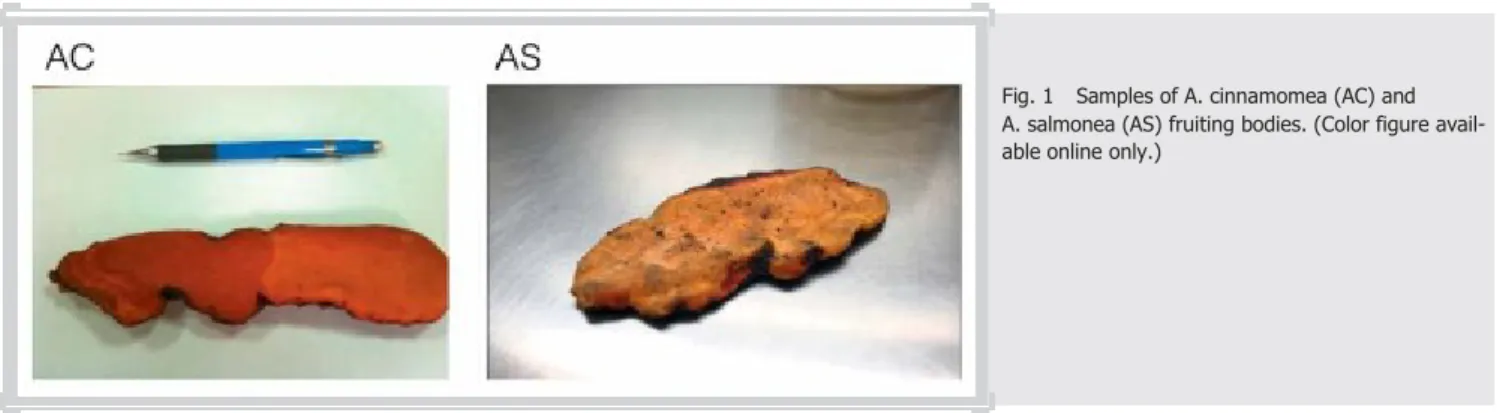
Fig. 1
Samples of A. cinnamomea (AC) and A. salmonea (AS) fruiting bodies. (Color figure avail-able online only.)
A. cinnamomea is that it only grows on the inner surface of the heartwood cavity of the evergreen tree Cinnamomum kanehirai Hayata (Lauraceae), which is an endangered species endemic to Taiwan. Antrodia salmonea (l Fig. 1) is a new species of the genus Antrodia, which was originally hosted by the indigenous conifer-ous tree Cunninghamia konishii Hayata (Cupressaceae) [4]. This mushroom is similar to A. cinnamomea. Both A. salmonea and A. cinnamomea have a strong bitter taste; however, the color of A. cinnamomea is cardinal red whereas A. salmonea is salmon-pink, and it is often easy to confuse the two species. Because of the difficulty in producing A. cinnamomea, as well as its high market price, A. salmonea is often used as a substitute for A. cinnamomea in the marketplace. So far, more than 250 scien-tific papers have been published on A. cinnamomea [1, 5–7], but literature on A. salmonea is rare. To our knowledge, around ten research articles have been published on A. salmonea to date. For the development of functional foods or phytomedicines, quality control, efficacy approbation, and safety are the three most im-portant requirements. Since A. salmonea now is commonly used as a substitute for A. cinnamomea, verification of the chemical in-gredients and bioactivity of A. cinnamomea and A. salmonea is an In the present study, the chemical compositions of A. salmonea and A. cinnamomea were analyzed by using SPME-GC/MS and HPLC profiling, and the cytotoxicity and anti-inflammation activity of these two mushrooms were also studied.
Results and Discussion
!
Odor is a significant distinguishing characteristic of the fruiting bodies of A. cinnamomea and A. salmonea. The volatile com-pounds of A. cinnamomea and A. salmonea were collected by us-ing SPME to obtain the volatiles, and analysis was done by GC/MS.
l Table 1 shows the compositions of volatile compounds emitted from A. cinnamomea and A. salmonea fruiting bodies. In the A. salmonea fruiting body, 3,4,5-trimethoxybenzaldehyde (30.04 %) was the most abundant compound, followed by δ-guaiene (9.88 %), isolongifolene (8.00 %), 1-octen-3-ol (7.08%), 4-terpine-nol (5.99 %), α-guaiene (5.99%), and p-cymene (4.96 %). In con-trast, the main compositions of volatiles of A. salmonea were α-cedrene (14.68 %), 1-octen-3-ol (9.31 %), D-limonene (9.21 %), ca-dinadiene (7.65 %), germacrene D (7.22 %), isolongifolene (6.72 %), and α-muurolene (5.31 %).
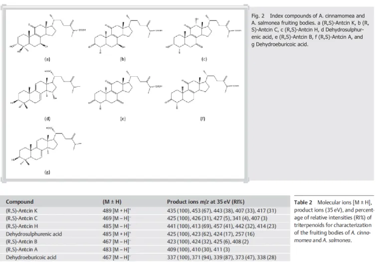
To date, more than 80 compounds have been identified from A. cinnamomea, including triterpenoids, benzolics, and poly-acetylenes [5]. In our previous study, we selected 13 index com-pounds to establish a comprehensive profile of the ethanol extract
of A.
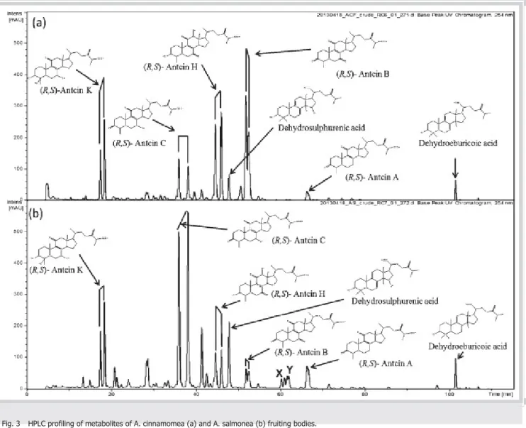
cinnamomea fruiting body [8]. Triterpenoids in both the my-celium and fruiting body are now considered to be the most bio-logically active components of A. cinnamomea [9]. Thus, five ergo-stane-type triterpenoids, i.e., antcin K, antcin C, (R,S)-antcin H, (R,S)-(R,S)-antcin B, and (R,S)-(R,S)-antcin A, as well as two lano-stane-type triterpenoids, dehydrosulphurenic acid and dehy-droeburicoic acid, were used as the index compounds to charac-terize the A. cinnamomea and A. salmonea extracts in this study. The structure and MS analysis data are shown in l Fig. 2 and l Ta-ble 2. Compounds a to g in l Fig. 2 were then used as index com-pounds to profile metabolites in A. cinnamomea and A. salmonea extracts. l Fig. 3 shows the HPLC metabolite profiles for
" " " " " "
Fig. 2
Index compounds of A. cinnamomea and A. salmonea fruiting bodies. a (R,S)-Antcin K, b (R, S)-Antcin C, c (R,S)-Antcin H, d Dehydrosulphur-enic acid, e (R,S)-Antcin B, f (R,S)-Antcin A, and g Dehydroeburicoic acid.
Some of
the compounds, including (R,S)-antcin K, (R,S)-antcin C, (R,S)-antcin H, and (R,S)-antcin B, showed two peaks, for R and S configurations, as these compounds contain a chiral center at the C25 position. We were not able to characterize the absolute
con-figuration for this group of compounds directly by NMR; so fur-ther R/S derivatives will be needed to determine their absolute configuration. Thus, both R-form and S-form triterpenoids were treated as the same compound in this study. To measure the con-tent of the index compounds in A. cinnamomea and A. salmonea samples, calibration curves of the index compounds were estab-lished using five dilution standards from 10 to 1000 µg/mL. The contents of eight index compounds were determined by the peak area in the HPLC profile and calculated by using the calibration curves of index compounds (purity > 99.5 %; l Table 3). The ergo-stane-type triterpenoid (R,S)-antcin B was the most abundant compound in the A. cinnamomea fruiting body (75.18 ± 0.11 µg/ mg) followed by (R,S)-antcin H (48.77 ± 0.31 µg/mg), (R,S)-antcin C (43.36 ± 0.76 µg/mg), and (R,S)-antcin A (19.86 ± 0.12 µg/mg). However, (R,S)-antcin C was the dominant triterpenoid in the A. salmonea fruiting body with the content of (R,S)-antcin C in the extract being up to 184.85 ± 0.96 µg/mg. (R,S)-antcin A was the second most abundant triterpenoid in A. salmonea with the content being 57.85 ± 0.11 µg/mg, followed by (R,S)-antcin H (19.86 ± 0.28 µg/mg) and (R,S)-antcin K (18.61 ± 0.33 µg/mg). A comparison of the quantity of the lanostane-type triterpenoids with ergostane-type-triterpenoids (dehydrosulphurenic acid and dehydroeburicoic acid) showed that the amounts of
lano-stane-type triterpenoids in A. cinnamomea and A. salmonea were lower than ergostane-type triterpenoids.
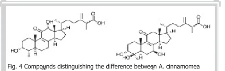
The results of the triterpenoid analysis for A. cinnamomea and A. salmonea showed that all the index compounds selected in this study could be detected in both A. cinnamomea and A. salmonea fruiting bodies, but the content of each triterpenoid varied in the two mushrooms. A comparison of the metabolite profiles of A. cinnamomea and A. salmonea (l Fig. 3) showed that there were two more peaks (X and Y) present in A. salmonea at the re-tention time of 60 to 62 min (l Fig. 3 B). The MS, 1HNMR, and 13CNMR spectral data were in good agreement with antcin M (X)
and methyl antcinate K (Y), which were reported previously [10]. Antcin M and methyl antcinate K (l Fig. 4) might be important constituents for distinguishing A. cinnamomea and A. salmonea fruiting bodies.
To evaluate the anti-inflammation activity of the extracts from A. cinnamomea and A. salmonea fruiting bodies, an LPS-stimulat-ed murine macrophage assay system was usLPS-stimulat-ed. l Table 4 shows the inhibitory effects of the extracts of A. cinnamomea and A. salmonea fruiting bodies. IC50 values (50 % inhibitory
concen-tration) of A. cinnamomea and A. salmonea were 73.89 µg/mg and 66.3 µg/mg, respectively. Test cells were healthy and viable at doses ranging from 10 to 80 µg/mL, as determined by the MTT colorimetric assay (data not shown). In addition to anti-inflam-mation activity, the cytotoxicity was evaluated for A. cinnamo-mea and A. salmonea extracts. As shown in the results presented in l Table 4, the A. cinnamomea extract possessed stronger
cyto-Fig. 3
HPLC profiling of metabolites of A. cinnamomea (a) and A. salmonea (b) fruiting bodies.
toxicity than A. salmonea. The IC50 value against MCF-7 cells was
59.18 µg/mL for A. cinnamomea and 91.45 µg/mL for A. salmonea. A. cinnamomea grown on its original host, C. kanehirai, has the highest market value [4]. However, excessive felling of C. kanehir-ai is prohibited by the government of Tkanehir-aiwan. A. salmonea is a similar mushroom to A. cinnamomea, and because the cultivation of wood for A. salmonea is easy to obtain, A. salmonea has become a common substitute for A. cinnamomea in the marketplace. Nu-merous studies have discussed the metabolites of A. cinnamomea and its bioactivities; however, to date, investigations of A. salmo-nea are rare.
Although some studies have analyzed the volatile compounds emitted from the mycelium of A. cinnamomea, no volatiles have been characterized for A. cinnamomea and A. salmonea fruiting bodies until now [11]. In the current study, first we characterized the volatiles of A. cinnamomea and A. salmonea fruiting bodies. According to our analysis, 3,4,5-trimethoxybenzaldehyde was the most abundant compound in the A. cinnamomea fruiting body. The other abundant compounds were δ-guaiene, isolongi-folene, 1-octen-3-ol, 4-terpinenol, α-guaiene, and p-cymene. In the case of A. salmonea, the most abundant volatiles were α-ce-drene, 1-octen-3-ol, D-limonene, cadinadiene, germacrene D, isolongifolene, and α-muurolene. The odor of the A. cinnamomea and A. salmonea fruiting bodies is quite different. A. cinnamomea
has a relatively rich woody odor, while A. salmonea has a lighter odor. δ-guaiene, isolongifolene, 4-terpinenol, and α-guaiene have distinguished woody odors and, therefore, the volatile compound composition might determine the different odors of A. cinnamo-mea and A. salmonea.
In addition to a comparison of the chemicals of A. cinnamomea and A. salmonea, the metabolite fingerprints of A. cinnamomea and A. salmonea were established by using HPLC. We purified the major triterpenoids, including five ergostane-type triterpe-noids and two lanostane-type triterpetriterpe-noids, the compound
"
Fig. 4 Compounds distinguishing the difference between A. cinnamomea and A. salmonea fruiting bodies; antcin M (X) and methyl antcinate K (Y).
structures of which were confirmed by UPLC-MS and NMR anal-ysis. These index compounds were used as the index compounds to identify and quantify the triterpenoids in the A. cinnamomea and A. salmonea metabolite fingerprints. Overall, the ergostane-type and lanostane-ergostane-type triterpenoids were detected both in the fingerprints of A. cinnamomea and A. salmonea. The A. cinnamo-mea fingerprint was similar to the metabolite profiling, which we established previously [8]. All of the index compounds shown in the A. salmonea fingerprint were observed in A. cinnamomea; however, the content of each compound was different in the two mushrooms. For the A. cinnamomea fruiting body, (R,S)-antcin B, (R,S)-antcin H, and (R,S)-antcin C were abundant compounds at 75.18 ± 0.11, 48.77 ± 0.31, and 43.36 ± 0.76 µg/mg, respectively. Interestingly, (R,S)-antcin C was the dominant compound in A. salmonea with levels of up to 184.85 ± 0.96 µg/mg. The second most abundant triterpenoid in A. salmonea was (R,S)-antcin A (57.85 ± 0.11 µg/mg) followed by (R,S)-antcin H (19.86 ± 0.28 µg/ mg) and (R,S)-antcin K (18.61 ± 0.33 µg/mg). In addition, we found a further two compounds in A. salmonea that were absent in A. cinnamomea. According to the spectral analysis, the pounds were antcin M and methyl antcinate K. These two com-pounds might be important for distinguishing A. cinnamomea and A. salmonea fruiting bodies.
Obviously, the compositions of A. cinnamomea and A. salmonea were dissimilar. We speculate that these different compositions might affect the bioactivity of A. cinnamomea and A. salmonea. In our previous study, the ethanolic extracts of A. cinnamomea exhibited potent anti-inflammatory activity in vitro and in vivo [2]. To evaluate the anti-inflammatory activity of A. cinnamomea and A. salmonea extracts, an LPS-stimulated murine macrophage assay system was used. The anti-inflammatory activity of A. salmonea extract (IC50 = 66.3 µg/mg) was slightly higher than
that of A. cinnamomea (IC50 = 73.89 µg/mg). (R,S)-antcin C is the
strongest anti-inflammatory triterpenoid in the A. cinnamomea fruiting body with an IC50 = 5.48 µg/mL [8]. However, A. salmonea
contained a higher amount of (R,S)-antcin C than A. cinnamomea. This might be the reason why A. salmonea possessed higher anti-inflammatory activity than A. cinnamomea. In addition to evalu-ating the anti-inflammatory activity, we examined the cytotoxic-ity of A. cinnamomea and A. salmonea against MCF-7 cell lines. The results showed that A. cinnamomea (IC50 = 59.18 µg/mg) was
more cytotoxic than A. salmonea (IC50 = 91.45 µg/mg). According
to the results of a cytotoxicity assay by Du and his coworkers, (R, S)-antcin H and (R,S)-antcin B exhibited stronger cytotoxicity against human leukemia cell lines in comparison with other er-gostane-type triterpenoids in the A. cinnamomea fruiting body [9]. (R,S)-antcin H and (R,S)-antcin B were abundant in A. cinna-momea. This might be why A. cinnamomea possessed stronger cytotoxicity than A. salmonea.
According to this study, we can distinguish between A. cinnamo-mea and A. salmonea by fingerprints, especially since two com-pounds, antcin M and methyl antcinate K, were only present in the A. salmonea fingerprint. A. salmonea had more anti-inflam-matory activity than A. cinnamomea, however, A. salmonea was more cytotoxic than A. cinnamomea. Nowadays, A. salmonea is often used as a substitute for A. cinnamomea. However, the com-position and levels of bioactivity are different in these two mush-rooms. Therefore, for safety reasons, we recommend a toxicolog-ical evaluation and biologtoxicolog-ical activity investigation of A. salmonea for use as an alternative to A. cinnamomea.
Materials and Methods
!
Antrodia cinnamomea and Antrodia salmonea fruiting bodies
The A. cinnamomea and A. salmonea fruiting bodies were col-lected by Mr. Wen-Wei Hsiao who is an assistant research fellow in Experimental Forest, National Taiwan University. A. cinnamo-mea was collected from the remained wood of C. kanehirai dis-tributed at the secondary growth of forest, which is located at Suili countryside, Nantou County, Taiwan. A. salmonea was also collected from its host wood, C. konishii. The remained wood of C. konishii was located at Sun-Link-Sea region in Nantou County, Taiwan. The samples were identified by Dr. Sheng-Yang Wang (Professor of Department of Forestry, Nation Chung-Hsing Uni-versity). The voucher specimens (TCFAC0001 and TCFAS0001) were deposited in the herbarium of the same university. l Fig. 1 shows the morphology of the fruiting bodies analyzed in this study.
Volatile metabolite analysis of Antrodia salmonea and Antrodia cinnamomea fruiting bodies by using solid-phase microextraction combined with gas chromatography coupled to mass spectrometry An SPME holder and carboxen-polydimethylsiloxane-coated fi-bers (75 mm) were purchased from Supelco (Bellefonte). The sample bottle was placed in a water bath (40 ± 2 °C) and condi-tioned (15 min, without fiber). Before use, SPME fibers were con-ditioned by heating in a hot injection port of a GC at 200 °C for 15 min to remove contaminants. After the equilibration time, the fiber was introduced into the sample bottle and exposed to the gases in the headspace of the A. salmonea and A. cinnamomea powder for 15 min. After 15 min, the SPME fiber was inserted in-to the injection port of the GC using an SPME liner for desorption at 120 °C for a 5-s splitless period. The analysis of all samples was performed using a Thermo Scientific ITQ 900 GC/MS equipped with a DB-5 column (30 m × 0.25 mm × 0.25 µm, Agilent J&W Sci-entific). The GC oven temperature was programmed from 40 °C, held 1 min, raised to 100 °C at 4 °C/min, then raised to 250 °C at
15 °C/min and held for 3 min. The sample injection was in the splitless mode. The flow rate of the carrier gas, helium, was at 1.0 mL/min. The Kovats indices were calculated for all volatile constituents using a homologous series of n-alkanes C9-C24. The
major components were identified by coinjection with standards (wherever possible), confirmed with Kovats indices using the Wi-ley (Ver. 8.0) and National Institute of Standards and Technology (NIST) Ver. 2.0 GC/MS libraries.
Metabolite profiling and quantification of ethanol extract of A. cinnamomea and A. salmonea fruiting bodies The protocol for the preparation of the ethanol (EtOH) extract of A. salmonea and A. salmonea fruiting bodies was according to our previous method [8]. Briefly, fresh material was lypholized for 72 h and A. cinnamomea and A. salmonea were ground to a pow-der (particle diameter < 0.7 mm), accurately weighed (around 5 g), placed in an Erlenmeyer flask (250 mL) with 100 mL EtOH, and sonicated in an ultrasonicator (Branson 5510, Branson Ultra-sonic) for 60 min. The extracts were then decanted, filtered under vacuum, concentrated in a rotary evaporator, and lyophilized. The metabolite profile of the ethanol extract prepared from the A. cinnamomea and A. salmonea were established by using seven index compounds, namely antcin K, antcin C, (R,S)-antcin H, dehydrosulphurenic acid, (R,S)-(R,S)-antcin B, (R,S)-(R,S)-antcin A, and dehydroeburicoic acid, which were identified by us previ-ously [8] and the structure was reconfirmed by using UPLC-MS analysis. An amaZon speed ion trap (Bruker) was set to an ion source temperature 250 °C with N2 as dry gas at 9.0 L min-1 with
the capillary voltage at 4500 V, the end plate offset at 500 V, and a scan range of 70 to 600 m/z in the positive and negative ioniza-tion modes. The unknown peaks presented in the metabolite profiling of A. salmonea were collected and identified by spectra analysis. UV spectra were recorded on a Jasco V-550 spectropho-tometer and IR spectra were recorded on a Bio-Rad FTS-40 spec-trometer. Electrospray ionization-mass spectrometric spectrom-etry data were collected with a Finnigan MAT-95 S mass spec-trometer, and NMR spectra were recorded with Bruker Avance 400 MHz FT-NMR spectrometers at 400 MHz (1 H) and 100 MHz
(13 C). d-Chloroform (CDCl3) was used for NMR analysis. All
spec-troscopic analysis data were in good agreement with the litera-ture. The standard calibration curves (peak area vs. concentra-tion) of each index compound were determined at the range of compound concentrations of 10, 25, 50, 100, 250, 500, and 1000 µg/mL. Quantification of the content of each index com-pound in fruiting bodies from A. cinnamomea and A. salmonea was then performed by HPLC analysis. The peak areas of the in-dex compounds in the chromatogram of the EtOH extracts (with known loading concentration) were then defined, and their con-tents in the extracts were calculated on the basis of the quantity calibrated from the standard calibration curves. The analyses were performed in triplicate and the results are presented as mean ± SE.
Nitric oxide inhibitory assay
The effect of A. cinnamomea and A. salmonea extracts on NO production was measured indirectly by analysis of nitrite levels using the Greiss reaction [12, 13]. Briefly, RAW 264.7 cells grown in a 75-cm2 culture dish and were seeded in 96-well plates at a
density of 2 × 10⁵ cells/well. Cells were cultured at 37 °C in DMEM
supplemented with 10 % FBS, 100 units/mL penicillin, and 100 µg/ mL streptomycin in a 5 % CO2 incubator as recommended by the
American Type Culture Collection (ATCC). Adherent cells were
then incubated with or without 1 µg/mL of LPS for 24 h in the presence or absence of A. cinnamomea and A. salmonea extracts. The nitrite concentration (as an estimate of NO production) was measured using the supernatant from the RAW 264.7 cells by the Griess reaction [14]. Curcumin (> 95 %, Sigma Co.) was used as a positive control.
Cytotoxicity analysis
MCF-7 (human breast adenocarcinoma, BCRC 60 436) was pur-chased from the Bioresource Collection and Research Center (BCRC), Food Industry Research, and Development Institute, Tai-wan. MCF-7 cells were cultured in DMEM supplemented with 10 % FBS, 1 % penicillin–streptomycin, and 1 mM sodium pyru-vate, and were maintained at 37 °C and 5 % CO2. All cells (1 × 10³
per well) were seeded in 96-well plates and incubated for 24 h, and different dosages of extracts of A. cinnamomea and A. salmo-nea extracts were added to each well in triplicate for 24 h. The cell viability was determined by the MTT assay [15]. Plumbagin (> 95%, Sigma Co.) was used as a positive control. Statistical analysis
Data are expressed as means ± SE. The significance of the differ-ences between group means was determined by analysis of var-iance (ANOVA) using Dunnettʼs test. Mean values within each col-umn with different labels (a, b, c, d) are significantly different (p < 0.05).
Acknowledgements
!
This study was supported by the Ministry of Science and Tech-nology, Taiwan (MOST-104–2911-I-005–301), and the Ministry of Education, Taiwan, under the ATU plan.
Conflict of Interest
!
The authors declare that they have no competing interests.
Affiliations
1 Department of Forestry, National Chung-Hsing University, Taichung, Taiwan 2 Experimental Forest, College of Bio-Resources and Agriculture, National
Taiwan University, Nantou, Taiwan
3 The Experimental Forest Management Office, National Chung-Hsing
University, Taichung, Taiwan
4 School of Forestry and Resource Conservation, National Taiwan University,
Taipei, Taiwan
5 Department of Chinese Pharmaceutical Sciences and Chinese Medicine
Resources, China Medical University, Taichung, Taiwan
6 Department of Biotechnology, Asia University, Taichung, Taiwan
7 Agricultural Biotechnology Research Center, Academia Sinica, Taipei, Taiwan,
Taiwan
References
1 Lu MC, El-Shazly M, Wu TY, Du YC, Chang TT, Chen CF, Hsu YM, Lai KH, Chiu CP, Chang FR, Wu YC. Recent research and development of Antro-dia cinnamomea. Pharmacol Ther 2013; 139: 124–156
2 Hsieh YH, Chu FH, Wang YS, Chien SC, Chang ST, Shaw JF, Chen CY, Hsiao WW, Kuo YH, Wang SY. Antrocamphin A, an anti-inflammatory princi-pal from the fruiting body of Taiwanofungus camphoratus, and its mechanisms. J Agric Food Chem 2010; 58: 3153–3158
3 Wang HC, Chu FH, Chian SC, Liao JW, Hsieh HW, Li WH, Lin CC, Shaw JF, Kuo YH, Wang SY. Establishment of the metabolite profile for an Antro-dia cinnamomea health food product and investigation of its chemo-prevention activity. J Agric Food Chem 2013; 61: 8556–8564
4 Chang TT, Chou WN. Antrodia cinnamomea reconsidered and A. salmo-nea sp. nov. on Cunninghamia konishii in Taiwan. Bot Bull Acad Sin 2004; 45: 347–352
5 Geethangili M, Tzeng YM. Review of pharmacology effects of Antrodia camphorata and its bioactivities compounds. Evid Based Complement Alternat Med 2011; 2011: 212641
6 Ao ZH, Xu ZH, Lu ZM, Xu HY, Zhang XM, Dou WF. Niuchangchih (Antrodia camphorata) and its potential in treating liver diseases. J Ethnophar-macol 2009; 121: 194–212
7 Yue PY, Wong YY, Wong KY, Tso YK, Leung KS. Current evidence for the hepatoprotective activities of the medicinal mushroom Antrodia cin-namomea. Chin Med 2013; 8: 21
8 Lin TY, Chen CY, Chien SC, Hsiao WW, Chu FH, Li WH, Lin CC, Shaw JF, Wang SY. Metabolite profiles for Antrodia cinnamomea fruiting bodies harvested at different culture ages and substrates from different wood. J Agric Food Chem 2011; 59: 7626–7635
9 Du YC, Wu TY, Chang FR, Lin WY, Hsu YM, Cheng FT, Lu CY, Yen MH, Tsui YT, Chen HL, Hou MF, Lu MC, Wu YC. Chemical profiling of the cytotoxic triterpenoid-concentrating fraction and characterization of ergostane stereo-isomer ingredients from Antrodia camphorate. J Pharm Biomed Anal 2012; 58: 182–192
10 Shen CC, Wang YH, Chang TT, Lin LC, Don MJ, Hou YC, Liou KT, Chang S, Wang WY, Ko HC, Shen YC. Anti-inflammatory ergostanes from the ba-sidiomata of Antrodia salmonea. Planta Med 2007; 73: 1208–1213 11 Xia Y, Zhang B, Li W, Xu G. Changes in volatile compound composition
of Antrodia camphorata during solid state fermentation. J Sci Food Agric 2011; 91: 2463–2670
12 Wang SY, Chang HN, Lin KT, Lo CP, Yang NS, Shyur LF. Antioxidant prop-erties and phytochemical characteristics of extracts from Lactuca indi-ca. J Agric Food Chem 2003; 51: 1506–1512
13 Hsieh YH, Kuo PM, Chien SC, Shyur LF, Wang SY. Effects of Chamaecyparis formosensis Matasumura extractives on lipopolysaccharide-induced release of nitric oxide. Phytomedicine 2007; 14: 675–780 14 Schmidt HHHW, Kelm M. Determination of nitrite and nitrate by the
Griess reaction. In: Feelisch M, Stamler J, editors. Methods in nitric ox-ide research. Chichester: Wiley; 1996: 491–497
15 Chen WL, Lin TY, Tseng YH, Chu FH, Chueh PJ, Kuo YH, Wang SY. Inhib-itory effect of human breast cancer cell proliferation via p 21-mediated G1 cell cycle arrest by araliadiol isolated from Aralia cordata Thunb. Planta Med 2011; 77: 164–168