Anti-inflammatory Spirostanol and Furostanol Saponins from Solanum macaonense
Chia-Lin Lee,†,‡ Tsong-Long Hwang,§ Juan-Cheng Yang,†,‡ Hao-Ting Cheng,‡ Wan-Jung He,‡
Chiao-Ting Yen,┴ Chao-Lin Kuo,║ Chao-Jung Chen,,○ Wen-Yi Chang,§,Δ and Yang-Chang Wu*,†,‡,┴
† School of Pharmacy, College of Pharmacy, China Medical University, Taichung 40402, Taiwan
‡ Chinese Medicine Research and Development Center, China Medical University Hospital, Taichung
40447, Taiwan
§ Graduate Institute of Natural Products, Chang Gung University, Taoyuan 33302, Taiwan
┴ Graduate Institute of Natural Products, Kaohsiung Medical University, Kaohsiung 80708, Taiwan
║ Department of Chinese Pharmaceutical Sciences and Chinese Medicine Resources, College of
Pharmacy, China Medical University, Taichung 40402, Taiwan
Graduate Institute of Integrated Medicine, China Medical University, Taichung 40402, Taiwan
○ Proteomics Core Laboratory, Department of Medical Research, China Medical University Hospital,
Taichung 40447, Taiwan
Δ Research Center for Industry of Human Ecology, Chang Gung University of Science and Technology,
ABSTRACT: Eight new spirostanol saponins, macaosides A–H (1–8) and ten new furostanol saponins,
macaosides I–R (9–18), together with six known spirostanol compounds (19–24) were isolated from
Solanum macaonense. The structures of the new compounds were determined from their spectroscopic
data, and the compounds were tested for in vitro antineutrophilic inflammatory activity. It was found that both immediate inflammation responses including superoxide anion generation and elastase release were significantly inhibited by treatment with compounds 20, 21, and 24 (superoxide anion generation:
IC50 7.0, 7.6, 4.0 µM; elastase release: IC50 3.7, 4.4, 1.0 µM, respectively). However, compounds 1 and 4
exhibited effects on the inhibition of elastase release only, with IC50 values of 3.2 and 4.2 µM,
respectively, while 19 was active against superoxide anion generation only, with an IC50 value of 6.1
µM. Accordingly, spirostanols may be promising lead compounds for further neutrophilic inflammatory
The plant genus Solanum (Solanaceae) is distributed in tropical and subtropical regions of the world and has about 1200 species. Eighteen species are found in Taiwan, including Solanum macaonense Dunal
that grows in the southwestern plains.1 As a part of an ongoing investigation on anti-inflammatory
agents from Taiwanese Solanum species, it was found that the 95% MeOH-soluble and n-BuOH-soluble extracts of the aerial parts of S. macaonense showed activity against superoxide anion generation and
elastase release induced by formyl-L-methionyl-L-leucyl-L-phenylalanine (FMLP)/cytochalasin B (CB)
in human neutrophils.However, neither phytochemical nor biological studies of S. macaonense have
been reported to date.
In the current study, chromatographic fractionation of the two above-mentioned soluble extracts of S.
macaonense led to the isolation of 24 compounds. Among these secondary metabolites, eight new
spirostanol saponins, macaosides A–H (1–8), ten new furostanol saponins, macaosides I–R (9–18), and six known spirostanol compounds, chlorogenin (19),2 saponin Sc 4 (20),2 saponin Sc 3 (21),2
chlorogenin 6-O-β-D-glucopyranoside (22),3 chlorogenin 6-O-β-D-xylopyranosyl-(1→3)-β-D
-glucopyranoside (23),4 and 6α-O-β-D-xylopyranosyl-(1→3)-β-D-
quinovopyranosyl-(25R)-5α-spirostan-3β-ol (24)2 were identified. Also, their antineutrophilic inflammatory effects and preliminary
structure-activity relationship (SAR) properties were investigated.
RESULTS AND DISCUSSION
The MeOH extract of the aerial parts of S. macaonense was partitioned into n-hexane-, 95% MeOH-,
n-BuOH- and H2O-soluble fractions, using an initial EtOAc and H2O (1:1) extract. Fractionation of the
six known compounds (19–24).
HRESIMS of 1 showed a [M+Na]+ ion at m/z 731.3982, indicating a molecular formula of
C38H60O12Na. The IR spectrum showed absorptions for hydroxy group (3401 cm-1) and carbonyl (1705
cm-1) functionalities. Thirty-eight carbon signals, including five methyls, eleven methylenes, eighteen
methines, and four quaternary carbons, were observed in the 1D-NMR spectra of 1 (Tables 1 and 2).
One quaternary carbon was identified as a carbonyl carbon on the basis of the chemical shift at C 210.6.
The 1D- and HSQC NMR spectra displayed two anomeric [δH 4.75 (d, J = 7.7 Hz)/δC 105.4, δH 5.25 (d,
J = 7.7 Hz)/δC 106.2] and two oxymethylene [δH 3.48 (t, J = 10.6 Hz), 3.57 o/δC 66.8, δH 3.69 o, 4.29
(dd, J = 11.5, 4.9 Hz)/δC 67.3] signals. The 1H and 13C NMR data of 1 were similar to those of 24, a
known spirostanol glycoside,2 with differences apparent only in the chemical shifts at positions 2, 3 and
4 in the A ring [ 1: δH 2.31, 2.41 (H2-2)/δC 38.1 (C-2), δC 210.6 (C-3), δH 2.46, 3.57 (H2-4)/δC 39.8 (C-4);
24: δH 1.74, 2.03 (H2-2)/δC 32.1 (C-2), δH 3.77 (H-3)/δC 70.6 (C-3), δH 1.68, 3.21 (H2-4)/δC 33.2 (C-4)].
The HMBC data (Figure 1) indicated that a carbonyl group (δC 210.6) was present at C-3 in 1 rather than
the hydroxy group (δC 70.6) found in 24. The 25R configuration of 1 was deduced from small
differences in chemical shift values (ab = δa − δb; 25S: ab > 0.35 ppm; 25R: ab < 0.2 ppm)5 between the
H2-23 (ab = 0.05), H2-24 (ab = 0) and H2-26 (ab = 0.09) geminal protons, as well as the H2-26 signal
pattern [δ 3.48 (t, J26eq,26ax = 10.6 Hz, J26eq,25ax = 10.6 Hz) and δ 3.57, overlapped with H-4 and H-4′].2,5-7
The absolute configurations of the sugar units, quinovose andxylose, were both determined to be D by
GC analysis. Figure 1 shows key COSY and HMBC correlations observed for 1. ROESY correlations
absence of ROESY correlations between Me-19/H-5, H-9, or Me-18/H-14, H-16, H-17 indicated that the C-6 sugar group is α-oriented and the four A/B, B/C, C/D, and D/E ring junctions are trans, trans,
trans, cis, respectively. Macaoside A (1) was elucidated, therefore, as
(25R)-6α-hydroxy-5α-spirostan-3-one 6-O-β-D-xylopyranosyl-(1→3)-β-D-quinovopyranoside.
Compound 2 gave the molecular formula, C40H66O13, as established by HRESIMS (m/z 777.4399
[M+Na]+). The 1D-NMR data of 2 were also similar to those of 1 except that the chemical shift of one
quaternary carbon was identified at C 100.7 instead of 210.6 and two methoxy group signals appeared
at δH 3.20 s/δC 47.3 and δH 3.24 s/δC 47.5, respectively. The HMBC 3J correlations of H-1 (δ 1.21, 1.50)
and OCH3 (δ3.20, 3.24) with C-3 (δ 100.7) as well as a long-range W-type COSY correlation of H-1
with OCH3 suggested that the two methoxy groups were both at C-3. Key HMBC, COSY, and ROESY
correlations are shown in Figures 1 and 3. Thus, compound 2 (macaoside B) was elucidated as
(25R)-3,3-dimethoxy-5α-spirostan-6α-ol 6-O-β-D-xylopyranosyl-(1→3)-β-D-quinovopyranoside, which is a
ketal form of 1. The dimethoxy group functionality may be produced from a carbonyl group under
anhydrous conditions and acid catalysis.3 However, compound 2 is not considered to be an extraction
artifact from 1 since acidic or alkaline reagents were not applied during the extraction, partition, and isolation procedures.
The molecular formula of 3 was deduced as C38H62O13 due to the appearance of a [M+Na]+ ion at
m/z 749.4086 in the HRESIMS. Furthermore, compound 3 showed similar spectroscopic data to five
known 23-hydoxyspirostanols, including (22R,23S,25R)-3β,6α,23-trihydroxy-5α-spirostane 6-O-β-D
6-O-β-D-xylopyranosyl-(1→3)-O-β-D-quinovopyranoside, (22R,23R,25S)-3β,6α,23-trihydroxy-5α-spirostane
6-O-β-D-xylopyranosyl-(1→3)-O-β-D -quinovopyranoside,8 6α-O-[β-D-xylopyranosyl-(1→3)-β-D
-quinovopyranosyl]-(25S)-5α-spirostan- 3β,23β-ol, and 6α-O-[β-D-xylopyranosyl-(1→3)-β-D
-quinovopyranosyl]-(25S)-5α-spirostan-3β,23α-ol.6 As shown in Figure 1, compound 3 has the same
planar structure as the above-mentioned known glycosides. The configuration at C-22 in 3 was determined as S (22-α-O), since the signal of H-20 was shifted to 3.05 owing to the presence of equatorial hydroxy group at C-23. Additionally, a 22S configuration was confirmed by the chemical shifts of H3-21 (δ 1.20) and C-20 (δ 35.8) (22-α-O-23-hydoxyspirostanol: H3-21 δ 1.07−1.26; C-20 δ
35.0−36.2; 22-β-O-23-hydoxyspirostanol: H3-21 δ 1.53−1.54; C-20 δ 43.1−44.1).7 ROESY correlations
were observed between H-23 and H-25 (Figure 3), indicating H3-27 to be in an equatorial position with
a 25R configuration. The new compound 3 (macaoside C) was therefore identified as
(22S,23S,25R)-3β,6α,23-trihydroxy-5α-spirostan 6-O-β-D-xylopyranosyl-(1→3)-β-D-quinovopyranoside.
The HRESIMS of 4 showed a [M+Na]+ ion at m/z 719.3987 (C
37H60O12Na), and hydroxy group
(3242 cm-1) absorption was observed in the IR spectrum. Based on its NMR data, compound 4 could be
assigned as a having a C27 steroidal skeleton with two sugar moieties, resembling the spirostanol saponin
24.2 The 1D- and HSQC NMR spectra displayed two anomeric [δ
H 4.80 (d, J = 7.3 Hz)/δC 106.0, δH 5.27
(d, J = 7.6 Hz)/δC 106.3] and three oxymethylene [δH 3.44 (t, J = 10.5 Hz), 3.54 (dd, J = 11.0, 3.0 Hz)/δC
66.8, δH 3.63 m, 4.30 m/δC 66.3, δH 3.74 m, 4.34 m/δC 67.4] signals. The key correlations shown in
Figure 1 suggested that a hydroxyl group substituent and a disaccharide unit were positioned at C-3 (δ
7.3 Hz) and H-1″ (δ 5.27, d, J = 7.6 Hz) exhibited 3J interactions with C-6 (δ 78.9) and C-3′ (δ 87.2),
respectively. HMBC and COSY correlations (Figure 1) indicated that the glycosidic portion of 4 was a xylopyranosyl-(1→3)-xylopyranosyl unit, connected to C-6 of the aglycone moiety via an O-linkage.
The absolute configuration of xylose was identified to be D by GC analysis. Key ROESY correlations
are shown in Figure 3. Consequently, 4 (macaoside D) was characterized as
(25R)-5α-spirostan-3β,6α-diol 6-O-β-D-xylopyranosyl-(1→3)-β-D-xylopyranoside.
Compounds 5 and 6 gave the same molecular formula (C39H64O13Na, m/z 763.4239 for 5;
C39H64O13Na, 763.4235 for 6) in the HRESIMS. The 1D-NMR (Tables 1 and 2) and key COSY, HMBC,
and ROESY correlations (Figures 1−3) supported the assignment of compounds 5 and 6 as chlorogenin
3β-O-[4-O-(α-L-rhamnopyranosyl)-β-D-glucopyranoside] and chlorogenin 3β-O-[2- O-(α-L
-rhamnopyranosyl)-β-D-glucopyranoside], respectively. Both structures were synthesized by Song and
associates in 2009 as H5N1 entry inhibitors.9 Compounds 5 and 6, which were isolated as new natural
products have been named macaosides E and F, respectively.
A [M+Na]+ ion at m/z 763.4238 (C
39H64O13) was found in the HRESIMS of 7. Compound 7 gave
the same molecular formula and showed similar spectroscopic data. On the basis of the COSY and HMBC data of 7 (Figure 1), glucopyranosyl and quinovopyranosyl groups could be located at C-3 and
C-6, in turn. Compound 7 (macaoside G) was elucidated as (25R)-5α-spirostan-3β,6α-diol 3-O-β-
D-glucopyranosyl-6-O-β-D-quinovopyranoside.
Compound 8 (macaoside H) exhibited a [M+Na]+ ion at m/z 909.4820 (C
45H74O17Na) in the
102.1, δH 5.84 s/δC 102.9], one oxymethylene [δH 3.50 o, 3.57 o/δC 66.9] and six methyl [δH 0.69 (d, J =
5.0 Hz)/δC 17.3, δH 0.84 s/δC 16.5, δH 0.94 s/δC 13.6, δH 1.14 (d, J = 6.5 Hz)/δC 15.0, δH 1.63 (d, J = 6.0
Hz)/δC 18.5, δH 1.78 (d, J = 6.0 Hz)/δC 18.6] signals. Using data shown in Figures 1 and 3, 8 was
assigned as chlorogenin 3β-O-[2,4-di-O-(α-L-rhamnopyranosyl)-β-D-glucopyranoside], which was
synthesized in 2005. 9,10 This is the first record of compound 8 from a natural source and it was named
macaoside H.
In their 13C NMR spectra, compounds 9–18 showed a downfield-shifted signal at
C74.9–75.5 in
place of the spirostanol oxymethylene carbon signal (C-26) at C 66.0–66.9 found in 1–8. Based on
these data, compounds 9–18 were suggested as furostanol derivatives having an O-linkage with a
glycopyranosyl group at C-26.11,12
With a [M+H]+ ion at m/z 873.4842 in the HRESIMS, compound 9 showed the same molecular
formula, C44H72O17, as the furostanol glycoside torvoside P, 26-O-β-D
-glucopyranosyl-25(S)-5α-furost-22(20)-en-3β,6α,26-triol 6-O-[β-D-xylopyranosyl-(1→3)-O-β-D-quinovopyranoside].13 Likewise, the
two compounds were found to exhibit almost identical NMR spectroscopic data, with the only difference between 9 and torvoside P being their respective 25R and 25S configurations. For either a 25R or a 25S orientation of the Me-27 group in furostane-type steroidal saponins, a difference in chemical shift value between the H2-26 geminal protons (ab = δa − δb; 25S: ab > 0.57 ppm; 25R: ab <
0.48 ppm)11 indicated a 25R configuration (
ab = 0.33) for 9. As supported by the data shown in Figures
2 and 3, compound 9 (macaoside I) was elucidated as 26-O-β-D
The molecular formula of 10 was shown to be C44H72O18, from the HRESIMS peak [M+Na]+ at m/z
911.4620, so this compound contains an additional oxygen atom in comparison with 9. Its 1D- and 2D-NMR data (Table 3) showed that 10 is a furostanol glycoside like 9. The main differences between 9
and 10 were the 1H and 13C NMR chemical shifts at H-18, and in the F ring (C-17, C-20, C-21), and the
C-22 side chain (C-22, H-23/C-23, H-24/C-24, C-25). According to the HMBC correlations (Figure 2) (H-17/C-18, C-20, C-22, H-21/C-17, C-20, C-22, H-23/C-22, C-25, H-24/C-22, C-23, C-25, C-27 and H-25/C-26), the double bond could be located between C-22 and C-23 and the hydroxy group at C-20, respectively. The ROESY spectrum showed correlations between Me-21/H-23 (Figure 3), while no
correlations were observed between Me-18/Me-21, and the small downfield shift of H3-18 (10: δH 0.86,
9: δH 0.69) indicated the double bond at 22 and 23 to be in the Z form and the hydroxy group at
C-20 in the β position, respectively. Compound 10 (macaoside J) was assigned structure as 26-O-β-D
-glucopyranosyl-25(R)-5α-furost-22(23)-en-3β,6α,20β,26-tetraol 6-O-[β-D-xylopyranosyl-(1→3)-O-β-D
-quinovopyranoside].
The molecular formula of 11 was determined to be C44H72O18 on the basis of HRESIMS (m/z
911.4614 [M+Na]+). As shown in Table 3, the chemical shifts of C-22, H-23/C-23, H-24/C-24,
H-25/C-25 of this compound showed differences from the analogous signal, in 9. The key COSY and HMBC correlations (Figure 2) indicated that a carbinol proton (δH 4.90/δC 63.7) occurs at C-23 in 11 instead of a
methylene group (δH 2.20, 2.24/δC 23.7) in 9. The ROESY data obtained are shown in Figure 3, and
compound 11 (macaoside K) was assigned as 26-O-β-D
The HRESIMS of 12 showed a [M+Na]+ ionat m/z 913.4773 (C
44H74O18Na). The 1D-NMR data
were very similar to those of torvoside B,14 25(S)-26-O-(β-D-glucopyranosyl)-
22α-methoxy-5α-furostan-3β,6α,26-triol 6-O-[β-D-xylopyranosyl-(1→3)-O-β-D-quinovopyranoside], except for the loss
of one methoxy unit at C-22. Additionally, thesmall difference in chemical shift values of the geminal
protons H2-26 indicated a 25R configuration11 (ab = 0.32) for 12. The key COSY, HMBC and ROESY
correlations observed for 12 are shown in Figures 2 and 3. However, the orientation of the C-22 hydroxy group could not be determined from the ROESY spectrum. Acid hydrolysis of 12 gave the spirostanol aglycone, chlorogenin (19), so that it was considered that the C-22 hydroxy group in 12 is in an
α-orientation. Compound 12 (macaoside L) was elucidated as 25(R)-26-O-(β-D
-glucopyranosyl)-5α-furostan-3β,6α,22α,26-tetraol 6-O-[β-D-xylopyranosyl-(1→3)-O-β-D-quinovopyranoside].
HRESIMS of 13 gave a [M+Na]+ ion at m/z 797.4295, indicating a molecular formula of
C39H66O15Na. The 1D- and HSQC NMR spectra displayed two anomeric [δH 4.82 (d, J = 8.0 Hz)/δC
104.9, δH 5.09 (d, J = 7.5 Hz)/δC 102.2] and three oxymethylene [δH 3.63 m, 3.95 o/δC 75.2, δH 4.39 m,
4.56 (brd, J = 11.5 Hz)/δC 62.9, δH 4.39 m, 4.56 (brd, J = 11.5 Hz)/δC 62.8] signals. In the HMBC
spectrum, the anomeric protons H-1′ (δ 5.09, d, J = 7.5 Hz) and H-1″ (δ 4.82, d, J = 8.0 Hz) exhibited 3J
interactions with C-3 (δ77.6) and C-26 (δ 75.2), respectively. HMBC and COSY correlations (Figure 1)
indicated that the glycosidic portion of 13 is constituted by two individual glucopyranosyl groups, connected, in turn, to C-3 and C-26 of the aglycone moiety via an O-linkage. The differences (ab =
0.32) of the chemical shift values between the H2-26 geminal protons (ab = δa − δb; 25S: ab > 0.57 ppm;
elucidated as 25(R)-26-O-(β-D-glucopyranosyl)-5α-furostan-3β,6α,22α,26-tetraol 3-O-β-D -quinovopyranoside.
Compounds 14 and 15 were found to be the 26-glycosylated furostanol derivatives of macaoside F (6) and macaoside G (7), and have been named macaosides N and O, respectively. The 1D- and 2D-NMR assignments obtained are detailed in Tables 4 and 5, and in Figures 2 and 3.
The HRESIMS of 16 showed an [M+Na]+ ionat m/z 927.4927 (C
45H76O18Na). The 1D-NMR data
was very similar to those of a known furostanol torvoside B.14 The only difference was the R
configuration (H2-26: ab = δa − δb = 0.36)11 of C-25 in 16 rather than S configuration at this position in
torvoside B.14 The key COSY, HMBC, and ROESY correlations are shown in Figures 2 and 3. In the
ROESY spectrum, correlations between H-20/H-23, OCH3-22/H-21, and OCH3-22/H-16 were not
observed. Also, the spirostanol aglycone, chlorogenin (19), was obtained after acid hydrolysis of 16.
Therefore, the OCH3 group at C-22 was proposed as being α-orientation. Compound 16 (macaoside P)
was elucidated, therefore, as 25(R)-26-O-(β-D-glucopyranosyl)-5α-furostan-22α-methoxy-3β,6α,26-triol
6-O-[β-D-xylopyranosyl-(1→3)-O-β-D-quinovopyranoside].
Macaosides Q (17) and R (18) were found to be the 26-glycosylated furostanol derivatives of macaosides A (1) and D (4), respectively. The 1D- and 2D-NMR assignments are detailed in Tables 4 and 5, and in Figures 2 and 3.
Consequently, twenty-four steroidal saponins including fourteen spirostanol compounds (1–8 and
19–24) and ten furostanol glycosides (9–18) were isolated from S. macaonense. Chlorogenin (19) is the
sapogenin of spirostanol saponins 1–8 and 20–24. Among the thirteen spirostanol glycosides, 1–6 and
rhamnopyranosyl-(1→2)-glucopyranosyl, rhamnopyranosyl-(1→4)-glucopyranosyl or xylopyranosyl-(1→3)-glucopyranosyl disaccharide unit, while 7 has two individual glucopyranosyl and quinovopyranosyl groups connected to different positions of the aglycone. The other four spirostanol glycosides, 8 and 20–22, contain dirhamnopyranosyl-(1→2, 1→4)-glucopyranosyl, quinovopyranosyl, xylopyranosyl and glucopyranosyl units, respectively. These sugar moieties are usually attached at C-6 of the aglycone, with the exception of compounds 5–8 (C-3). Compounds 9–18 were found to be 26-glycosylated furostanol derivatives, and the sugar moieties and their positions of attachment on the aglycone were similar to those of the above-mentioned spirostanol derivatives. Most of the naturally occurring furostanol glycosides usually have a glucopyranosyl group at C-26 along with an OH/OMe group at C-22 (OH: 12–15; OMe: 16–18) or unsaturation between C-20 and C-22 (9 and 11). A rare unsaturation between C-22 and C-23 was found in this study in furostanol 10. The OMe-22 furostanol saponins have been considered as genuine compounds by some researchers, but others have regarded
them as artifacts from MeOH extraction.11 The orientations of the 22-hydroxy and 22-methoxy groups
present in certain compounds were not able to be determined using ROESY spectra in this investigation, but since the known aglycone chlorogenin was obtained on hydrolysis, the α-oriented configuration of
OH-22 and OCH3-22 was assigned for these furostanols (9–18). Both the spirostanol (1–8 and 19–24),
and the furostanol saponins (9–18) isolated have 25R configurations and their A/B, B/C, C/D, and D/E ring junctions were determined as trans, trans, trans, cis, respectively in this investigation.
Superoxide anion and elastase are produced from the activated neutrophils in response to FMLP/CB and are mediators of neutrophilic inflammation. Most of all isolates obtained were evaluated
for antineutrophilic inflammatory effects (Table 6), although, the quantities of spirostanols 2, 5, 6, and
23 isolated were insufficient for biological testing. The furostanol glycosides (9–18) did not show
anti-neutrophilic inflammatory activity (Table 6). Among the ten spirostanol compounds tested, 1 and 4
showed inhibition against elastase release with IC50 values of 3.2 and 4.2 µM, respectively, while 19 was
active against superoxide anion generation with an IC50 value of 6.1 µM. In turn, compounds 20, 21, and
24 inhibited both of these inflammatory mediators (superoxide anion generation: IC50 7.0, 7.6, 4.0 µM;
elastase release: IC50 3.7, 4.4, 1.0 µM, respectively). Compounds 1, 4, 20, and 21 (up to 10 µM) did not
affect neutrophilic cell viability, as measured by LDH release (Supporting Information). However, compounds 19 and 24 showed anti-inflammatory activity with toxicity toward neutrophils.
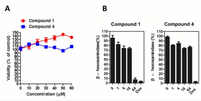
Furthermore, to confirm whether the anti-inflammatory effect of 1 and 4 is reliant on the inhibition of degranulation, a β-hexosamindase degranulation assay was conducted to examine the anti-degranulation effects on the basophils (RBL-2H3 cells). Compounds 1 and 4 did not exhibit significant cytotoxicity on cell viability as assessed by a MTT assay (Figure 4A). As shown in Figure 4B, RBL-2H3 cells treated with 1 at doses of up to 64 μM inhibited over 90% of DNP-BSA-stimulated degranulation in comparison with a control group. However, 4 only showed the slight effects on anti-degranulation even in the presence of high dose used.
A preliminary SAR study conducted showed that the spirostanol compounds have greater potential antineutrophilic inflammatory activities than the furostanol glycosides. For the active spirostanols 1, 4,
19, 20, 21, and 24, the composition of the sugar moieties do not seem to influence the resultant of
the sugar units are attached to C-6 than at C-3. Compounds 1 (C-3: C=O, C-23: H), 3 (C-3: OH, C-23: OH), and 24 (C-3: OH, C-23: H) contain the same sugar moiety at C-6, but 3 was inactive, 1 was only active against elastase release and 24 showed significant inhibition on both inflammatory mediators. In a previous study, neochlorogenin-type spirostanols (25S) isolated from S. torvum also showed significant
activities against superoxide anion generation and elastase release induced by human neutrophils.15 In
order to establish a more meaningful SAR, a great diversity of spirostanols and spirostanosides and their antineutrophilic inflammatory effects should be further assembled and evaluated.
EXPERIMENTAL SECTION
General Experimental Procedures. Optical rotations and IR spectra were recorded on a
JASCO-P-1020 polarimeter (cell length 10 mm) and a Shimadzu model IR Prestige-21 Fourier- transform infrared
spectrophotometer, respectively. 1D- and 2D-NMR spectra were measured on a Bruker UltrashieldTM
500 Plus instrument. Chemical shift (δ) values are in ppm and coupling constants (J) are in Hz with
C5D5N used as the internal standard. Low- and high-resolution ESIMS were measured on a Bruker
Daltonics Esquire HCT ultra high capacity trap mass spectrometer and an Orbitrap mass spectrometer
(LTQ Orbitrap XL, Thermo Fisher Scientific), respectively. TLC was performed on Kieselgel 60 F254
(0.25 mm, Merck) and/or RP-18 F254S (0.25 mm, Merck), coated plates, and then stained by spraying
with 10% H2SO4 and heating on a hot plate. Silica gel (Silicycle 70–230 and 230–400 mesh), RP-18
(LiChroprep® 40–63 µm, Merck), SephadexTM LH-20 (GE Healthcare, Uppsala, Sweden) and Diaion®
RID-10A refractive index detector (Shimadzu Inc., Kyoto, Japan) along with a Supelco Ascentis® (250
× 10 mm i.d., 5 µm, C18) column were used for HPLC. Sugar reagents, including D-xylose (Acros
Organics, Fair Lawn, NJ, USA), L-xylose (Alfa Aesar, Heysham, England), D-rhamnose (Carbosynth
Limited, Berkshire, UK), L-rhamnose (Acros Organics), D-glucose (MP Biomedicals, LLC, Illkirch,
France), L-glucose (Alfa Aesar) and D-quinovose (Sigma, St. Louis, MO, USA) were used for GC/MS
analysis.
Plant Material. Aerial parts of S. macaonense were collected in Kaohsiung, Taiwan, in March, 2011
and identified by C.-L. K. A voucher specimen (SM201103) was deposited at the Natural Medicinal Products Research Center, CMUH, Taichung, Taiwan.
Extraction and Isolation. The aerial parts of S. macaonense (1.23 kg) were extracted four times with
MeOH (8.0 L each) at room temperature to obtain a crude extract. The MeOH extract was partitioned
between EtOAc and H2O (1:1, v/v) to give an EtOAc-soluble fraction (25.09 g) and an aqueous phase
(113.27 g), which were partitioned with n-hexane/95% MeOH and n-BuOH/H2O (1:1), respectively, to
give four fractions.
Fractionation of the 95% MeOH-soluble fraction (16.26 g) was subjected to open column
chromatography on silica gel (70–230 mesh, column: 8 × 20 cm), using gradients of CHCl3–MeOH–
H2O (100:0:0, 5.0 L; 50:1:0.1, 3.5 L; 30:1:0.1, 4.2 L; 20:1:0.1, 2.0 L; 10:1:0.1, 2.0 L; 8:1:0.1, 1.4 L;
0:1:0, 2.0 L), and gave ten subfractions (SME1–SME10). Subfraction SME5 (2.04 g) was fractionated
into seven fractions by Sephadex LH-20 (column: 5 × 57 cm; CHCl3–MeOH, 1:1). Fraction SME5-3
400 mesh, column: 2 × 26 cm; CHCl3–MeOH, 40:1) chromatography to give fraction SME5-3-3-6 (20.0
mg). Fraction SME5-3-3-6 was further purified by Sephadex LH-20 (column: 2 × 30 cm; CHCl3–
MeOH, 1:1) and preparative silica TLC (CHCl3–MeOH, 45:1) to give 19 (13.0 mg). SME7 (1.40 g) was
chromatographed over Sephadex LH-20 (column: 5 × 57 cm; CHCl3–MeOH, 1:1), with subfraction
SME7-3 (226.0 mg) then subjected to RP-18 column chromatography (column: 2.5 × 29 cm; MeOH–
H2O 70:30 to 90:10), to give six subfractions. Subfraction SME7-3-4 (65.9 mg) was purified by passage
over Sephadex LH-20 (column: 2 × 28 cm; MeOH) to furnish 1 (52.4 mg). Subfraction SME7-3-5 (13.9
mg) was purified using Sephadex LH-20 (column: 2 × 28 cm; MeOH) and RP-HPLC (MeOH–H2O,
93:7; flow rate: 2.0 mL/min) to give 2 (2.6 mg, tR = 30 min).
Subfraction SME8 (3.05 g) was subjected to separation over Sephadex LH-20 (column: 5 × 53 cm;
CHCl3–MeOH, 1:1) to obtain six subfractions. Subfraction SME8-3 (808.2 mg) was separated by silica
gel chromatography (230–400 mesh, column: 3 × 23 cm; CHCl3–MeOH, 15:1 to 10:1), and its
subfraction SME8-3-3 (165.0 mg) was further subjected to RP-18 chromatography (column: 2 × 28 cm;
MeOH–H2O, 85:15, 90:10, 100:0), to give six additional subfractions (SME8-3-3-1–SME8-3-3-6). Of
these, SME8-3-3-4 (35.5 mg) was subjected to RP-HPLC (MeOH–H2O, 85:15; flow rate: 2.0 mL/min)
to give 20 (17.0 mg, tR = 56 min). SME8-3-4 (182.4 mg) was subjected to RP-18 chromatography
(column: 2 × 28 cm; MeOH–H2O, 90:10) to give seven subfractions (SME8-3-4-1–SME8-3-4-7), as
well as, pure compound 4 (20.2 mg). Subfraction SME8-3-4-3 (7.5 mg) was purified further with 75%
MeOH by RP-HPLC (MeOH–H2O, 73:27; flow rate: 2.0 mL/min) to give 3 (4.9 mg, tR = 46 min).
mL/min) to give 22 (3.5 mg, tR = 29 min). In turn, subfraction SME8-3-5 (117.8 mg) was subjected to
RP-18 chromatography (column: 2 × 28 cm; MeOH–H2O, 90:10) to give 24 (42.6 mg). Subfraction
SME8-4 (1.18 g) was chromatographed on a silica gel column (230–400 mesh, column: 3 × 23 cm),
using CHCl3–MeOH mixtures (10:1 to 8:1)for eluting. Further subfraction, SME8-4-3 (267.4 mg), was
purified further using Sephadex LH-20 (column: 2 × 29 cm) with MeOH, along with RP-18
chromatography (column: 2.5 × 26 cm; MeOH–H2O, 95:5) and RP-HPLC (MeOH–H2O, 90:10; flow
rate: 2.0 mL/min) to obtain 21 (6.0 mg, tR = 24 min).
Subfraction SME9 (3.41 g) was subjected to separation over a Sephadex LH-20 (column: 5 × 58 cm;
CHCl3–MeOH, 1:1) and then silica gel chromatography (230–400 mesh, column: 3 × 28 cm; CHCl3–
MeOH, 10:1 to 6:1) to obtain six subfractions. SME9-3-3 (55.7 mg) was further separated into three
subfractions by RP-18 chromatography (column: 2 × 27 cm; MeOH–H2O, 80:20). Subfractions
SME9-3-3-2 (4.0 mg) and SME9-3-3-3 (3.7 mg) were purified by RP-HPLC (MeOH–H2O, 80:10 and 82:18;
flow rate: 2.0 mL/min) to obtain 5 (2.2 mg, tR = 43 min) and 23 (2.4 mg, tR = 46 min), respectively.
SME9-3-4 (38.9 mg) was subjected to RP-18 chromatography (column: 2 × 26 cm; MeOH–H2O, 80:20)
and purified by RP-HPLC (MeOH–H2O, 80:20; flow rate: 2.0 mL/min) to obtain 6 (1.7 mg, tR = 50
min). Subfraction SME9-3-5 (47.1 mg) was subjected to RP-18 chromatography (column: 2 × 26 cm;
MeOH–H2O, 80:20) to give 7 (6.4 mg).
The n-BuOH-soluble fraction (34.4 g) was chromatographed over a Diaion HP-20 column (7 × 14.5
cm; H2O–MeOH–acetone, 100:0:0, 1.9 L; 80:20:0, 2.0 L; 60:40:0, 2.0 L; 40:60:0, 2.3 L; 20:80:0, 2.2 L;
was purified over Sephadex LH-20 (column: 5 × 58 cm; MeOH) to produce seven subfractions. Subfraction SMB4-2 (2.92 g) was separated by silica gel chromatography (230–400 mesh, column: 4.5
× 30 cm; CHCl3–MeOH, 5:1, 4:1, 3:1, 2:1, 1:1), and its subfraction SMB4-2-6 (217.0 mg) was further
subjected to Sephadex LH-20 chromatography (column: 2 × 25 cm; MeOH–H2O, 50:50) to give three
additional subfractions (SMB4-2-6-1–SMB4-2-6-3). Subfraction SMB4-2-6-2 (192.0 mg) was purified
by RP-18 chromatography (column: 2.5 × 24 cm; MeOH–H2O, 70:30) and RP-HPLC (MeCN–H2O,
30:70; flow rate: 2.0 mL/min) to give 10 (9.0 mg, tR = 10 min), 11 (7.0 mg, tR = 12 min), and 13 (5.0 mg,
tR = 16 min). Subfraction SMB4-2-7 (367.0 mg) was separated into four subfractions by column
chromatography over Sephadex LH-20 (column: 2 × 25 cm; MeOH–H2O, 50:50), and subfraction
SMB4-2-7-1 (299.0 mg) was subjected to RP-18 chromatography (column: 2.5 × 24 cm; MeOH–H2O,
80:20). Subfraction SMB4-2-7-1-2 (221.0 mg) was purified by RP-18 chromatography (column: 2.5 ×
24 cm; MeOH–H2O, 65:35) followed by HPLC (MeOH–H2O, 60:40; flow rate: 2.0 mL/min) to obtain
subfraction SMB4-2-7-1-2-2-2 (31.0 mg), which was further purified by RP-HPLC (MeCN–H2O, 30:70;
flow rate: 2.0 mL/min) to give 15 (2.0 mg, tR = 16 min) and 14 (7.0 mg, tR = 18 min).
Subfraction SMB5 (7.7 g) was separated over a Sephadex LH-20 column (5 × 58 cm; MeOH–H2O,
100:0 to 80:20) to obtain six subfractions. Subfraction SMB5-2 (3.49 g) was purified by silica gel
chromatography (230–400 mesh, column: 4.5 × 23 cm; CHCl3–MeOH, 5:1), and its subfraction
SMB5-2-3 (431.0 mg) was subjected to RP-18 chromatography (column: 2.5 × 24 cm; MeOH–H2O, 70:30) and
then HPLC (MeOH–H2O, 70:30; flow rate: 2.0 mL/min) to give SMB5-2-3-2-4-1 (60.0 mg).
H2O, 35:65) to give 17 (46.0 mg). Subfraction SMB5-2-4 (1.19 mg) was subjected to RP-18
chromatography (column: 2.5 × 24 cm; MeOH–H2O, 80:20) to give SMB5-2-4-4 (661.0 mg), which was
separated into four fractions by RP-18 chromatography (column: 2.5 × 24.5 cm; MeOH–H2O, 80:20).
SMB5-2-4-4-2 (510.0 mg) was purified by RP-HPLC (MeOH–H2O, 80:20 and MeCN–H2O, 35:65) and
RP-18 chromatography (column: 2.5 × 28 cm; MeCN–H2O, 35:65) to give 18 (21.0 mg). Subfraction
SMB5-2-4-4-2-3 (315.0 mg) was subjected to RP-HPLC (MeCN–H2O, 35:65) to give two major
fractions, SMB-5-2-4-4-2-3-2 (80.0 mg) and SMB-5-2-4-4-2-3-3 (60.0 mg), which were purified by
RP-18 chromatography (column: 2.5 × 28 cm; MeCN–H2O, 30:70 and 35:65) to give 12 (37.0 mg) and 16
(47.0 mg), respectively. Subfraction SMB5-2-4-4-4 (37.0 mg) was subjected to RP-HPLC (MeOH–H2O,
80:20) to give 9 (22.0 mg, tR = 19 min).
Subfraction SMB7 (1.5 g) was subjected to Sephadex LH-20 (column: 5 × 48 cm; MeOH) and silica
gel chromatography (230–400 mesh, column: 3 × 24 cm; CHCl3–MeOH, 4:1) to obtain seven
subfractions. SMB7-4-4 (90.0 mg) was further purified by RP-18 chromatography (column: 2 × 20.5
cm; MeOH–H2O, 90:10) and RP-HPLC (MeOH–H2O, 85:15; flow rate: 2.0 mL/min) to afford pure
compound 8 (3.0 mg, tR = 26 min).
Macaoside A (1): [α]Com
b i n −20.7 (c 0.15, MeOH); IR (neat) νmax 3401 (OH), 2928 (CH), 1705 (C=O),
1452, 1379, 1240, 1169, 1074, 1049 (C-O-C), 982, 964, 918, 899, 866, 756 cm-1; 1H and 13C NMR
spectroscopic data, see Tables 1 and 2; HRESIMS m/z 731.3982 [M+Na]+ (calcd for C
38H60O12Na,
731.3977).
Macaoside B (2): [α]Com
1454, 1375, 1240, 1175, 1096, 1047 (C-O-C), 982, 920, 899, 878, 866, 756 cm-1; 1H and 13C NMR
spectroscopic data, see Tables 1 and 2; HRESIMS m/z 777.4399 [M+Na]+ (calcd for C
40H66O13Na,
777.4396).
Macaoside C (3): []Com
b i n +17.1 (c 0.07, MeOH); IR (neat) νmax 3389 (OH), 2951, 2930, 2874 (CH),
1452, 1379, 1244, 1169, 1070, 1049 (C-O-C), 1003, 961, 920, 897, 862, 756, 667 cm-1; 1H and 13C NMR
spectroscopic data, see Tables 1 and 2; HRESIMS m/z 749.4086 [M+Na]+ (calcd for C
38H62O13Na,
749.4083).
Macaoside D (4): [α]Com
b i n −25.5 (c 0.11, MeOH); IR (neat) νmax 3242 (OH), 2930, 2857 (CH), 1242,
1215, 1173, 1076, 1053 (C-O-C), 982, 957, 920, 899, 866, 756, 667 cm-1; 1H and 13C NMR
spectroscopic data, see Tables 1 and 2; HRESIMS m/z 719.3987 [M+Na]+ (calcd for C37H60O12Na,
719.3977).
Macaoside E (5): [] 28
D +35.0 (c 0.08, MeOH); IR (neat) νmax 3319 (OH), 2951, 2928, 2857 (CH),
1454, 1379, 1240, 1215, 1055 (C-O-C), 1038, 982, 920, 899, 866, 758, 665 cm-1; 1H and 13C NMR
spectroscopic data, see Tables 1 and 2; HRESIMS m/z 763.4239 [M+Na]+ (calcd for C39H64O13Na,
763.4239).
Macaoside F (6): []Com
b i n +43.8 (c 0.08, MeOH); IR (neat) νmax 3389 (OH), 2930, 2872 (CH), 1240,
1128, 1045 (C-O-C), 982, 955, 918, 897, 864, 814, 756 cm-1; 1H and 13C NMR spectroscopic data, see
Tables 1 and 2; HRESIMS m/z 763.4235 [M+Na]+ (calcd for C
39H64O13Na, 763.4239). Macaoside G (7): []Com
b i n +48.8 (c 0.08, MeOH); IR (neat) νmax 3381 (OH), 2924, 2850 (CH), 1449,
C39H64O13Na, 763.4239). Macaoside H (8): []
28
D −41.0 (c 0.10, MeOH); IR (neat) νmax 3356 (OH), 2930 (CH), 1452, 1379,
1215, 1128, 1043 (C-O-C), 982, 955, 918, 899, 866, 837, 812, 756, 667 cm-1; 1H and 13C NMR
spectroscopic data, see Tables 1 and 2; HRESIMS m/z 909.4820 [M+Na]+ (calcd for C
45H74O17Na,
909.4818).
Macaoside I (9): []Com
b i n +27.0 (c 0.10, MeOH); IR (neat) νmax 3360 (OH), 2914, 2874 (CH), 1692,
1643, 1449, 1379, 1304, 1215, 1167, 1074, 1045 (C-O-C), 939, 893, 856, 756, 665 cm-1; 1H and 13C
NMR spectroscopic data, see Table 3; HRESIMS m/z 873.4842 [M+H]+ (calcd for C
44H73O17, 873.4842). Macaoside J (10): []Com
b i n +39.0 (c 0.10, MeOH); IR (neat) νmax 3360 (OH), 2932, 2872 (CH), 1643,
1452, 1379, 1169, 1074, 1047 (C-O-C), 1016, 959, 895, 756 cm-1; 1H and 13C NMR spectroscopic data,
see Table 3; HRESIMS m/z 911.4620 [M+Na]+ (calcd for C
44H72O18Na, 911.4611). Macaoside K (11): []Com
b i n −28.0 (c 0.10, MeOH); IR (neat) νmax 3360 (OH), 2930, 2872 (CH), 1447,
1379, 1215, 1167, 1074, 1043 (C-O-C), 895, 758, 667 cm-1; 1H and 13C NMR spectroscopic data, see
Table 3; HRESIMS m/z 911.4614 [M+Na]+ (calcd for C
44H72O18Na, 911.4611). Macaoside L (12): []
28
D −82.5 (c 0.12, MeOH); IR (neat) νmax 3360 (OH), 2930 (CH), 1454, 1379,
1337, 1310, 1248, 1167, 1072, 1045 (C-O-C), 893, 754 cm-1; 1H and 13C NMR spectroscopic data, see
Tables 4 and 5; HRESIMS m/z 913.4773 [M+Na]+ (calcd for C
44H74O18Na, 913.4767). Macaoside M (13): []
28
D −83.8 (c 0.08, MeOH); IR (neat) νmax 3381 (OH), 2932, 2849 (CH), 1643,
1454, 1381, 1165, 1076, 1040 (C-O-C), 1018, 955, 897, 868, 756 cm-1; 1H and 13C NMR spectroscopic
data, see Tables 4 and 5; HRESIMS m/z 797.4295 [M+Na]+ (calcd for C
39H66O15Na, 797.4294). Macaoside N (14): []
28
1452, 1383, 1265, 1134, 1101, 1080, 1043 (C-O-C), 953, 912, 837, 816, 779, 669 cm-1; 1H and 13C NMR
spectroscopic data, see Tables 4 and 5; HRESIMS m/z 943.4879 [M+Na]+ (calcd for C
45H76O19Na,
943.4873).
Macaoside O (15): [] 28
D +25.0 (c 0.10, MeOH); IR (neat) νmax 3381 (OH), 2932 (CH), 1643, 1452,
1379, 1260, 1171, 1074, 1045 (C-O-C), 953, 895, 629 cm-1; 1H and 13C NMR spectroscopic data, see
Tables 4 and 5; HRESIMS m/z 943.4878 [M+Na]+ (calcd for C
45H76O19Na, 943.4873). Macaoside P (16): []
28
D −88.0 (c 0.10, MeOH); IR (neat) νmax 3304 (OH), 2934 (CH), 1452, 1379,
1215, 1169, 1072, 1047 (C-O-C), 935, 895, 756 cm-1; 1H and 13C NMR spectroscopic data, see Tables 4
and 5; HRESIMS m/z 927.4927 [M+Na]+ (calcd for C
45H76O18Na, 927.4924). Macaoside Q (17): []
28
D +21.0 (c 0.10, MeOH); IR (neat) νmax 3360 (OH), 2932 (CH), 1703 (C=O),
1452, 1379, 1167, 1072, 1045 (C-O-C), 893, 754 cm-1; 1H and 13C NMR spectroscopic data, see Tables 4
and 5; HRESIMS m/z 925.4770 [M+Na]+ (calcd for C
45H74O18Na, 925.4767). Macaoside R (18): []
28
D −62.2 (c 0.09, MeOH); IR (neat) νmax 3379 (OH), 2930 (CH), 1643, 1452,
1379, 1248, 1169, 1076, 1045 (C-O-C), 897, 754 cm-1; 1H and 13C NMR spectroscopic data, see Tables 4
and 5; HRESIMS m/z 913.4770 [M+Na]+ (calcd for C
44H74O18Na, 913.4767).
Acid Hydrolysis and GC/MS Analysis. Isolates (9.8 mg for 12, 9.7 mg for 16, 0.5–1 mg for the
remaining compounds) were hydrolyzed in 1 M HCl/1,4-dioxane (1:1, 1.4 mL) at 90 °C for 3 h. Each was cooled and partitioned with CHCl3/H2O (1:1, 14 mL), with the CHCl3 fraction providing an
aglycone moiety. Each aqueous layer was neutralized with saturated aqueous Na2CO3 solution and then
CHCl3 and H2O (1:1, 14 mL). The acetylated CHCl3 fraction was subjected to gas chromatography–
mass spectrometry (DSQ II GC/MS, Thermo Scientific, USA) under the following conditions: DB-5MS capillary column (30 m, 0.25 mm i.d., 0.25 µm film thickness); injector, 250 °C; injection volume, 1 µL; column oven temperature, 155 °C for 60 min; column flow, 1.0 mL/min; ion source temperature, 250
°C; EI, 70 eV; mass range, m/z 50–800. Under these conditions, the retention times (tR) for the acetate
derivatives of standard sugars were: D-xylose (13.51 and 14.76 min), L-xylose(13.50 and 14.78 min), D
-rhamnose (14.09 and 14.31 min), L-rhamnose (14.06 and 14.32 min), D-glucose (49.27 and 49.95 min),
L-glucose (49.15 and 49.83 min), and D-quinovose (13.13 and 13.30 min). Co-injection studies of the
hydrolysates with the different standards were also conducted to confirm the presence of D-xylose, L
-rhamnose, D-glucose, and D-quinovose in compounds 1–18.
Isolation of human neutrophils. The study was approved by the Institutional Review Board of
Chang Gung Memorial Hospital (IRB 102-1595A3). All healthy donors (20-30 years old) did not take any medicine within the week before blood sample collection. Human neutrophils were isolated from heparinized peripheral venous blood by the dextran sedimentation and Ficoll-Hypaque centrifugation.
After hypotonic lysis of contaminating erythrocytes, neutrophils isolated were suspended in Ca2+-free
HBSS at pH 7.4 and maintained at 4 °C before use. Wright-Giemsa stain was used to confirm the purity of the neutrophil suspension, and greater than 98% viability was determined by trypan blue exclusion.
Measurement of Superoxide Anion Generation.15,16 Neutrophils were equilibrated with 0.5 mg/mL
ferricytochrome c and 1 mM Ca2+ at 37 °C for 2 min and then incubated with each test compound for 5
-methionyl-L-leucyl-L-phenylalanine (FMLP, 100 nM) for 10 min. FMLP/CB was used as a stimulant to activate neutrophils to produce superoxide anion. The changes in absorbance with reduction of ferricytochrome c at 550 nm were continuously monitored in a double-beam, six-cell positioned spectrophotometer (Hitachi U-3010, Tokyo, Japan) with constant stirring. Calculations were based on differences in the reactions with and without superoxide dismutase (100 U/mL) divided by the
extinction coefficient for the reduction of ferricytochrome c (ε = 21.1/mM/10 mm). The positive control
was diphenyleneiodonium (DPI), a NADPH oxidase inhibitor.
Elastase Release Assay.15,16 Neutrophils were equilibrated in an elastase substrate,
MeO-Suc-Ala-Ala-Pro-Val-p-nitroanilide (100 µM), at 37 °C for 2 min and then incubated with test compounds for 5 min. Cells were activated by 100 nM FMLP and 0.5 µg/mL CB, and the changes in absorbance at 405 nm were monitored continuously to assay elastase release. The results were expressed as the percentage of elastase release in the FMLP/CB-activated, drug-free control system. Sivelestant, an inhibitor of human neutrophil elastase, was used as the positive control.
Lactate Dehydrogenase (LDH) Release. This assay was carried out according to an established
protocol.17 Release of LDH into the cell medium indicated cell membrane damage. Neutrophilic
cytotoxicity was determined by LDH release in the cell-free medium as a percentage of the total LDH release.
Degranulation Assay. The cell viability was tested using a MTT assay.18 Analysis of
β-hexosaminidase release is a well-known assay for measuring the degree of degranulation in RBL-2H3
containing 10% FBS and then seeded into a 96-well plate at a concentration of 104 cells/well. The cells
were treated with compounds 1, 4 (0, 1, 4, 16, 64 μM), and the positive control dexamethasone (100 nM), respectively, for 48 h. The treated RBL-2H3 cells were incubated overnight with anti-DNP IgE (500 μg/mL) for sensitization. Cells were then washed with Tyrode’s buffer and stimulated with 2,4-dinitrophenylated albumin from bovine serum (DNP-BSA, 0.1 μg/mL in Tyrode’s buffer) for 1 h to induce the immediate-phase antigen-stimulated degranulation assay. The reaction of degranulation was stopped by cooling in ice bath, and the supernatant was collected for β-hexosaminidase release measurement. The degree of β-hexosaminidase release was assessed by the enzymatic activity observed.
The supernatant was incubated with substrate (0.1 M p-nitrophenyl-N-acetyl-D-glucosaminide in 0.1 M
citrate buffer) for 1 h and was stopped by adding sodium bicarbonate stopping buffer. The activity of the test samples was measured by the absorbance observed at 405 nM. The ratio of β-hexosaminidase release was calculated by the following equation: the release % = (OD sample – OD background)/(OD total lysate – OD background) × 100.
ASSOCIATED CONTENT
Supporting Information The 1D- and 2D-NMR data of all new compounds are available free of
charge via the Internet at http://pubs.acs.org.
AUTHOR INFORMATION Corresponding Author
Notes
The authors declare no competing financial interest.
ACKNOWLEDGMENTS
This work was supported by the grants from National Science Council (NSC 101-2325-B-039-004) and National Health Research Institutes (NHRI-EX102-10241BI), awarded to C.L. Lee and Y.C. Wu, and CMU under the Aim for Top University Plan of the Ministry of Education, Taiwan.
REFERENCES
(1) Boufford, D. E.; Hsieh, C.-F.; Huang, T.-C.; Lowry, P. P.; Ohashi, H.; Peng, C.-I.; Yang, S.-Y.; Hsiao, A.; Lin, H.-W.; Yu, C.-L. Flora of Taiwan; Editorial Committee of the Flora of Taiwan: Taipei, 1998; Vol. 4, pp 563, 569−570.
(2) Zamilpa, A.; Tortoriello, J.; Navarro, V.; Delgado, G.; Alvarez, L. J. Nat. Prod. 2002, 65, 1815−1819.
(3) Mimaki, Y.; Sashida, Y.; Kawashima, K. Phytochemistry 1991, 30, 3721−3727. (4) Mimaki, Y.; Sashida, Y.; Kawashima, K. Chem. Pharm. Bull. 1992, 40, 148−152. (5) Agrawal, P. K. Magn. Reson. Chem. 2003, 41, 965−968.
(6) González, M.; Zamilpa, A.; Marquina, S.; Navarro, V.; Alvarez, L. J. Nat. Prod. 2004, 67, 938−941.
(7) Iida, Y.; Yanai, Y.; Ono, M.; Ikeda, T.; Nohara, T. Chem. Pharm. Bull. 2005, 53, 1122−1125. (8) Lu, Y.; Luo J.; Kong, L. Phytochemistry 2011, 72, 668−673.
Chem. 2009, 52, 7368−7371.
(10) Zhang, Y.; Li, Y.; Guo, T.; Guan, H.; Shi, J.; Yu, Q.; Yu, B. Carbohydr. Res. 2005, 340, 1453−1459.
(11) Agrawal, P. K. Steroids 2005, 70, 715−724.
(12) Agrawal, P. K.; Jain, D. C.; Gupta, R. K.; Thakur, R. S. Phytochemistry 1985, 24, 2479−2496. (13) Lu, Y.; Luo, J.; Kong, L. Magn. Reson. Chem. 2009, 47, 808−812.
(14) Yahara, S.; Yamashita, T.; Nozawa, N.; Nohara, T. Phytochemistry 1996, 43, 1069−1074.
(15) Lee, C. L.; Hwang, T. L.; He, W. J.; Tsai, Y. H.; Yen, C. T.; Yen, H. F.; Chen, C. J.; Chang, W. Y.; Wu, Y. C. Phytochemistry 2013, 95, 315−321.
(16) Hwang, T. L.; Wang, C. C.; Kuo, Y. H.; Huang, H. C.; Wu, Y. C.; Kuo, L. M.; Wu, Y. H.
Biochem. Pharmacol. 2010, 80, 1190−1200.
(17) Wu, Y. C.; Sureshbabu, M.; Fang Y. C.; Wu, Y. H.; Lan, Y. H.; Chang, F. R.; Chang, Y. W.; Hwang, T. L. Toxicol. Appl. Pharmacol. 2013, 266, 399−407.
(18) Yang, J. C.; Lu, M. C.; Lee, C. L.; Chen, G. Y.; Lin, Y. Y.; Chang, F. R.; Wu, Y. C. Free Radic.
Biol. Med. 2011, 51, 641–657.
(19) Chen, B. H.; Hung, M. H.; Chen, J. Y. F.; Chang, H. W.; Yu, M. L.; Wan, L.; Tsai, F. J.; Wang, T. P.; Fu, T. F.; Chiu, C. C. Food Chem. 2012, 132, 968–974.
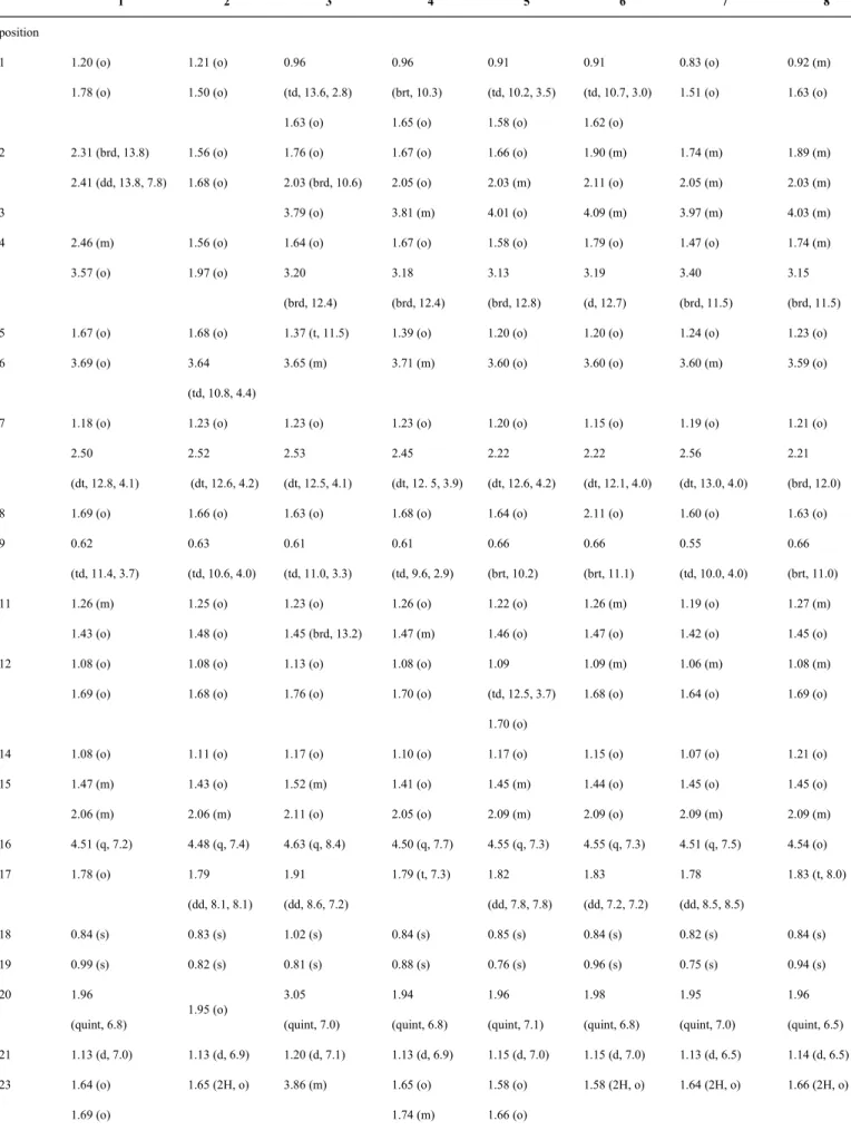
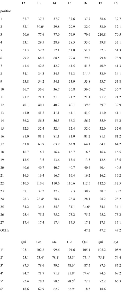
Table 1. 1H NMR Spectroscopic Data of Compounds 1–8 (500 MHz, C 5D5N)a 1 2 3 4 5 6 7 8 position 1 1.20 (o) 1.78 (o) 1.21 (o) 1.50 (o) 0.96 (td, 13.6, 2.8) 1.63 (o) 0.96 (brt, 10.3) 1.65 (o) 0.91 (td, 10.2, 3.5) 1.58 (o) 0.91 (td, 10.7, 3.0) 1.62 (o) 0.83 (o) 1.51 (o) 0.92 (m) 1.63 (o) 2 2.31 (brd, 13.8) 2.41 (dd, 13.8, 7.8) 1.56 (o) 1.68 (o) 1.76 (o) 2.03 (brd, 10.6) 1.67 (o) 2.05 (o) 1.66 (o) 2.03 (m) 1.90 (m) 2.11 (o) 1.74 (m) 2.05 (m) 1.89 (m) 2.03 (m) 3 3.79 (o) 3.81 (m) 4.01 (o) 4.09 (m) 3.97 (m) 4.03 (m) 4 2.46 (m) 3.57 (o) 1.56 (o) 1.97 (o) 1.64 (o) 3.20 (brd, 12.4) 1.67 (o) 3.18 (brd, 12.4) 1.58 (o) 3.13 (brd, 12.8) 1.79 (o) 3.19 (d, 12.7) 1.47 (o) 3.40 (brd, 11.5) 1.74 (m) 3.15 (brd, 11.5)
5 1.67 (o) 1.68 (o) 1.37 (t, 11.5) 1.39 (o) 1.20 (o) 1.20 (o) 1.24 (o) 1.23 (o)
6 3.69 (o) 3.64
(td, 10.8, 4.4)
3.65 (m) 3.71 (m) 3.60 (o) 3.60 (o) 3.60 (m) 3.59 (o)
7 1.18 (o) 2.50 (dt, 12.8, 4.1) 1.23 (o) 2.52 (dt, 12.6, 4.2) 1.23 (o) 2.53 (dt, 12.5, 4.1) 1.23 (o) 2.45 (dt, 12. 5, 3.9) 1.20 (o) 2.22 (dt, 12.6, 4.2) 1.15 (o) 2.22 (dt, 12.1, 4.0) 1.19 (o) 2.56 (dt, 13.0, 4.0) 1.21 (o) 2.21 (brd, 12.0)
8 1.69 (o) 1.66 (o) 1.63 (o) 1.68 (o) 1.64 (o) 2.11 (o) 1.60 (o) 1.63 (o)
9 0.62 (td, 11.4, 3.7) 0.63 (td, 10.6, 4.0) 0.61 (td, 11.0, 3.3) 0.61 (td, 9.6, 2.9) 0.66 (brt, 10.2) 0.66 (brt, 11.1) 0.55 (td, 10.0, 4.0) 0.66 (brt, 11.0) 11 1.26 (m) 1.43 (o) 1.25 (o) 1.48 (o) 1.23 (o) 1.45 (brd, 13.2) 1.26 (o) 1.47 (m) 1.22 (o) 1.46 (o) 1.26 (m) 1.47 (o) 1.19 (o) 1.42 (o) 1.27 (m) 1.45 (o) 12 1.08 (o) 1.69 (o) 1.08 (o) 1.68 (o) 1.13 (o) 1.76 (o) 1.08 (o) 1.70 (o) 1.09 (td, 12.5, 3.7) 1.70 (o) 1.09 (m) 1.68 (o) 1.06 (m) 1.64 (o) 1.08 (m) 1.69 (o)
14 1.08 (o) 1.11 (o) 1.17 (o) 1.10 (o) 1.17 (o) 1.15 (o) 1.07 (o) 1.21 (o)
15 1.47 (m) 2.06 (m) 1.43 (o) 2.06 (m) 1.52 (m) 2.11 (o) 1.41 (o) 2.05 (o) 1.45 (m) 2.09 (m) 1.44 (o) 2.09 (o) 1.45 (o) 2.09 (m) 1.45 (o) 2.09 (m) 16 4.51 (q, 7.2) 4.48 (q, 7.4) 4.63 (q, 8.4) 4.50 (q, 7.7) 4.55 (q, 7.3) 4.55 (q, 7.3) 4.51 (q, 7.5) 4.54 (o) 17 1.78 (o) 1.79 (dd, 8.1, 8.1) 1.91 (dd, 8.6, 7.2) 1.79 (t, 7.3) 1.82 (dd, 7.8, 7.8) 1.83 (dd, 7.2, 7.2) 1.78 (dd, 8.5, 8.5) 1.83 (t, 8.0) 18 0.84 (s) 0.83 (s) 1.02 (s) 0.84 (s) 0.85 (s) 0.84 (s) 0.82 (s) 0.84 (s) 19 0.99 (s) 0.82 (s) 0.81 (s) 0.88 (s) 0.76 (s) 0.96 (s) 0.75 (s) 0.94 (s) 20 1.96 (quint, 6.8) 1.95 (o) 3.05 (quint, 7.0) 1.94 (quint, 6.8) 1.96 (quint, 7.1) 1.98 (quint, 6.8) 1.95 (quint, 7.0) 1.96 (quint, 6.5) 21 1.13 (d, 7.0) 1.13 (d, 6.9) 1.20 (d, 7.1) 1.13 (d, 6.9) 1.15 (d, 7.0) 1.15 (d, 7.0) 1.13 (d, 6.5) 1.14 (d, 6.5)
24 1.56 (2H, o) 1.49 (2H, o) 1.78 (o) 2.10 (o)
1.54 (2H, o) 1.66 (2H, o) 1.68 (2H, o) 1.57 (2H, o) 1.57 (2H, o)
25 1.56 (o) 1.56 (o) 1.83 (m) 1.54 (o) 1.58 (o) 1.62 (o) 1.57 (o) 1.57 (o)
26 3.48 (t, 10.6) 3.57 (o) 3.47 (t, 10.6) 3.57 (brd, 9.8) 3.47 (t, 10.9) 3.54 (dd, 10.9, 3.3) 3.44 (t, 10.5) 3.54 (dd, 11.0, 3.0) 3.50 (t, 10.6) 3.59 (o) 3.50 (t, 10.5) 3.59 (o) 3.48 (t, 10.5) 3.57 (brd, 10.0) 3.50 (o) 3.57 (o) 27 0.68 (d, 5.4) 0.69 (d, 5.3) 0.73 (d, 6.2) 0.66 (d, 5.9) 0.70 (d, 5.4) 0.70 (d, 5.2) 0.69 (d, 4.5) 0.69 (d, 5.0) OCH3 3.20 (s) OCH3 3.24 (s)
Qui Qui Qui Xyl Glc Glc Glc Glc
1′ 4.75 (d, 7.7) 4.79 (d, 7.4) 4.82 (d, 7.4) 4.80 (d, 7.3) 5.00 (o) 5.10 (d, 7.7) 5.14 (d, 8.0) 4.96 (o)
2′ 4.03 (o) 4.02 (o) 4.05 (o) 4.04 (m) 3.99 (o) 4.28 (t, 8.6) 4.04 (t, 8.0) 4.20 (o)
3′ 4.08 (t, 9.0) 4.09 (t, 8.9) 4.11 (m) 4.10 (o) 4.18 (t, 9.0) 4.21 (t, 8.8) 4.29 (o) 4.14 (m)
4′ 3.57 (o) 3.58 (o) 3.62 (m) 4.10 (o) 4.46 (t, 9.3) 4.15 (o) 4.29 (o) 4.35 (o)
5′ 3.73 (m) 3.72 (m) 3.74 (o) 3.63 (m) 4.30 (m) 3.64 (o) 3.79 (m) 3.86 (m) 3.50 (o) 6′ 1.56 (d, 6.0) 1.56 (d, 6.0) 1.60 (d, 6.1) 4.13 (brd, 11.6) 4.27 (brd, 11.6) 4.36 (o) 4.50 (brd, 11.4) 4.35 (dd, 11.9, 5.3) 4.43 (bd, 11.9, 2.3) 4.07 (m) 4.20 (o)
Xyl Xyl Xyl Xyl Rha Rha Qui Rha
1″ 5.25 (d, 7.7) 5.27 (d, 7.7) 5.26 (d, 7.7) 5.27 (d, 7.6) 5.90 (s) 6.36 (s) 4.79 (d, 7.5) 6.36 (s)
2″ 4.06 (o) 4.02 (o) 4.05 (o) 4.07 (m) 4.71 (brs) 4.76 (brs) 4.00 (t, 8.5) 4.78 (brs)
3″ 4.14 (o) 4.14 (o) 4.18 (o) 4.19 (o) 4.59
(dd, 9.2, 3.1) 4.62 (dd, 9.2, 4.0)
4.13 (t, 8.5) 4.60 (brd, 9.0)
4″ 4.14 (o) 4.15 (o) 4.20 (o) 4.19 (o) 4.35 (t, 9.3) 4.34 (o) 3.72 (t, 9.0) 4.33 (o)
5″ 3.69 (o) 4.29 (dd, 11.5, 4.9) 3.68 (m) 4.29 (dd, 11.2, 5.0) 3.73 (o) 4.33 (dd, 11.3, 4.5) 3.74 (m) 4.34 (m) 5.00 (o) 5.05 (m) 3.76 (m) 4.96 (o) 6″ 1.74 (d, 6.2) 1.79 (d, 6.3) 1.63 (d, 6.0) 1.78 (d, 6.0) Rha 1′′′ 5.84 (s) 2′′′ 4.68 (brs) 3′′′ 4.54 (o) 4′′′ 4.33 (o) 5′′′ 4.90 (o) 6′′′ 1.63 (d, 6.0)
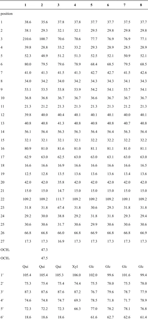
Table 2. 13C NMR Spectroscopic Data of Compounds 1–8 (125 MHz, C 5D5N)a-c 1 2 3 4 5 6 7 8 position 1 38.6 35.6 37.8 37.8 37.7 37.7 37.5 37.7 2 38.1 29.3 32.1 32.1 29.5 29.8 29.8 29.8 3 210.6 100.7 70.6 70.6 77.7 76.9 76.9 77.1 4 39.8 28.8 33.2 33.2 29.3 28.9 28.5 28.9 5 52.3 48.9 51.2 51.3 52.5 52.1 50.9 52.1 6 80.0 79.5 79.6 78.9 68.4 68.5 79.5 68.5 7 41.0 41.3 41.5 41.3 42.7 42.7 41.5 42.6 8 34.0 34.2 34.0 34.2 34.3 34.3 34.1 34.3 9 53.1 53.5 53.8 53.9 54.2 54.1 53.7 54.1 10 36.8 36.8 36.7 36.7 36.6 36.7 36.7 36.7 11 21.3 21.2 21.3 21.3 21.3 21.3 21.2 21.3 12 39.8 40.0 40.4 40.1 40.1 40.1 40.0 40.1 13 40.8 40.8 41.3 40.8 40.8 40.8 40.7 40.8 14 56.1 56.4 56.3 56.3 56.4 56.4 56.3 56.4 15 32.1 32.1 32.1 32.1 32.2 32.2 32.2 32.2 16 80.9 81.0 81.6 81.0 81.1 81.1 81.0 81.1 17 62.9 63.0 62.5 63.0 63.0 63.1 63.0 63.0 18 16.6 16.6 16.9 16.6 16.6 16.6 16.6 16.5 19 12.5 12.8 13.5 13.6 13.6 13.6 13.4 13.6 20 42.0 42.0 35.8 42.0 42.0 42.0 42.0 42.0 21 15.0 15.0 14.7 15.0 15.0 15.0 15.0 15.0 22 109.2 109.2 111.7 109.2 109.2 109.2 109.1 109.2 23 31.8 31.8 67.4 31.8 30.6 29.3 31.8 31.8 24 29.2 30.0 38.8 29.2 31.8 31.8 29.3 29.4 25 30.6 30.6 31.7 30.6 29.9 30.6 30.6 30.6 26 66.8 66.8 66.0 66.8 66.9 66.8 66.8 66.9 27 17.3 17.3 16.9 17.3 17.3 17.3 17.3 17.3 OCH3 47.3 OCH3 47.5
Qui Qui Qui Xyl Glc Glc Glc Glc
1′ 105.4 105.4 105.3 106.0 102.0 99.6 101.6 99.4 2′ 75.3 75.4 75.4 74.4 75.5 78.0 75.5 78.0 3′ 87.3 87.6 87.6 87.2 76.7 79.6 78.7 77.9 4′ 74.6 74.8 74.7 69.3 78.5 71.8 71.7 78.9 5′ 72.3 72.2 72.3 66.3 77.0 78.2 78.1 76.8 6′ 18.6 18.6 18.6 61.6 62.7 62.6 61.4
Xyl Xyl Xyl Xyl Rha Rha Qui Rha 1″ 106.2 106.2 106.5 106.3 102.7 102.2 106.0 102.1 2″ 74.6 74.6 74.8 75.5 72.7 72.5 75.9 72.4a 3″ 78.2 78.2 78.3 78.2 72.8 72.8 78.1 72.7b 4″ 70.9 70.9 70.9 71.0 74.0 74.3 76.8 73.9c 5″ 67.3 67.4 67.4 67.4 70.5 69.5 72.6 69.4 6″ 18.6 18.7 18.8 18.6 Rha 1′′′ 102.9 2′′′ 72.5a 3′′′ 72.8b 4′′′ 74.2c 5′′′ 70.4 6′′′ 18.5
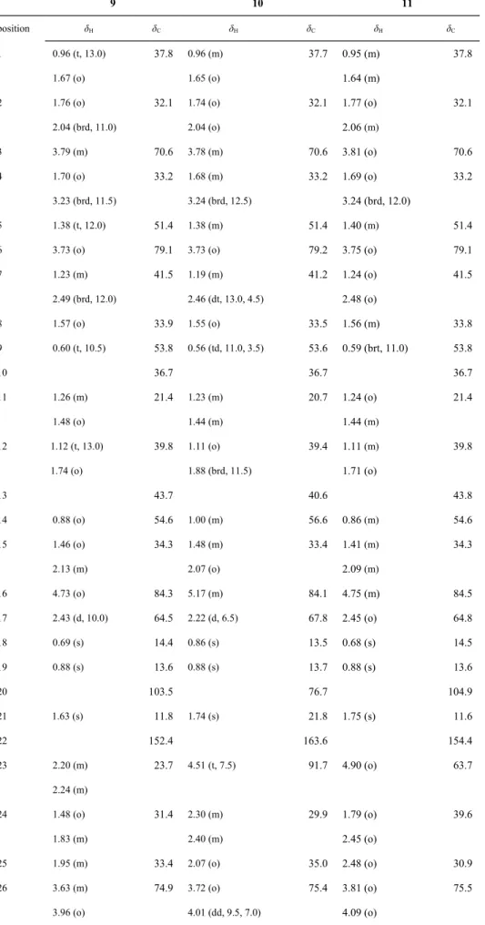
Table 3. 1H and 13C NMR Spectroscopic Data of Compounds 9–11 (500 MHz; 125 MHz, C 5D5N)a, b 9 10 11 position δH δC δH δC δH δC 1 0.96 (t, 13.0) 1.67 (o) 37.8 0.96 (m) 1.65 (o) 37.7 0.95 (m) 1.64 (m) 37.8 2 1.76 (o) 2.04 (brd, 11.0) 32.1 1.74 (o) 2.04 (o) 32.1 1.77 (o) 2.06 (m) 32.1 3 3.79 (m) 70.6 3.78 (m) 70.6 3.81 (o) 70.6 4 1.70 (o) 3.23 (brd, 11.5) 33.2 1.68 (m) 3.24 (brd, 12.5) 33.2 1.69 (o) 3.24 (brd, 12.0) 33.2 5 1.38 (t, 12.0) 51.4 1.38 (m) 51.4 1.40 (m) 51.4
6 3.73 (o) 79.1 3.73 (o) 79.2 3.75 (o) 79.1
7 1.23 (m) 2.49 (brd, 12.0) 41.5 1.19 (m) 2.46 (dt, 13.0, 4.5) 41.2 1.24 (o) 2.48 (o) 41.5 8 1.57 (o) 33.9 1.55 (o) 33.5 1.56 (m) 33.8 9 0.60 (t, 10.5) 53.8 0.56 (td, 11.0, 3.5) 53.6 0.59 (brt, 11.0) 53.8 10 36.7 36.7 36.7 11 1.26 (m) 1.48 (o) 21.4 1.23 (m) 1.44 (m) 20.7 1.24 (o) 1.44 (m) 21.4 12 1.12 (t, 13.0) 1.74 (o) 39.8 1.11 (o) 1.88 (brd, 11.5) 39.4 1.11 (m) 1.71 (o) 39.8 13 43.7 40.6 43.8 14 0.88 (o) 54.6 1.00 (m) 56.6 0.86 (m) 54.6 15 1.46 (o) 2.13 (m) 34.3 1.48 (m) 2.07 (o) 33.4 1.41 (m) 2.09 (m) 34.3 16 4.73 (o) 84.3 5.17 (m) 84.1 4.75 (m) 84.5 17 2.43 (d, 10.0) 64.5 2.22 (d, 6.5) 67.8 2.45 (o) 64.8 18 0.69 (s) 14.4 0.86 (s) 13.5 0.68 (s) 14.5 19 0.88 (s) 13.6 0.88 (s) 13.7 0.88 (s) 13.6 20 103.5 76.7 104.9 21 1.63 (s) 11.8 1.74 (s) 21.8 1.75 (s) 11.6 22 152.4 163.6 154.4 23 2.20 (m) 2.24 (m) 23.7 4.51 (t, 7.5) 91.7 4.90 (o) 63.7 24 1.48 (o) 1.83 (m) 31.4 2.30 (m) 2.40 (m) 29.9 1.79 (o) 2.45 (o) 39.6 25 1.95 (m) 33.4 2.07 (o) 35.0 2.48 (o) 30.9 26 3.63 (m) 74.9 3.72 (o) 75.4 3.81 (o) 75.5
27 1.07 (d, 6.5) 17.4 1.11 (d, 6.5) 17.7 1.20 (d, 6.0) 17.8
Qui Qui Qui
1′ 4.87 (o) 105.1 4.87 (d, 7.0) 105.2 4.88 (o) 105.2
2′ 4.06 (o) 75.3b 4.07 (o) 75.4 4.10 (o) 75.4b
3′ 4.08 (o) 87.6 4.09 (o) 87.6 4.10 (o) 87.6
4′ 3.62 (o) 74.9 3.62 (td, 9.0, 2.5) 74.9 3.62 (brt, 8.0) 74.7
5′ 3.74 (o) 72.3 3.76 (o) 72.4 3.79 (o) 72.4
6′ 1.54 (d, 6.0) 18.6 1.59 (d, 6.0) 18.7 1.58 (d, 5.5) 18.6
Xyl Xyl Xyl
1″ 5.25 (d, 7.5) 106.4 5.26 (d, 8.0) 106.5 5.26 (d, 7.5) 106.5
2″ 4.06 (o) 74.7b 4.07 (o) 74.7 4.07 (o) 74.9b
3″ 4.24 (o) 78.2 4.21 (m) 78.2 4.19 (o) 78.3 4″ 4.18 (o) 70.9 4.23 (m) 70.9 4.19 (o) 70.9 5″ 3.70 (o) 4.31 (brd, 11.5) 67.4 3.73 (o) 4.33 (dd, 11.0, 4.0) 67.4 3.70 (o) 4.33 (brd, 11.5) 67.4 6″ Glc Glc Glc 1′′′ 4.87 (o) 104.9 4.84 (d, 8.0) 104.9 4.88 (o) 105.0
2′′′ 4.06 (o) 75.2b 4.07 (o) 75.2 4.07 (o) 75.2b
3′′′ 4.18 (o) 78.6 4.25 (o) 78.6 4.24 (m) 78.7
4′′′ 4.24 (o) 71.7 4.25 (o) 71.7 4.22 (o) 71.8
5′′′ 3.96 (o) 78.5 3.95 (m) 78.5 3.97 (m) 78.6 6′′′ 4.40 (m) 4.57 (brd, 11.0) 62.8 4.41 (m) 4.56 (brd, 12.0) 62.8 4.31 (o) 4.57 (brd, 10.5) 62.9
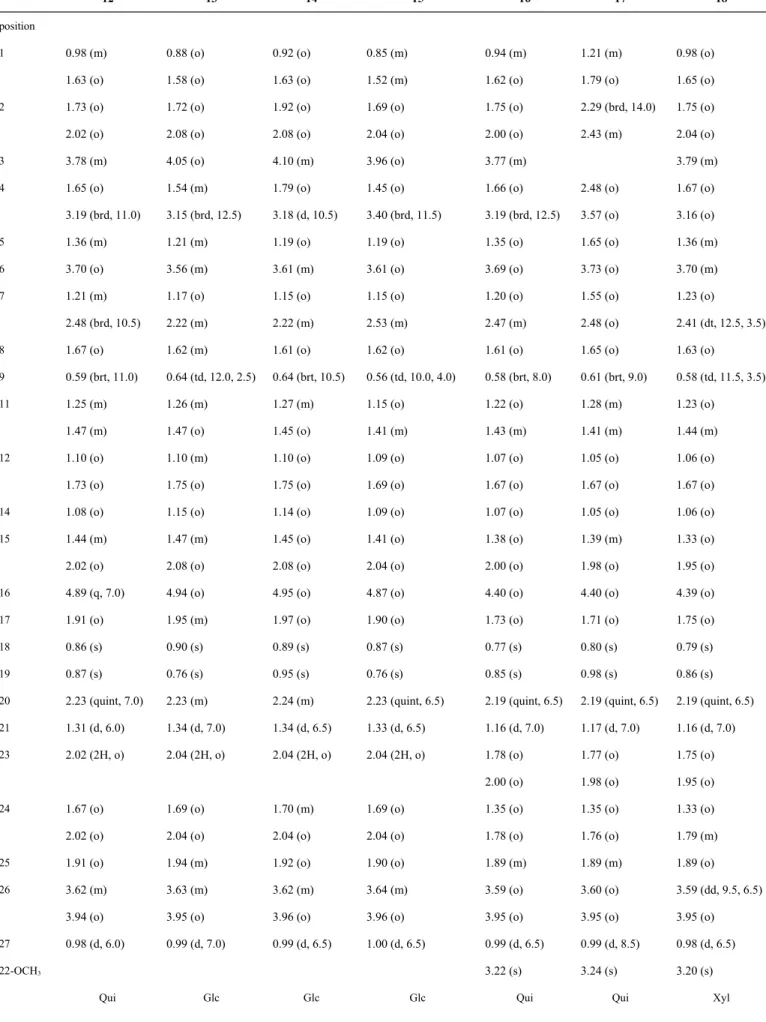
Table 4. 1H NMR Spectroscopic Data of Compounds 12–18 (500 MHz, C 5D5N)a-c 12 13 14 15 16 17 18 position 1 0.98 (m) 1.63 (o) 0.88 (o) 1.58 (o) 0.92 (o) 1.63 (o) 0.85 (m) 1.52 (m) 0.94 (m) 1.62 (o) 1.21 (m) 1.79 (o) 0.98 (o) 1.65 (o) 2 1.73 (o) 2.02 (o) 1.72 (o) 2.08 (o) 1.92 (o) 2.08 (o) 1.69 (o) 2.04 (o) 1.75 (o) 2.00 (o) 2.29 (brd, 14.0) 2.43 (m) 1.75 (o) 2.04 (o) 3 3.78 (m) 4.05 (o) 4.10 (m) 3.96 (o) 3.77 (m) 3.79 (m) 4 1.65 (o) 3.19 (brd, 11.0) 1.54 (m) 3.15 (brd, 12.5) 1.79 (o) 3.18 (d, 10.5) 1.45 (o) 3.40 (brd, 11.5) 1.66 (o) 3.19 (brd, 12.5) 2.48 (o) 3.57 (o) 1.67 (o) 3.16 (o)
5 1.36 (m) 1.21 (m) 1.19 (o) 1.19 (o) 1.35 (o) 1.65 (o) 1.36 (m)
6 3.70 (o) 3.56 (m) 3.61 (m) 3.61 (o) 3.69 (o) 3.73 (o) 3.70 (m)
7 1.21 (m) 2.48 (brd, 10.5) 1.17 (o) 2.22 (m) 1.15 (o) 2.22 (m) 1.15 (o) 2.53 (m) 1.20 (o) 2.47 (m) 1.55 (o) 2.48 (o) 1.23 (o) 2.41 (dt, 12.5, 3.5)
8 1.67 (o) 1.62 (m) 1.61 (o) 1.62 (o) 1.61 (o) 1.65 (o) 1.63 (o)
9 0.59 (brt, 11.0) 0.64 (td, 12.0, 2.5) 0.64 (brt, 10.5) 0.56 (td, 10.0, 4.0) 0.58 (brt, 8.0) 0.61 (brt, 9.0) 0.58 (td, 11.5, 3.5) 11 1.25 (m) 1.47 (m) 1.26 (m) 1.47 (o) 1.27 (m) 1.45 (o) 1.15 (o) 1.41 (m) 1.22 (o) 1.43 (m) 1.28 (m) 1.41 (m) 1.23 (o) 1.44 (m) 12 1.10 (o) 1.73 (o) 1.10 (m) 1.75 (o) 1.10 (o) 1.75 (o) 1.09 (o) 1.69 (o) 1.07 (o) 1.67 (o) 1.05 (o) 1.67 (o) 1.06 (o) 1.67 (o)
14 1.08 (o) 1.15 (o) 1.14 (o) 1.09 (o) 1.07 (o) 1.05 (o) 1.06 (o)
15 1.44 (m) 2.02 (o) 1.47 (m) 2.08 (o) 1.45 (o) 2.08 (o) 1.41 (o) 2.04 (o) 1.38 (o) 2.00 (o) 1.39 (m) 1.98 (o) 1.33 (o) 1.95 (o)
16 4.89 (q, 7.0) 4.94 (o) 4.95 (o) 4.87 (o) 4.40 (o) 4.40 (o) 4.39 (o)
17 1.91 (o) 1.95 (m) 1.97 (o) 1.90 (o) 1.73 (o) 1.71 (o) 1.75 (o)
18 0.86 (s) 0.90 (s) 0.89 (s) 0.87 (s) 0.77 (s) 0.80 (s) 0.79 (s)
19 0.87 (s) 0.76 (s) 0.95 (s) 0.76 (s) 0.85 (s) 0.98 (s) 0.86 (s)
20 2.23 (quint, 7.0) 2.23 (m) 2.24 (m) 2.23 (quint, 6.5) 2.19 (quint, 6.5) 2.19 (quint, 6.5) 2.19 (quint, 6.5)
21 1.31 (d, 6.0) 1.34 (d, 7.0) 1.34 (d, 6.5) 1.33 (d, 6.5) 1.16 (d, 7.0) 1.17 (d, 7.0) 1.16 (d, 7.0) 23 2.02 (2H, o) 2.04 (2H, o) 2.04 (2H, o) 2.04 (2H, o) 1.78 (o) 2.00 (o) 1.77 (o) 1.98 (o) 1.75 (o) 1.95 (o) 24 1.67 (o) 2.02 (o) 1.69 (o) 2.04 (o) 1.70 (m) 2.04 (o) 1.69 (o) 2.04 (o) 1.35 (o) 1.78 (o) 1.35 (o) 1.76 (o) 1.33 (o) 1.79 (m)
25 1.91 (o) 1.94 (m) 1.92 (o) 1.90 (o) 1.89 (m) 1.89 (m) 1.89 (o)
26 3.62 (m) 3.94 (o) 3.63 (m) 3.95 (o) 3.62 (m) 3.96 (o) 3.64 (m) 3.96 (o) 3.59 (o) 3.95 (o) 3.60 (o) 3.95 (o) 3.59 (dd, 9.5, 6.5) 3.95 (o) 27 0.98 (d, 6.0) 0.99 (d, 7.0) 0.99 (d, 6.5) 1.00 (d, 6.5) 0.99 (d, 6.5) 0.99 (d, 8.5) 0.98 (d, 6.5) 22-OCH3 3.22 (s) 3.24 (s) 3.20 (s)
1′ 4.82 (d, 7.5) 5.09 (d, 7.5) 5.09 (d, 7.5) 5.14 (d, 7.5) 4.83 (d, 7.5) 4.76 (d, 7.5) 4.80 (d, 7.5)
2′ 4.04 (o) 4.02 (o) 4.25 (o) 4.07 (o) 4.01 (o) 4.02 (o) 4.05 (o)
3′ 4.09 (o) 4.28 (o) 4.18 (o) 4.28 (o) 4.06 (o) 4.07 (o) 4.12 (o)
4′ 3.58 (o) 4.23 (o) 4.16 (o) 4.25 (o) 3.57 (o) 3.60 (o) 4.12 (o)
5′ 3.70 (o) 3.89 (o) 3.79 (m) 3.87 (o) 3.73 (o) 3.73 (o) 3.68 (m)
4.39 (m) 6′ 1.54 (d, 5.0) 4.39 (m) 4.56 (brd, 11.5) 4.34 (o) 4.50 (brd, 11.0) 4.39 (dd, 12.0, 5.0)b 4.56 (dd, 12.0, 2.0)c 1.54 (d, 6.0) 1.55 (d, 6.0)
Xyl Glc Rha Qui Xyl Xyl Xyl
1″ 5.23 (d, 7.5) 4.82 (d, 8.0) 6.35 (s) 4.80 (d, 7.5) 5.21 (d, 7.5) 5.22 (d, 7.5) 5.27 (d, 8.0)
2″ 4.04 (o) 4.02 (o) 4.76 (brs) 4.03 (o) 4.01 (o) 4.02 (o) 4.05 (o)
3″ 4.16 (o) 4.28 (o) 4.61 (brd, 9.0) 4.28 (o) 4.15 (o) 4.13 (o) 4.19 (o)
4″ 4.16 (o) 4.23 (o) 4.32 (o) 3.72 (m) 4.15 (o) 4.13 (o) 4.19 (o)
5″ 3.70 (o)
4.30 (brd, 11.0)
3.89 (o) 5.02 (o) 3.76 (m) 3.73 (o)
4.29 (dd, 12.0, 4.5) 3.69 (o) 4.27 (o) 3.72 (m) 4.39 (m) 6″ 4.39 (m) 4.56 (brd, 11.5) 1.79 (d, 5.5) 1.62 (d, 6.0) Glc Glc Glc Glc Glc Glc 1′′′ 4.80 (d, 7.5) 4.82 (d, 7.5) 4.82 (d, 8.0) 4.83 (d, 7.5) 4.83 (d, 7.5) 4.84 (d, 7.5)
2′′′ 4.04 (o) 4.04 (m) 4.03 (o) 4.01 (o) 4.02 (o) 4.05 (o)
3′′′ 4.22 (o) 4.25 (o) 4.28 (o) 4.21 (o) 4.21 (o) 4.24 (o)
4′′′ 4.22 (o) 4.25 (o) 4.25 (o) 4.18 (o) 4.21 (o) 4.24 (o)
5′′′ 3.92 (o) 3.96 (o) 3.96 (o) 3.93 (o) 3.93 (o) 3.95 (o)
6′′′ 4.37 (brd, 11.5) 4.53 (brd, 11.5) 4.39 (o) 4.55 (brd, 11.5) 4.33 (dd, 12.0, 5.5)b 4.44 (dd, 12.0, 2.5)c 4.36 (o) 4.53 (dd, 12.0, 2.0) 4.37 (o) 4.54 (brd, 10.0) 4.39 (o) 4.56 (dd, 11.5, 2.0) a o: Overlapped with other signals; m: Multiplet signals. b,cData under the same entry are interchangeable.
Table 5. 13C NMR Spectroscopic Data of Compounds 12–18 (125 MHz, C 5D5N)a-e 12 13 14 15 16 17 18 position 1 37.7 37.7 37.7 37.6 37.7 38.6 37.7 2 32.1 30.0a 29.8 29.9 32.0 38.0 32.1 3 70.6 77.6 77.0 76.9 70.6 210.8 70.5 4 33.1 29.5 28.9 28.5 33.0 39.8 33.1 5 51.3 52.2 52.1 51.0 51.2 52.3 51.3 6 79.2 68.5 68.5 79.4 79.2 79.8 78.9 7 41.4 42.8 42.7 41.5 41.3 40.9 41.3 8 34.1 34.3 34.3 34.3 34.1a 33.9 34.1 9 53.8 54.2 54.1 53.9 53.8 53.7 53.8 10 36.7 36.6 36.7 36.8 36.6 36.7 36.7 11 21.2 21.3 21.3 21.2 21.1 21.2 21.2 12 40.1 40.1 40.2 40.1 39.8 39.7 39.9 13 41.0 41.2 41.1 41.1 41.0 41.0 41.1 14 56.2 56.3 56.3 56.3 56.2 55.9 56.2 15 32.3 32.4 32.4 32.4 32.0 32.0 32.0 16 81.0 81.1 81.1 81.0 81.2 81.1 81.2 17 63.8 63.9 63.9 63.9 64.1 64.1 64.2 18 16.7 16.7 16.4 16.7 16.5 16.4 16.5 19 13.5 13.5 13.6 13.4 13.5 12.5 13.5 20 40.6 40.7 40.7 40.7 40.4 40.4 40.5 21 16.3 16.4 16.7 16.4 16.2 16.2 16.2 22 110.5 110.6 110.6 110.6 112.5 112.5 112.5 23 37.1 37.2 37.2 37.3 30.7 30.7 30.7 24 28.3 28.4a 28.4 28.4 28.1 28.2 28.2 25 34.2 34.3 34.3 34.1 34.0a 34.1 34.1 26 75.4 75.2 75.2 75.2 75.2 75.2 75.2 27 17.4 17.4 17.4 17.5 17.1 17.1 17.1 OCH3 47.2 47.2 47.2
Qui Glc Glc Glc Qui Qui Xyl
1′ 105.1 102.2 99.6 101.6 105.1 105.2 105.9 2′ 75.1 75.4b 78.1a 75.5a 75.1b 75.1a 74.4 3′ 87.5 78.6 79.5 78.6b 87.5 87.3 87.2 4′ 74.7 71.7 71.8 71.8c 74.6c 74.5 69.2 5′ 72.4 78.3 78.5 78.5d 72.2 72.2 66.3 6′ 18.6 62.9 62.7 62.9e 18.5 18.6
Xyl Glc Rha Qui Xyl Xyl Xyl 1″ 106.3 104.9 102.1 105.9 106.3 106.1 106.3 2″ 74.8 75.3b 72.5 76.0a 74.8c 74.6 75.2 3″ 78.1 78.6 72.8 78.1b 78.1 78.1 78.3 4″ 70.8 71.7 74.2 76.9 70.8 70.8 70.9 5″ 67.3 78.5 69.4 72.6 67.3 67.3 67.4 6″ 62.8 18.7 18.8 Glc Glc Glc Glc Glc Glc 1′′′ 104.8 104.9 104.9 104.8 104.8 104.9 2′′′ 75.3 75.3 75.3a 75.2b 75.2a 75.4 3′′′ 78.4 78.6a 78.7b 78.5 78.5 78.6 4′′′ 71.6 71.7 71.8c 71.7 71.7 71.7 5′′′ 78.5 78.2 78.5d 78.4 78.4 78.5 6′′′ 62.7 62.8 62.7e 62.8 62.8 62.9
Table 6. Effects of Compounds 1–24 on Superoxide Anion Generation and Elastase Release in FMLP/CB-induced Human Neutrophilsa
compound superoxide anion elastase release
IC50 (µM)b IC50 (µM)b 1 > 10 3.2 ± 0.2*** 4 > 10 4.2 ± 0.4*** 19 6.1 ± 0.5*** > 10 20 7.0 ± 0.8*** 3.7 ± 0.3*** 21 7.6 ± 0.4*** 4.4 ± 0.6*** 24 4.0 ± 0.1*** 1.0 ± 0.2*** DPIc 0.7 ± 0.4 Sivelestatc (nM) 50.2 ± 0.2
aResults are presented as means ± S.E.M. (n = 3 or 4) (*** p < 0.001 compared with the control value).
Compounds 2, 5, 6 and 23 were not tested. Compounds 3, 7–18 and 22 were inactive in both assays (IC50 > 10 µM). bConcentration necessary for 50 % inhibition (IC50). cDiphenyleneiodonium (DPI) and
sivelestat were used as positive controls for superoxide anion generation and elastase release, respectively.
Figure 4. Effects of compounds 1 and 4 on the inhibition of immediate inflammation degranulation in
RBL-2H3 cells. (A) The cytotoxicities of 1 and 4 were tested using a MTT viability assay. (B) The effects of anti-degranulation were examined by a β-hexosaminidase release assay. Dexamethasone (Dex) was the positive control. The RBL-2H3 cells were treated with 1 and 4 at the indicating concentrations and with dexamethasone at the concentration of 100 nM.
Graphic Abstract
Anti-inflammatory Spirostanol and Furostanol Saponins from Solanum macaonense
Chia-Lin Lee, Tsong-Long Hwang, Juan-Cheng Yang, Hao-Ting Cheng, Wan-Jung He, Chiao-Ting Yen, Chao-Lin Kuo, Chao-Jung Chen, Wei-Yi Chang, and Yang-Chang Wu