Multiphoton fluorescence and second harmonic generation
microscopy of different skin states
Sung-Jan Lin
1,2,3, Wen Lo
4, Yen Sun
4, Shiou-Hwa Jee
2,5, Chen-Yuan Dong
4 1Institute of Biomedical Engineering, College of Medicine and College of
Engineering, National Taiwan University, Taipei, Taiwan
2
Department of Dermatology, National Taiwan University Hospital, Taipei, Taiwan
3Department of Dermatology, Yulin Branch, National Taiwan University Hospital,
Douliu, Yulin, Taiwan
4
Department of Physics, National Taiwan University, Taipei, Taiwan
5Department of Dermatology, National Taiwan University College of Medicine,
Taipei, Taiwan
Abstract
In recent years, non-linear imaging techniques such as multiphoton fluorescence and harmonic generation microscopy have been successfully applied to dermatological imaging. Confocal-like image quality, enhanced depth penetration, and non-linear spectral signatures are among the main advantages of this family of techniques. In this presentation, we will focus on the applications of multiphoton microscopy to skin specimens in different physiological states. Images of normal and diseased tissue specimens will be presented and spectrally characterized. Our work has potential applications in developing multiphoton microscopy into a clinically applicable diagnostic tool.
Keywords: multiphoton fluorescence microscopy, second-harmonic generation, skin imaging Address correspondence to: [email protected]
1. Introduction
Fungal infection is of great significance in the medical practice. For example, dermatophytosis has a prevalence of at least 10-20% among the world’s population. Corneal fugal infection may cause visual loss. Systemic fungal infection, especially in the immunocompromised patients, can be life threatening. The diagnosis of fungal infection can be very difficult. Dermatologists rely on a combination of fungal culture and direct microscopy examination of skin and nail specimens after potassium hydroxide (KOH) treatment for the diagnosis of dermatophytosis.1 However, even in
field of pharmacology, the efficiency of an antifungal agent can only be assessed by clinical symptoms because repeated biopsy and fungal cultures hinder continuous observation of a treatment response.2
Cutaneous photoaging is a filed of great interest in dermatology. Photoaging is caused by cumulative damage from sun light exposure.3 The ultraviolet spectrum is believed to play a major role
in causing photoaging. Dryness, irregular pigmentation, and wrinkling are the clinical features of photoaging. Histologically, severe photoaging shows increased disorganized elastic fibers in the superficial dermis (solar elastosis). The optical property of solar elastosis is currently unknown. A number of surgical procedures and topical medication, including laser resurfacing,4 intense pulse light
treatment,5 chemical peeling,6 and topical tretinoids,3 aimed at remodeling collagen and elastic fibers in
the superficial dermis are used to rejuvenate photoaged skin. However, there is lack of an appropriate non-invasive clinical method to quantify the cutaneous photoaging and to monitor the treatment response. Non-invasive method for monitoring the chronological changes of photoaging has been demonstrated using fluorescence spectroscopy.7 However, spatial discrimination can not be obtained
using fluorescence spectroscopy.
In recent years, mutliphoton fluorescence microscopy has gained much popularity in the biomedical imaging.8,9 The non-linear excitation of fluorescence photons using ultrafast, near- infrared
excitation sources have important advantages in its ability to acquire enhanced axial depth discrimination images, reduced overall specimen photodamage, and increased imaging penetration depths. In addition to multiphoton fluorescence imaging, non- linear polarization effects from a special class of biological materials also have biomedical significance. In biological structures lacking an inversion symmetry, a non-vanishing second order susceptibility can contribute to a second harmonic generation signal. In addition to morphological information, characteristic autofluorescence and second harmonic generation from different cells and components of extracellular matrix can help to specifically feature subjects of interest.10
In this work, we demonstrate that the application of multiphoton fluorescence and second harmonic generation microscopy can be a useful tool in the investigation and diagnosis of skin photoaging and fungal infection.
2. Experimental Method
The multiphoton imaging system used in this study is a modified version of a home-built laser scanning microscopic imaging system based on an upright microscope (E800, Nikon, Japan) described previously.11 A diode-pumped (Millennia X, Spectra Physics, Mountain View, CA), titanium-sapphire
(ti-sa, Tsunami, Spectra Physics) was used as the excitation source. The 760 nm output of the ti-sa laser is scanned in the focal plane by a galvanometer-driver x-y mirror scanning system (Model 6220, Cambridge Techno logy, Cambridge, MA). Prior to entering the upright microscope, the laser is beam expanded to ensure overfilling of the objective’s back aperture. For high resolution imaging, a high
numerical aperture, oil immersion objective (S Fluor 40x, NA 1.3, Nikon) is selected. To direct the expanded laser spot to the sample, a short-pass dirochoic mirror (700DCSPXRUV-3p, Chroma Technology, Brattleboro, VT) is used to reflect the incident excitation laser source. To ensure even excitation of fungi at different orientations, a λ/4 wave plate was used to convert the linearly polarized ti-sa laser beam into one with circularly polarization. The generated MPF signal is collected in the back-scattering geometry where a band-pass filter (HQ460/50, Chroma Technology) is used to isolate the fluorescence signals and a band pass filter (HQ380/20, Chroma Technology) is used to isolate second harmonic generation signals. The signal photons are processed by a single-photon counting PMT (R7400P, Hamamatsu, Japan) and a home-built discriminator.
Microculturing of Microsporum canisi is performed on Sabourauds agar at 25oC using a N.O. 1.5
coverlip. After 14 days in culture, the coverslip attached with fungi is removed from the agar and covered onto a new microscope slide with the fungus-containing surface oriented to the slide for viewing. The average MPF signal per pixel is computed and plotted. For comparison, Microsporum
canis is also stained by lactophenol cotton blue for light microscopic examination. Cutaneous fungal
infection is investigated by inoculating Microsporum canis to nude mice as previously described.2 A
skin fold chamber is used for imaging the fungi in vivo as we previously described.12
To investigate the optical properties of solar elastosis, a facial skin specimen obtained from the cheek of a 70-year-old patient undergoing facial cosmetic surgery is used for imaging. A thin slice of formalin-fixed samples is mounted on the slide and covered with a N.O. 1.5 coverslip for the imaging. For high resolution imaging, a high numerical aperture, oil immersion objective (S Fluor 40x, NA 1.3, Nikon) is selected. A large area scan along the dermal-epidermal junction composed of 3 by 3 array of neighboring images is serially acquired and assembled. For comparison, the specimens are further processed for histological examination using hematoxylin and eosin stain and elastic stain (Verhoeff-van Gieson stain). In hematoxylin and eosin stain, elastin and collagen have varying degrees of pink staining and severe solar elastosis is characterized by bluish gray curvy fibers in the superficial dermis. In elastic stain, elstic fibers are stained blue-black and collagen fibers are red.
3. Results
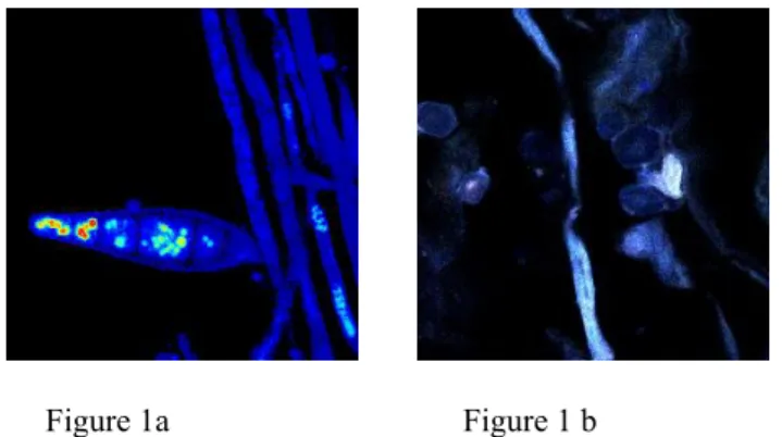
Figure 1a shows the multiphoton fluorescence images of the Microsporum canis in vitro and figure 1b shows the fungal images in vivo. Microsporum canis is intensely autofluorescent. Both the hyphae and spores are intensely autofluorescent and second harmonic signals are not detected.
Morphological analysis of fungi can be obtained using multiphoton images without further fixation and staining of fungi. In figure 1b, the intense autofluorescence of Microsporum canis provides high contrast to the surrounding stratum corneum.
Traditionally, clinicians reply on direct microscopy examination of skin and nail specimens after potassium hydroxide (KOH) treatment. Potassium hydroxide solution disintegrates stratum corneum
Figure 1a Figure 1 b
Figure 1. (a) Multiphoton autofluorescence of Micorsoprum canis in vitro. (b) Multiphoton autofluorescence images of cutaneous Micorsoprum canis infection in vivo.
and nail but fungal walls are resistant to this treatment. Hence, fungi penetrating in the horny layers can be released and visualized. However, this examination has a low sensitivity of diagnosis even in experienced hands. In this work, we demonstrate that fungi are strongly autofluorescent. Its autofluorescence provides contrast to the surrounding stratum corneum cells. Hence, fungi can be visualized without further treatment of the skin. It suggests that multiphoton microscopy can be a very useful tool for the clinical diagnosis of cutaneous fungal infection. This is of great significance in evaluating efficiency of antifungal agents. Currently, we depend on continuous observation of gross cutaneous symptoms and signs, such as erythema, scaling and itching, to evaluate the treatment response of antifungal agents. In the cases free of gross symptoms, microscopic persistence of fungi can not be ruled out because repeated biopsy is not performed. Microscopic observation without damaging the host skin and fungi can be achieved by use of multiphoton microscopy. The continuous process of attachment of fungi to skin and its subsequent invasion to stratum corneum cal also be visualized using multiphoton microscopy. Further, fungi are ineffective in producing second harmonic generation signals. In cases of cutaneous deep fungal infection in which fungi invade the dermis, multiphoton microscopy can help to differentiate fungi from the surrounding dermis which generates second harmonic generation signals due to its high collagen content. This method can also be applied to other organ which is subjective to fungal infection.
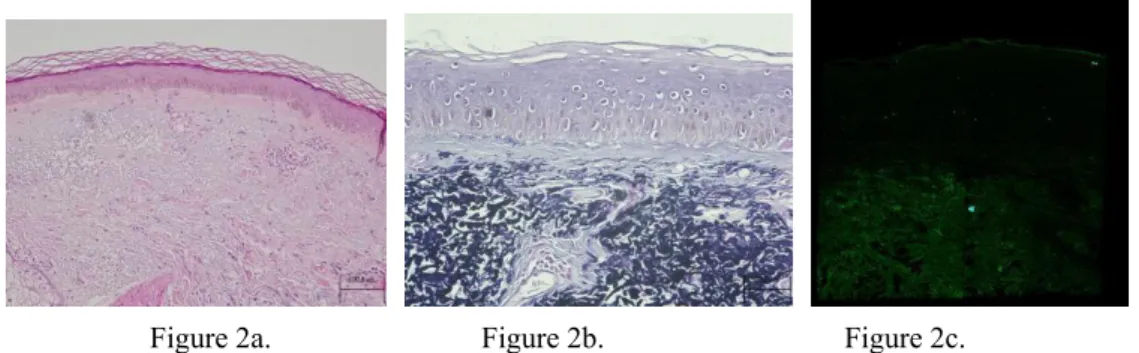
Figure 2a shows the hematoxylin and eosin stain, figure 2b shows the elastic stain and figure 2c shows merged image of autofluorescence and second harmonic generation of the facial skin of a 75-year-old patient. In figure 2a, solar elastosis is featured by thick curvy bluish gray fibers in the dermis. The elastotic change is confirmed by the elastic stain in figure 2b in which elastic fibers are stained black-blue and collagen fibers are stained red. In figure 2c, the dermis is almost composed of autofluorescent signals and only scanty second harmonic generation signals are detected in the area just below epidermis.
Figure 2a. Figure 2b. Figure 2c.
Figure 2. Histology and merged autofluorescence and second harmonic generation images of facial skin of a 75-year-old patient. (a) Hematoxylin and eosin stains (b) Elastic stain (c) Merged autofluorescence and second harmonic generation image (green: autofluorescence; blue: second harmonic generation)
Careful comparison of figure 2a, 2b and 2c reveals that second harmonic generation comes from collagen and solar elastosis is devoid of SHG. Instead, solar elastosis is effective in generating autofluorescence. In our setting, solar elastosis can be easily differentiated from the surrounding collagen. This can be of great help in clinical research of photoaging. Various treatments are currently used to rejuvenate photoaged skin, aimed at remodeling collagen and elastic fibers in the dermis. The treatment response usually depends on subjective clinical assessment by the physicians and patients, because there is lack of a reliable non-invasive method to quantify the aging changes. Our work shows that multiphoton fluorescence and second harmonic generation microscopy can be further developed into a non-invasive tool for the assessment of photoaging and also the treatment response. Solar elastosis can be characterized by the presence of curvy autofluorescent fibers in the superficial dermis. The remodeling process of collagen and elastic fibers in response to treatment can be non-invasively visualized.
4. Conclusion
Multiphoton fluorescence and second harmonic generation microscopy can be a very useful tool for non-invasive monitoring of cutaneous fungal infection and photoaging. Our work has potential applications in developing multiphoton and second harmonic generation microscopy into a clinically applicable diagnostic tool.
ACKNOWLEDGMENT
We like to acknowledge the support of National Research Program for Genomic Medicine, Taiwan ( NSC 93-3112-B-002-034) for this work.
REFERRENCES
1. Lawry A, Haneke E, Strobeck K, Martin S, Zimmer B, Romano PS, "Methods for diagnosing onychomycosis,"Archives of Dermatology, 136: 1112–16 , 2000.
2. Weber J, and Balish E, "Antifungal therapy of dermatophytosis in guinea pigs and congenitally athymic rats, " Mycopathologia, 90: 47-54, 1985.
3. Yarr M, Gilchrest BA: Aging of skin, in Fitzpatrick’s Dermatology in General Medicine, 6th edition, edited by Freedberg IM, Eisen AZ, Wolff K, Austen KF, Goldsmith LA, Katz SI. New York, McGraw Hill, 2003, p1386-1398.
4. Manuskiatti W. Fitzpatrick RE. Goldman MP. Long-term effectiveness and side effects of carbon dioxide laser resurfacing for photoaged facial skin. Journal of the American Academy of
Dermatology. 40:401-11, 1999.
5. Kligman DE. Zhen Y. Intense pulsed light treatment of photoaged facial skin. Dermatologic Surgery. 30:1085-90, 2004.
6. Butler PE. Gonzalez S. Randolph MA. Kim J. Kollias N. Yaremchuk MJ. Quantitative and qualitative effects of chemical peeling on photo-aged skin: an experimental study. Plastic & Reconstructive Surgery 107(1):222-8, 2001.1G.S. Bulmer, "Fungus Diseases in the Orient". 3rd ed., Manila, Philipines: REX Book Store; 1998.
7. Kollias N. Gillies R. Moran M. Kochevar IE. Anderson RR. Endogenous skin fluorescence includes bands that may serve as quantitative markers of aging and photoaging. Journal of Investigative Dermatology. 111:776-80, 1998
8. Denk W, Strickler JH, and Webb WW. Two-photon laser scanning fluorescence microscopy.
Science. 248: 73-76, 1990.
9. So PTC, Dong CY, Masters BR, and Berland KM, "Two-photon excitation fluorescence microscopy," Annual Review of Biomedical Engineering 2: 399, 2000.
10. W. R. Zipfel, R. M. Williams, R. Christie, A.Y. Nikitin, B. T. Hyman, W. W. Webb, " Live tissue intrinsic emission microscopy using multiphoton-excited native fluorescence and second harmonic generation," Proceedings of the National Academy of Sciences of the United States of America, 100:7075-80, 2003.
11. Sun Y, Su JW, Lo W, Lin SJ, Jee SH, and Dong CY, "Multiphoton polarization imaging of the stratum corneum and the dermis in ex-vivo human skin, " Optics Express, 11: 3377, 2003. 12. Li FC, Wang CC, Lin SJ, Jee SH, Dong CY, " Dorsal skinfold titanium chamber for non-invasive
imaging in nude mice using multiphoton and harmonic generation microscopy," Proceedings of the Second Asian and Pacific rim Symposium on Biophotonics 2004, p185-186.