行政院國家科學委員會專題研究計畫 期中進度報告
利用奈米光電生醫感測與分子影像分析 WWOX/WOX1 之功能 (2/3)
期中進度報告(完整版)
計 畫 類 別 : 個別型
計 畫 編 號 : NSC 98-3112-B-006-009-
執 行 期 間 : 98 年 05 月 01 日至 99 年 04 月 30 日 執 行 單 位 : 國立成功大學工程科學系(所)
計 畫 主 持 人 : 陳顯禎
處 理 方 式 : 本計畫可公開查詢
中 華 民 國 99 年 03 月 08 日
目 錄
一、 計畫摘要
1a. 中文摘要 (一頁為限) --- page 2 1b. 英文摘要 (一頁為限) --- page 3 二、 背景介紹 (至多不超過三頁)
2a. 研究目的與文獻探討--- page 4 2b. 研究方法--- page 5 三、 研究成果 (至少二頁)
3a. 具體研究成果 --- page 11 3b. 分析與討論 --- page 11 四、 未來研究構想 --- page 17
五、 成果自評 (整合型計畫之總計畫主持人需另針對整體計畫評估)
5a. 研究成果與原設定目標之相符程度 --- page 18 5b. 達成預期目標情形 --- page 18 5c. 研究成果之學術或應用價值 --- page 18 5d. 學術期刊發表情形 --- page 18 六、 參考文獻 --- page 19 七、 附件
7a. 學術論文 --- page 21 7b. 可供推廣之研發成果資料表 --- page 22
一、計畫摘要 1a. 中文摘要
表面電漿共振(surface plasmon resonance, SPR)感測器可量測在固體與氣體或固體與液 體界面之奈米膜層厚度與介電常數之微量變化。有別於一般單點或區域性之量測,利用表 面電漿共振相位影像系統(phase image system)不僅可以提供大量平行空間資訊,且可獲得高
靈敏之相位資訊。表面電漿共振相位偵測是利用同時包含有 p-wave 與 s-wave 之入射光激
發表面電漿子(surface plasmons, SPs),由於在 SPR 角時只有 p-wave 會因待測物條件改變而 造成相位劇烈變化,s-wave 則否,因此以量測反射光 p-wave 與 s-wave 之間相位差變化來 獲得感測界面上之變化資訊。本論文中提出一共光程之表面電漿共振移相干涉術來觀測空 間平面上之相位資訊,配合五步相位還原演算法來完成相位的重建,在此量測奈米膜層之 相位影像差異並且討論此干涉方式之長時間相位穩定與空間解析度。此影像系統具有高靈
敏度之特性可以量到2x10⁻⁷ RIU 的折射率變化,並且由於其為一共光程干涉術所以具有相
當高之相位穩定,於長時間下之相位穩定可達 2.5x10⁻⁴π。此系統提供了高解析與高通量之
DNA 為陣列量測之能力,且不需要對其作額外的螢光標定不僅如此還可提供量化的依據。
而在蛋白質的量測上還結合了微機電製程之微流道系統,實現多通道蛋白質生物分子量 測。為了觀察細胞之貼附與爬行之情形,本論文中發展了兩表面電漿子顯微影像系統,並 且結合衰逝全反射螢光顯微鏡於細胞貼附之研究。一為菱鏡偶合方式另一為利用高數值孔 徑之顯微油鏡偶合。文中比較了此兩系統之優缺點及其限制之外並將其運用於細胞貼附之 學習。最後再藉由利用超快雷射於細胞外間質的結構製造,此方式可於特定地方製造出二 維甚至三維之生物結構,本論文中創造了一具有線性濃度梯度之纖維蛋白,並藉此控制纖 維母細胞之爬行與貼附型態之改變。
關鍵字:表面電漿子、內全反射、螢光顯微術、雙光子、細胞膜。
一、計畫摘要
1b. 英文摘要 (一頁為限)
Surface plasmon resonance (SPR) sensor can measure the thickness or dielectric constant change of nanolayer between solid-gas or solid-liquid interface. SPR phase image system not only can obtain high sensitivity phase information but also can provide high-throughout space information. Using the incident wave which including p-wave and s-wave to excite surface plasmon, p-wave has a violent phase jump and shift by the sample change in the resonance angle, and s-wave not. According to above, measure the phase difference of reflection wave between p-wave and s-wave to obtain SPR phase information. We setup a common-path phase-shift SPs image system to measure the nano layer at the surface. The system uses five-step phase reconstruct algorithm to obtain the spatial phase information. It has very high sensitivity and the limitation of measurement can achieve 2x10⁻⁷ reflect index change. Besides that, The phase stability can achieve 2.5x10⁻⁴π in long-term measurement due to the common-path interfermetry.
The imaging system offers high resolution and high-throughout screening capabilities for microarray DNA image without the need for additional labeling, and provides valuable quantitative information. It also combines with micro fluid which is fabricated by MEMS to achieve the measurement of multichannel protein chip. In order to study the behavior of cell adhesion and migration which interaction with surface, two kinds of system configuration for wide-field SPP microscopy is reported. One is using the prism to couple surface plasmon and the other one is using the high-numerical-aperture oil-immersion objective and the features are discussed. Finally, we use femtosecond laser to fabrication extra cellular matrix (ECM). It can fabricate two-dimension or even three-dimension biometrical specifically. In this project we fabricate fibronectin with a linear gradient concentration and control the migration, adhesion and differentiation of fibroblast cell.
Keywords: Surface plasmons, total internal reflection, fluorescence microscopy, two photon, cell membrane.
二、背景介紹 (至多不超過三頁) 2a. 研究目的與文獻探討
Numerous biochemical functions of proteins are mediated on cell membranes. Therefore, how to develop a microscope that can image the cell-substrate contact region more efficiently is a crucial research topic for concluding the role of signaling proteins in cell spreading and migration.
By inducing the evanescent field from incident light with an incident angle greater than the critical angle to selectively excite fluorescent molecules on or near a surface in a cellular environment only, total internal reflection fluorescence microscopy (TIRFM) has been utilized to observe the cell-substrate contact region [1] and to study dynamics of proteins [2], endocytosis and exocytosis [3] near the cell membrane [1,4]. Nevertheless, fluorescent molecules are needed to dye or transfect into observed cells in the TIRFM. Intensity microscopy based on the enhancement of surface plasmons (SPs) has been utilized to observe cell-substrate contact localities without any addition of dye labels [5].
SPs are oscillations of the free electrons located on the surface of a metal film and can be usually excited by incident light based on the prism-coupled attenuated total reflection (ATR) or grating-coupled diffraction methods [6]. Conveniently, the prism-coupled excitation can be substituted via the excitation of a highly oblique collimating beam that is generated by focusing a light beam to the outer regions of the back focal plane (BFP) of a high numerical aperture (NA) objective [7]. When the wave vector of an incident evanescent transverse magnetic (TM) light matches that of the SPs, the so-called surface plasmon resonance (SPR) phenomenon occurs and the SPR associated with the EM field is greatly enhanced [6]. The SPs induce a quantum of the photon-photon transverse wave field with a skin depth of less than a few hundred nanometers, which is suitable for the biomolecular scale. The two main features of SPs in bio-sensing and imaging are: 1) very high sensitivity to the interface change to detect tiny biomolecular interactions on the metal surface based on the intensity and phase variations of reflection light;
and 2) to enhance the local electro-magnetic field to improve fluorescence and Raman signals [6].
According to the first feature, intensity-based SPR microscopes with high NA objectives have been utilized to image the cell surface interface in fluid at improved lateral resolution.
Additionally, they can contrast [8] and provide the quantitative data of a refractive index map regarding interactions between cell membrane and extracellular matrix in the cell-substrate contact region [9]. To enhance the sensitivity for detecting the tiny changes of biomolecular interaction, we have integrated the SPR and common-path phase-shift interferometry (PSI) techniques to develop a phase microscope for imaging the two-dimensional (2D) spatial phase variation caused by biomolecular interactions on a sensing chip without the need for additional labeling [10,11]. The common-path PSI technique has the advantage of long-term stability, even when subjected to external disturbances. The SP phase microscope has demonstrated a phase shift stability of 2.5×10⁻⁴π over four hours and a resolution of 2×10⁻⁷ refraction index difference.
However, either the SPR intensity or phase microscopes cannot provide fluorescence signals, which are sometimes regarded as the information for molecular interactions near the bio-surface and to identify the specific protein binding areas [12]. Therefore, to apply the second feature, the
prism-based SP-enhanced fluorescence image can be enhanced at least 10 fold compared to that of the TIRFM [12,13]. Although prism-based SPR images of cell-substrate interactions have previously been elucidated with the corresponding fluorescence images, the cells were fixed by paraformaldehyde after SPR experiments [14]. Furthermore, the physical restraint of the prism confines the NA and magnification of the imaging system. Hence, a simultaneous and real-time observation of objective-based SP-enhanced fluorescence and phase image will make the biochemical responses of living cells near the cell-substrate contacts to be clearly identified according to the brighter fluorescence and more sensitive phase information under the high NA imaging system.
This project reports the first combination of high NA oil-immersion objective-based wide-field SP-enhanced fluorescence microscopy and SP phase microscopy. To diminish the need of a very high incident angle associated with SP images in buffer solution, we adopt a reduction in the thickness of thin metal film and do not attempt to find the highest NA objective. According to theoretical investigations, the decrease of thin silver film thickness not only decreases the propagation length of SPs, so that more detailed intensity and phase features can be recognized owing to improved lateral resolution, but also decreases the SPR angle so as to resolve the restriction associated with the maximum angle imposed by finite NA value of the objective [6,15,16]. In addition, the sensitivity in the index of refraction is not seriously compromised for cell-substrate contacts detection. The SP-enhanced fluorescence imaging of living cells is also able to be achieved by making the use of backward oil-immersion objective (collecting the fluorescence from the bottom of the slide) as well as forward water-immersion objective (from the upper side of the slide). The employment of the silver film as an SP excitation candidate for imaging cells transfected with enhanced green fluorescent protein (eGFP) can enhance the fluorescence via SPs excited by a blue laser [12]. Through a moderate spacer to adjust the metal-fluorophore distance, the SPs obviously contribute to the increase in the quantum yield for achieving brighter a fluorescent signal and a reduction in the fluorescence lifetime for attaining better photostability [17,18]. A candidate tumor suppressor WW domain-containing oxidoreductase, known as marine WOX1 or WWOX, and monkey kidney COS7 fibroblasts are used to demonstrate the imaging capabilities of the developed microscopy [19]. WOX1 proteins play an important role in the regulation of a wide variety of cellular functions such as protein degradation, transcription, and RNA splicing [20].
2b. 研究方法
A. Surface plasmons excited by light and simulation
SPR is an optical phenomenon in which an incident TM wave excites SPs, i.e. a surface charge density oscillation, at the interface between a thin metal film and a dielectric sample medium. SPR occurs when the parallel component of the wave vector of the incident TM wave,
k
x, matches the wave vector of the SP traveling over a semi-infinite structure,k
sp0, i.e., sin
1 2
1 0 2
sp
0
k c
kx c (1) where 0, 1, and 2 are the wavelength-dependent complex dielectric constants of the 1.51 refractive index matching oil, the metal layer, and the dielectric sample, respectively, and
is at the SPR angle. The SPR angle depends on the dielectric constant and thickness of the nanolayer over the sensing metal layer. By simplifying the SPR configuration to the current three-layer 0/1/2 system, the optical coupling of the SP sensor can be analyzed by calculating the reflectivity,R
012, using the Fresnel equation, i.e.2, 1, , 0 , for with
) 2 exp(
1
) 2
exp(
21 1 12
01
1 1 12
012 01
k k k k i j
d r jk r
r
d jk r
R r
j zj i zi j
zj i ij zi z
z (2)
where iand
k
zi i
c
2 k
x2
12 are the dielectric constants and the wave vector components
perpendicular to the interface in medium i, respectively, and d1 is the finite thickness of the metal
film.
In order to unravel the constraint relevant to the maximum incident angle imposed by a finite NA value of the objective in the objective-based SP microscope, the SPR angle based on the three-layer configuration (substrate (BK7)/metal (Ag)/dielectric (H2O)) can be diminished from 83.9o to 78.3o by decreasing the thickness of the thin silver film from 50 nm to 30 nm at the wavelength of 473 nm. The angle associated with the maximum fluorescence enhancement, i.e.
77.5o, is the incident angle of the excitation which slightly lower than the SPR angle at 78.3o. An incident angle smaller than the SPR angle is usually employed to optimize the contrast of reflectivity [7]. Also, in the visible light and near infrared ranges, the longer the incident wavelength used, the smaller the SPR angle is. Therefore, the constraints regarding the range of available angles can be solved with the use of the 30 nm silver film excited by 473 nm or longer wavelength light. However, a shorter SP excitation wavelength will broaden the SPR reflectivity spectrum, which would then clearly reduce the contrast of the SPR image [21]. Also, a thinner silver film would reduce the sensitivity of the SPR image; meanwhile, a shorter incident wavelength could decrease the penetration depth into cell samples.
As a result of the higher sensitivity attained by means of phase measurement rather than intensity detection, in this resport the common-path PSI is adopted to obtain the phase information of living cell observations [10]. Also, a 473 nm blue laser with a shorter incident wavelength is utilized due to the lateral resolution issue and to excite the eGFP simultaneously.
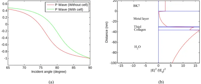
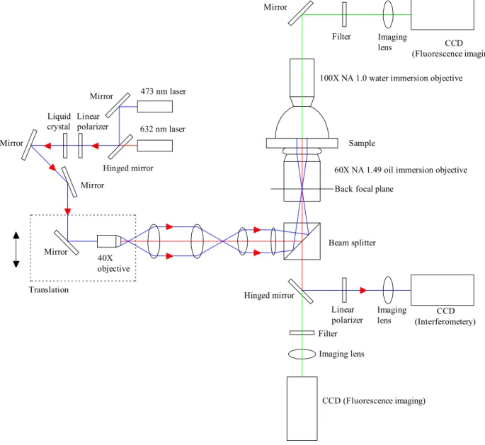
Figure 1(a) illustrates the angle-resolved phase reflectivity spectrum based on the substrate (BK7)/metal (Ag/30nm)/dielectric (H2O) at the wavelength of 473 nm. In this simulation for the SP phase microscopy, the green line and red line represent the simulated phase reflectivity spectrum of the SP chip with and without the cell adhered on the top of bio-surface, respectively, where the phase difference between the two lines is 0.38 π at the excitation angle. Figure 1(b) shows that the distribution of the enhancement factor of the electric field intensity is perpendicular to the interface of the SP chip at 77.5o with two additional layers of thiol and
collagen. The thicknesses of the collagen and thiol layers are approximately 5.5 nm and 1.1 nm, respectively, and the corresponding refractive indexes are 1.46 and 1.49 [12]. The enhancement factor at the interface between the collagen and the buffer has a value around 16.5, as shown in Figure 1(b). Based on the simulation for the TIRFM chip, the maximum value of the enhancement factor is 1.1. From the calculations with the 30 nm silver film, an approximately 15 times higher local field enhancement of the SP chip, compared to that of the conventional TIRFM chip, could be estimated. Local field enhancement leads to an increased excitation rate, and is therefore expected to enhance the fluorescence intensity. However, the interaction of fluorophores with SPs under a short metal-fluorophore distance occurred, and hence the fluorescence quenching effect was counted [22]. Taking the competing phenomena into account, the enhancement in the fluorescence signal obtained from the proposed SP chip is more likely to be no more than 5 times that of the TIRFM chip. To compensate for the competing effect and the thinner silver film issue, a spacer is inserted (a thiol SAM and a collagen layer) to increase the distance between live cell membranes and the 30 nm silver film such that the experimental results demonstrate a capability of producing an approximately 5 times brighter fluorescent live cell image via the excitation of SPs, as discussed in Section 3B.
65 70 75 80 85 90
-1 -0.8 -0.6 -0.4 -0.2 0 0.2 0.4 0.6
Incident angle (degree) Phase ()
P Wave (Without cell) P Wave (With cell)
-15 -10 -5 0 5 10 15
-100 -80 -60 -40 -20 0 20
|E|2 /|E
0|2
Distance (nm)
BK7
Metal layer Thiol
H2O Collagen
(a) (b)
Fig. 1. (a) Phase reflectivity spectrum based on a three-layer configuration: BK7/Ag (30nm)/H2O (Red line: bio-substrate without cell; Green line: bio-substrate with cell). (b) Enhancement factor distribution is perpendicular at sensing interface at incident angle of 77.5o.
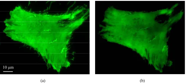
B. Overall system
Figure 2 shows the system configuration of the wide-field oil-immersion objective-based SP microscopy. The SP phase and fluorescence images are mainly obtained by utilizing a diode-pumped solid-state laser light source (20 mW, λ = 473 nm) to excite SPs on the thin silver film. A 632.8 nm He-Ne laser which can be switched by a flipper mirror is available as well. The laser beam passes through two linear polarizers to control the polarization and intensity. The liquid crystal adjusts the phase delay between the TM wave and transverse electric (TE) wave for the PSI which is mentioned above. Then, the light is focused onto the BFP of a high NA oil-immersion objective (60×, NA = 1.49, Nikon) through an objective (40×, Nikon) and a relay
lens pair, finally, emerging from the objective as a parallel beam. The incident angle is controlled by means of adjusting the focusing spot position on the BFP through a linear translation stage. A 0.17 mm cover slide (BK7) coated with metal film via a RF sputtering deposition process is coupled into the oil-immersion objective by adding index-matching oil. The range of available excitation angles of the setup is up to 79.5o. The SPs are excited and the reflection from the metal film comes into the objective, and then goes through a beam splitter, a linear polarizer, and an imaging lens; finally, they are imaged on to a regular CCD camera. The linear polarizer is adjusted at a suitable angle between its optical axis and the incident plane in order to make the TM wave and TE wave interference with better contrast [10]. In comparison with the 50 nm thin silver film, a coating with 30 nm is an adequate thickness for simultaneously achieving the SP phase imaging and fluorescence imaging with the NA 1.49 oil-immersion objective. At the same time, the SP fluorescence imaging of living cells is also able to be achieved by using the bottom oil-immersion objective and an upper water-immersion objective (100×, NA = 1.0, Olympus) as well. The eGFP fluorescence excited via SPs from the cell-substrate contact region is collected by the oil-immersion objective and the water-immersion objective, and individually imaged onto two high-speed frame rate EMCCD cameras (Luca and iXon DV885, Andor) by passing through band-pass filters (BPF, λ = 500 - 535 nm, Semrock) and imaging lenses.
As aforementioned, a nematic liquid crystal phase shifter is applied to produce PSI in the common optical path. It is a positive crystal of single optical axis. As a linear polarization beam with two polarization components in orthogonal directions enters the liquid crystal, they experience different optical paths resulting in different phase shifts. If the incident angle is fixed, the direction of the optical axis of the liquid crystal can be changed by altering the voltage applied to the liquid crystal. Shifts of the modulated phase between the fast axis and slow axis are thus created. Applying the voltages calibrated to produce the optical axis at these five-step phase shifts δ, δ+π/2, δ+π, δ+3π/2, and δ+2π, where δ is an initial phase, five interference images of different phase shifts can be displayed through the linear polarization and be captured by the regular CCD camera as digital image data (I1, I2, I3, I4, I5 in order). Applying a five-step phase shift reconstruction algorithm [23], the 2D wrapped phase distribution
( y x , )
can be obtained with high resolution as follows:
2 . tan 2 ,
1 5 3
4 1 2
I I I
I y I
x (3)Finally, the unwrapped phase
( y x , )
will be completely reconstructed by using our developed multichannel phase wrapping algorithm [24].473 nm laser
632 nm laser
Hinged mirror Mirror
Linear polarizer Liquid
crystal Mirror
Mirror
Mirror
Mirror
Hinged mirror 40X
objective Translation
Filter Imaging
lens CCD
(Fluorescence imaging)
Linear
polarizer Imaging
lens CCD
(Interferometery) Filter
Imaging lens
CCD (Fluorescence imaging) Beam splitter
60X NA 1.49 oil immersion objective Back focal plane
100X NA 1.0 water immersion objective
Sample
Fig. 2. Schematic illustration of experimental configuration employed for simultaneous live cell-substrate contacts imaging using a combination for wide-field oil-immersion objective based-on SP phase microscopy and SP-enhanced fluorescence microscopy.
C. Cell culture protocol
In this report, cultured monkey kidney COS-7 fibroblasts were suspended in a serum-free culture medium, containing 2 mg/ml bovine albumin, and electroporated with EGFP-WOX1 construct (BTX ECM 830 Electroporator, Genetronics; 5 μg DNA/3×106 cells, 220 volt and 50 msec). Albumin enhances both the transfection efficiency and gene expression 3-5 fold [25]. The cells were cultured 24-48 hrs, and then observed by the proposed wide-field oil-immersion objective-based SP microscope.
In order to observe the molecular interactions between cell membranes and extracellular matrix with the SP microscope, the cells were added to collagen immobilized by chemical self-assembly monolayers (SAMs) on a thin silver film BK7 slide containing a pre-warmed medium. In developing this SP chip, the metal film was immersed in 1 mM 2-aminoethanethiol hydrochloride solution to form a dense SAM on its surface. To immobilize the protein collagen, covalent activation was conducted by immersing the chip in a solution containing
EDC[N-(3-dimethylaminopropyl)-N’-ethylcarbodimide hydrochloride, 2 mM] and NHS(N-hydroxysuccinimide, 5 mM) for 6 hrs. One prior study for detecting fluorescent erythrocytes on metal surfaces to induce the quenching effect with a similar cell culture protocol can be referred to in [26].
三、研究成果 (至少二頁) 3a. 具體研究成果
This project has developed the first combination of SP phase microscopy and SP-enhanced fluorescence microscopy with a 1.49 NA oil-immersion objective by decreasing the thickness of a silver film to 30 nm. The distributions of WOX1-containing clusters and cell-substrate distance can be qualitatively illustrated at the same time through a comparison of the SP-enhanced fluorescence and phase images. The experimental results, consistent with the simulation, have shown that a 5-fold enhancement of the SP-enhanced fluorescence image and a better than 3 μm lateral resolution of the SP phase image could be achieved by the simultaneous observation of a living COS7 fibroblast transfected with the eGFP-WOX1 construct on the cell-substrate contact region.
3b. 分析與討論
A. Considerations of enhancement and lateral resolution
The complex dielectric constants of gold and silver have large negative real numbers in the visible light region. Therefore, they are good candidates as the medium of SP excitation. In this project, a 473 nm blue laser is adopted as a light source for the excitation of eGFP via SPs, the SP intensity and phase images. On account of preceding studies of SP chips with thin silver film coatings, it has been shown that not only the fluorescence intensity can be enhanced by the excitation of SPs, but also the photostability of fluorophores can be improved [12]. Compared with thin gold films, superior fluorescence enhancement is able to be obtained by the excitation of thin silver films with the blue laser beam.
The lateral resolution of SP microscopy is usually limited by the propagation length of SPs.
The SP field decays exponentially while traveling along a dielectric/metal interface with the propagation length determined by the imaginary part of the SP wave vector [6]. Usually, the propagation length of gold is shorter than that of silver. Also, the propagation length excited by a shorter wavelength is shorter than that by a longer wavelength. Furthermore, the propagation length excited under an optimal thickness is longer than that under others. For example, an around 45 nm thin gold film excited by a 632.8 nm laser in air is an optimal thickness for the excitation of the SPR. A 30 nm silver film excited by the 473 nm laser in air is not. Figure 3(a) shows E-beam lithographical PMMA removed square patterns at different sizes (1×1 μm2, 3×3 μm2, 5×5 μm2, & 7×7 μm2) on a BK7 substrate coated with a metal film. The space between the square holes is 5 μm and the thickness of the PMMA is controlled at 30 nm. The patterned SP images arise due to the refractive index difference between the PMMA and air. The incident angle is modulated to excite the SPR condition for the PMMA/metal interface, not for the air/metal interface. Figures 3(b) & (c) indicate that the SP intensity and phase images excited by the 632.8 nm He-Ne laser are based on an optimal SPR condition of 45 nm gold film, while Figures 3(d) &
(e) show a 30 nm silver film excited by the 473 nm laser at the incident angle of 77.5o. The phase images are inverted due to the phase reconstruction. Also, the incident light is from left to right.
The compromise based on the above issues is that the propagation length of SPs at the 30 nm
silver film excitation condition could be shorter than that of the 45 nm gold film excitation condition. As shown in Figures 3(b)-(e), the experimental results of the patterned SP intensity and phase images demonstrate that the spatial resolution of the 30 nm thin silver film condition is better than that of the 45 nm gold film condition according to the feature discrimination of the smallest square holes and the minimal coupling effect between the holes. The lateral resolution of the SP phase image based on the setup approaches to 1 μm, as checked in Figure 3(e).
As with any microscopy, lateral spatial resolution problems in SP microscopy can not be discussed without addressing the question of contrast. From Figures 3(b)-(e), the superior contrasted images with higher maximum-to-minimum ratios are found in the gold film condition due to the optimal thickness at a longer excitation wavelength. From the experimental results and prior studies, a non-absorbing dielectric spacer (a thiol SAM and a collagen layer) that is 6.1 nm thick not only reduces the effect of metallic surface-induced fluorophore quenching at very short distances in SP-enhanced fluorescence microscopy, but also promotes high contrast SP images without loss of lateral resolution [12,21,27]. Therefore, associated with the thinner silver film setup, the SP phase image shows an acceptable lateral resolution, while an SP-enhanced fluorescence image can also be enhanced significantly.
B. Appropriate setup for surface plasmon-enhanced fluorescence excitation
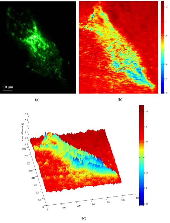
Recent investigations have supported the contention that thrombomodulin, an integral membrane glycoprotein, plays an important role in extravascular activities [28] and thrombomodulin-mediated cell adhesion [29]. As indicated in Figures 4(a) & (b), the SP-enhanced fluorescence imaging of a living melanoma-GFP-tagged thrombomodulin cell can be realized at an incident angle of 77.5o associated with the maximum fluorescence enhancement by making utilization of the bottom oil-immersion objective through the thinner silver film as well as the upper water-immersion objective. The fluorescence signal collected from the bottom objective is quenched when a thicker sliver film is adopted. A five times brighter fluorescent live cell image via the SPs can be observed by the bottom oil-immersion objective based on the appropriate microscopy setup. Compared to the observation of melanoma-GFP-tagged thrombomodulin cells through the culture medium with the water-immersion objective, better collection efficiency and spatial resolution of SP-enhanced fluorescence images can be obtained by the use of the oil-immersion objective with a higher NA value, as shown in Figures 4(a) & (b).
The decrease in thickness of the silver film solves the two problems regarding imaging in fluid via the 1.49 NA oil-immersion objective and improvement in the lateral resolution. Moreover, a simultaneous observation of phase image can be made with this setup.
C. Simultaneous fluorescence and phase imaging
Figures 5 (a), (b) and (c) demonstrates the simultaneous fluorescence and phase imaging of a single living COS7 fibroblast transiently transfected with the eGFP-WOX1 construct cultured on the collagen-coated SP chip based on the developed SP-enhanced fluorescence and phase microscopy. In contrast to the flat adhesion of the melanoma-GFP-tagged thrombomodulin cell illustrated in Figure 4, the expression of WOX1 proteins that possesses two N-terminal WW
domains (containing conserved tryptophan residues), a nuclear localization sequence, and a C-terminal short-chain alcohol dehydrogenase/reductase domain (which contains a mitochondria-targeting sequence) tagged with eGFP is imaged with the punctuated adhesion of the COS7 fibroblast near its plasma membrane area, as shown in Figure 5 (a) [20]. Also, it presents the localization to the perinuclear region and mitochondria of the WOX1-containing clusters. Simultaneously, Figures 5 (b) and (c) show the acquired 2D and three-dimensional (3D) SP phase images of the same cell, while the phase difference between the areas with and without the cell is seen to be approximately 0.35 π. The regions with large phase differences indicate the localities that are directly in contact with the bio-surface and the areas of greater density which previous research has explained reflect where the contraction of microfilaments in the cytoskeleton have sustained the adhesion of the cell [11]. Contrary to the dense perinuclear localization of the WOX1-containing clusters, the SP phase image corresponding to the cell’s footprint exhibited greater phase difference resulting from shorter cell-substrate distance and greater density in the cell cytoplasm close to the membrane. Further instrumental developments and theoretical modeling are planned to employ both the SP phase microscopy and SP-enhanced fluorescence microscopy to allow quantitative measurements of cell-substrate distances at the same time.
(a)
(b) (c)
(d) (e)
Fig. 3. (a) Epi-illumination image of E-beam lithographical 30 nm PMMA square patterns spaced 5μm apart on BK7 substrate with a metal film at different sizes (1×1μm2, 3×3μm2, 5×5μm2, 7×7μm2). The SP (b) intensity and (c) phase image of these PMMA patterns on a 45 nm thin gold film excited by a 632.8 nm laser in air. The SP (d) intensity and (e) phase image of these PMMA patterns located on a 30 nm thin silver film. The color bars indicate phase difference in π.
(a) (b)
10 μm
Fig. 4. Live melanoma-GFP-tagged thrombomodulin SP-enhanced fluorescence images by utilizing (a) the 1.49 NA oil-immersion objective and (b) the 1.0 NA water-immersion objective, both with an exposure time of 0.5 sec at an incident angle of 77.5o.
(a) (b)
(c) 10 μm
Fig. 5. Simultaneous (a) SP-enhanced fluorescence image, (b) 2D SP phase image, and (c) its 3D phase image of a living COS7 fibroblast transfected with an eGFP-WOX1 construct. The color bars indicate phase difference in π.
四、未來研究構想
This report demonstrates the first combination for wide-field surface plasmon (SP) phase microscopy and SP-enhanced fluorescence microscopy to image living cells’ contacts on the surface of a bio-substrate simultaneously. The phase microscopy with a phase-shift interferometry and common-path optical setup can provide high-sensitivity phase information in long-term stability. Simultaneously, the fluorescence microscopy with the enhancement of a local electromagnetic field can supply bright fluorescent images. The combined microscope imposes a high numerical aperture objective upon the excitation of surface plasmon through a silver film with a thickness of 30 nm. The developed SP microscope is successfully applied to the real-time bright observation of the transfected fluorescence of living cells localized near the cell membrane on the bio-substrate and the high-sensitivity phase image of the cell-substrate contacts at the same time. In the coming year, two specific aims are:
1) To optical image WOX1 translocation on living cell membranes in real time. A newly
developed plasmon-enhanced TIRFM with brighter fluorescent signal and improved photostability can dynamically image the migration pathway of WOX1 fused to green fluorescent protein (GFP). Moreover, a novel SPP phase microscope can be utilized to directly image living cell membranes without additional labeling and will provide more information for cell attachment and migration on biosurface.2) To track the translocation of WOX1 in whole cells. To examine WOX1 trafficking in living
cells, we propose to use quantum dots (QDs) labeling to image the translocation and real time tracking. Using QDs labeling on WOX1 signaling can greatly improve the resolution of molecular imaging and provide information on WOX1 trafficking.五、成果自評
Objective: This project are to develop the state-of-the-art nanophotonic biosensing and
molecular imaging for the BIA of living cells, and utilize them to analyze the translocation and conformational changes of WOX1 induced by external effects such as stress stimuli and chemical environment. Furthermore, the outcomes of the research subproject will benefit to understand the signaling network of WOX1 in living cells and identify the regulated disease genes that allow control of aberrant growth in cancer cells.Achievements:
- Advanced Surface-enhanced Raman Scattering (SERS) Biosensing
- Molecular Imaging: the state-of-the-art SP Phase and SP-enhanced Fluorescence Images - To Strengthen the Lab and International Collaborations
The research lab. (Adaptive Photonics Lab., NCKU) has a good standing in the international communities on the research of nanoplasmonic biosensing and molecular imaging. The PI is recognized as the expert in the research field of Taiwan and 5 papers were published, 2 papers were submitted, and 1 international collaborations in the area of the proposed research grant.
Publications based on work supported by this grant (
*corresponding)
Q. Hong, C.-I Sze, S.-R. Lin, M.-H. Lee, R.-Y. He, L. Schultz, L.-J. Hsu, S.-J. Chen, R. J.
Boackle, and N.-S. Chang, “Complement C1q activates tumor suppressor WWOX to kill prostate cancer cells: downregulation of C1q in hyperplasia and cancerous prostate,” PLoS ONE, vol. 4, no 6, e5755, June 2009.
C.-H. Lin, L. Jiang, Y.-H. Chai, H. Xiao, S.-J. Chen, and H.-L. Tsai, “One-step fabrication of nanostructures by femtosecond laser for surface-enhanced Raman scattering,” Optics Express, vol. 17, no. 24, pp. 21581-21589, November 2009. (Impact Factor: 3.880)
R.-Y. He, C.-Y. Lin, Y.-D. Su, K.-C. Chiu, N.-S. Chang, H.-L. Wu, and S.-J. Chen*, “Imaging live cell membranes via surface plasmon-enhanced fluorescence and phase microscopy,”
Optics Express, vol. 18, no. 4, pp. 3649-3659, February 2010. (Impact Factor: 3.880)
J.-Y. Chang, H.-P. Lin, L.-J. Hsu, F.-J. Lai, Q. Hong, S.-J. Chen, and N.-S. Chang, “Signaling from membrane receptors to tumor suppressor WWOX,” Experimental Biology and Medicine, 2010 (accepted). (Impact Factor: 2.202)
W.-S. Kuo, C.-N. Chang, Y.-H. Chien, Y.-T. Chang, S.-J. Chen, and C.-S. Yeh, “Near-infrared Au nanorods in imaging, photodynamic therapy, and hyperthermia of malignant cells,”
Angewandte Chemie Int. Ed., 2010 (accepted). (Impact Factor: 10.879)
International collaborations based on work supported by this grant:
Cooperated with Prof. H.-L. Tsai, UMR onto “Ultrashort-pulse Laser Micromachining.” The PhD student, Mr. Cheng-Hsin Lin, got the fellowship of US$54,000 from UMR and to study on UMR for three years (02/2007-01/2010).
六、參考文獻
1. G. A. Truskey, J. S. Burmeister, E. Grapa, and W. M. Reichert, “Total internal reflection fluorescence microscopy (TIRFM) II. Topographical mapping of relative cell/substratum separation distances,” J. Cell Sci. 103, 491-499 (1992).
2. S. E. Sund and D. Axelrod, “Actin dynamics at the living cell submembrane imaged by total internal reflection fluorescence photobleaching,” Biophys. J. 79, 1655-1669 (2000).
3. W. J. Betz, F. Mao, and C. B. Smith, “Imaging exocytosis and endocytosis,” Curr. Opin.
Neurobiol. 6, 365-371 (1996).
4. D. Axelrod, “Total internal reflection fluorescence microscopy in cell biology,” Methods Enzymol. 361, 1-33 (2003).
5. K. F. Giebel, C. Bechinger, S. Herminghaus, M. Riedel, P. Leiderer, U. Weiland, and M.
Bastmeyer, “Imaging of cell/substrate contacts of living cells with surface plasmon resonance microscopy,” Biophys. J. 76, 509-516 (1999).
6. H. Raether, Surface Plasmons on Smooth and Rough Surfaces and on Gratings (Springer, 1998).
7. B. Huang, F. Yu, and R. N. Zare, “Surface plasmon resonance imaging using a high numerical aperture microscope objective,” Anal. Chem. 79, 2979-2983 (2007).
8. M. M. A. Jamil, M. C. T. Denyer, M. Youseffi, S. T. Britland, S. Liu, C. W. See, M. G.
Somekh, and J. Zhang, “Imaging of the cell surface interface using objective coupled widefield surface plasmon microscopy,” J. Struct. Biol. 164, 75-80 (2008).
9. K. J. Moh, X. C. Yuan, J. Bu, S. W. Zhu, and B. Z. Gao, “Surface plasmon resonance imaging of cell-substrate contacts with radially polarized beams,” Opt. Express 25, 20734-20741 (2008).
10. Y. D. Su, S. J. Chen, and T. L. Yeh, “Common-path phase-shift interferometry surface plasmon resonance imaging system,” Opt. lett. 30, 1488-1490 (2005).
11. K. H. Lee, Y. D. Su, S. J. Chen, F. G. Tseng, and G. B. Lee, “Microfluidic systems integrated with two-dimensional surface Plasmon resonance phase imaging systems for microarray immunoassay,” Biosens. Bioelectron. 23, 466-472 (2007).
12. R. Y. He, G. L. Chang, H. L. Wu, C. H. Lin, K. C. Chiu, Y. D. Su, and S. J. Chen, “Enhanced live cell membrane imaging using surface plasmon-enhanced total internal reflection fluorescence microscopy,” Opt. Express 14, 9307-9316 (2006).
13. R.-Y. He, Y.-D. Su, K.-C. Cho, C.-Y. Lin, N.-S. Chang, C.-H. Chang, and S.-J. Chen,
“Surface plasmon-enhanced two-photon fluorescence microscopy for live cell membrane imaging,” Opt. Express 17, 5987-5997 (2009).
14. A. W. Peterson, M. Halter, A. Tona, K. Bhadriraju, and A. L. Plant, “Surface plasmon resonance imaging of cells and surface-associated fibronectin,” BMC Cell Biol. 10, 16 (2009).
15. B. Rothenhausler and W. Knoll, “Interferometric determination of the complex wave vector of plasmon surface polaritons,” J. Opt. Soc. Am. B 5, 1401-1405 (1988).
16. W. Knoll, “Optical characterization of organic thin films and interfaces with evanescent
waves,” Mat. Res. Soc. Bulletin. 16, 29-39 (1991).
17. C. D. Geddes and J. R. Lakowicz, “Metal-enhanced fluorescence,” J. Fluor. 12, 121-129 (2002).
18. J. R. Lakowicz, “Radiative decay engineering: biophysical and biomedical applications,”
Anal. Biochem. 298, 1-24 (2001).
19. N. S. Chang, N. Pratt, J. Heath, L. Schultz, D. Sleve, G. B. Carey, and N. Zevotek,
“Hyaluronidase induction of a WW domain-containing oxidoreductase that enhances tumor necrosis factor cytotoxicity,” J. Biol. Chem. 276, 3361-3370 (2001).
20. N. S. Chang, L. J. Hsu, Y. S. Lin, F. J. Lai, and H. M. Sheu, “WW domain-containing oxidoreductase: a candidate tumor suppressor,” Trends Mol. Med. 13, 12-22 (2006).
21. C. E. H. Berger, R. P. H. Kooyman, and J. Greve, “Resolution in surface plasmon microscopy,” Rev. Sci. Instrum. 65, 2829-2836 (1994).
22. P. Anger, P. Bharadwaj, and L. Novotny, “Enhancement and quenching of single-molecule fluorescence,” Phys. Rev. Lett. 96, 113002 (2006).
23. P. Hariharan, B. F. Oreb, and T. Eiju, “Digital phase-shifting interferometry: a simple error-compensating phase calculation algorithm,” Appl. Optics 26, 2504-2505 (1987).
24. J.-J. Chyou, S.-J. Chen, and Y.-K. Chen, “Two-dimensional phase unwrapping using a multichannel least-mean-square algorithm,” Appl. Optics 43, 5655-5661 (2004).
25. Q. Hong, L. J. Hsu, L. Schultz, N. Pratt, J. Mattison, and N. S. Chang, “Zfra affects TNF-mediated cell death by interacting with death domain protein TRADD and negatively regulates the activation of NF-κB, JNK1, p53 and WOX1 during stress response,” BMC Mol.
Biol. 8, 50 (2007).
26. R. M. Fulbright and D. Axelrod, “Dynamics of nonspecific adsorption of insulin to erythrocyte membranes,” J. Fluor. 3, 1-16 (1993).
27. B. Rothenhausler and W. Knoll, “Surface-plasmon microscopy,” Nature 332, 615-617 (1988).
28. M. C. Boffa, B. Burke, and C. C. Haudenschild, “Preservation of thrombomodulin antigen on vascular and extravascular surfaces,” J. Histochem. Cytochem. 35, 1267-1276 (1987).
29. H. C. Huang, G. Y. Shi, S. J. Jiang, C. S. Shi, C. M. Wu, H. Y. Yang, and H. L. Wu,
“Thrombomodulin-mediated cell adhesion,” J. Biol. Chem. 278, 46750-46759 (2003).
七、附件
7a. 學術論文 (2009.05~2010.04)
Q. Hong, C.-I Sze, S.-R. Lin, M.-H. Lee, R.-Y. He, L. Schultz, L.-J. Hsu, S.-J. Chen, R. J.
Boackle, and N.-S. Chang, “Complement C1q activates tumor suppressor WWOX to kill prostate cancer cells: downregulation of C1q in hyperplasia and cancerous prostate,” PLoS ONE, vol. 4, no 6, e5755, June 2009.
P.-J. Su, W.-L. Chen, J.-B. Hong, T.-H. Li, R.-J. Wu, C.-K. Chou, S.-J. Chen, C. Hu, S.-J.
Lin, and C.-Y. Dong, “Discrimination of collagen in normal and pathological skin dermis through second-order susceptibility microscopy,” Optics Express, vol. 17, no. 13, pp.
11161-11171, June 2009. (Impact Factor: 3.880)
C.-H. Lin, L. Jiang, H. Xiao, Y.-H. Chai, S.-J. Chen, and H.-L. Tsai, “Fabry-Perot
interferometer embedded in a glass chip fabricated by femtosecond laser,” Optics Letters, vol.
34, no. 16, pp. 2408-2410, August 2009. (Impact Factor: 3.772)
W. Lo, Y.-L. Chang, J.-S. Liu, C.-M. Hseuh, V. Hovhannisyan, S.-J. Chen, H.-Y. Tan, and C.-Y. Dong, “Multimodal, multiphoton microscopy and image correlation analysis for characterizing corneal thermal damage,” Journal of Biomedical Optics, vol. 14, no. 5, 054003, September 2009. (Impact Factor: 2.970)
C.-H. Lin, L. Jiang, Y.-H. Chai, H. Xiao, S.-J. Chen, and H.-L. Tsai, “One-step fabrication of nanostructures by femtosecond laser for surface-enhanced Raman scattering,” Optics Express, vol. 17, no. 24, pp. 21581-21589, November 2009. (Impact Factor: 3.880)
C. H. Lin, L. Jiang, Y. H. Chai, H. Xiao, S.-J. Chen, and H. L. Tsai, “Fabrication of microlens arrays in photosensitive glass by femtosecond laser direct writing,” Apply Physics A, vol. 97, no. 4, pp. 751-757, December 2009. (Impact Factor: 1.884)
R.-Y. He, C.-Y. Lin, Y.-D. Su, K.-C. Chiu, N.-S. Chang, H.-L. Wu, and S.-J. Chen*, “Imaging live cell membranes via surface plasmon-enhanced fluorescence and phase microscopy,”
Optics Express, vol. 18, no. 4, pp. 3649-3659, February 2010. (Impact Factor: 3.880)
C.-H. Lin, R. A. Powell, L. Jiang, H. Xiao, S.-J. Chen, and H.-L. Tsai, “Real-time depth measurement for micro-holes drilled by lasers,” Measurement Science & Technology, vol. 21, no. 2, pp. 025307, February 2010. (Impact Factor: 1.493)
C.-H. Lin, L. Jiang, Y.H. Chai, H. Xiao, S.-J. Chen, and H.-L. Tsai, “A method to fabricate 2D nanoparticle arrays,” Apply Physics A, vol. 98, no. 4, pp. 855-860, March 2010. (Impact Factor: 1.884)
J.-Y. Chang, H.-P. Lin, L.-J. Hsu, F.-J. Lai, Q. Hong, S.-J. Chen, and N.-S. Chang, “Signaling from membrane receptors to tumor suppressor WWOX,” Experimental Biology and Medicine, 2010 (accepted). (Impact Factor: 2.202)
W.-S. Kuo, C.-N. Chang, Y.-H. Chien, Y.-T. Chang, S.-J. Chen, and C.-S. Yeh, “Near-infrared Au nanorods in imaging, photodynamic therapy, and hyperthermia of malignant cells,”
Angewandte Chemie Int. Ed., 2010 (accepted). (Impact Factor: 10.879)
7b. 可供推廣之研發成果資料表 (無)
可供推廣之研發成果資料表
□ 可申請專利 □ 可技術移轉 日期:___年___月___日
國科會補助計畫
計畫名稱:
計畫編號:
計畫主持人:
(中文) 產品/技術名稱
(英文) 發明人/單位
(中文)
產品/技術說明
(100~500 字) (英文)
應用範圍
產品/技術優勢
市場潛力
(可能競爭之國內外廠商及其產品?可能技轉或合作之國內外廠商?)
產品/技術 保護狀況
(專利名稱?專利通過年度/申請日期?是否有技轉?)
1. 每項研發成果請填寫一式二份,一份隨成果報告送繳本會,一份送 貴單位研發成果推廣
單位(如技術移轉中心)。
2. 本項研發成果若尚未申請專利,請勿揭露可申請專利之主要內容。
3. 本表若不敷使用,請自行影印使用。