行政院國家科學委員會補助專題研究計畫 ■成果報告
□期中進度報告
新穎 DNA 傳輸載體之開發及其質子海綿效應與轉染效率關係之 研究
計畫類別:■個別型計畫 □整合型計畫 計畫編號:NSC 96 -2221-E-041-018-MY3 執行期間: 96 年 08 月 01 日至 99 年 07 月 31 日 執行機構及系所:嘉南藥理科技大學生物科技系
計畫主持人:蕭明達 共同主持人:程中玉
計畫參與人員:劉幸宜、林楫程、陳榮傑、張維揚、陳祉佑、陳曉慈、
張萬豐
成果報告類型(依經費核定清單規定繳交):□精簡報告 ■完整報告
本計畫除繳交成果報告外,另須繳交以下出國心得報告:
□赴國外出差或研習心得報告
□赴大陸地區出差或研習心得報告
□出席國際學術會議心得報告
□國際合作研究計畫國外研究報告
處理方式:除列管計畫及下列情形者外,得立即公開查詢
□涉及專利或其他智慧財產權,□一年□二年後可公開查詢 中 華 民 國 99 年 10 月 25
CONTENTS
Chapter One: Abstract ---I 中文摘要---II Chapter Two: Abstract---III 中文摘要--- IV Chapter Three: Abstract---V 中文摘要---VI Chapter Four: Abstract--- VII
中文摘要---VIII Chapter One: The Characteristics and Transfection Efficiency
of CationicPoly (ester-co-urethane) - Short Chain
PEI Conjugates Self-assembled with DNA--- 1
Introduction--- 3
Materials and methods--- 4
Results and discussion--- 9
Conclusion--- 13
Acknowledgement--- 13
References--- 13
Chapter Two: The characteristics and transfection efficiency of PEI modified by biodegradable poly(β -amino ester)--- 28
Introduction--- 30
Materials and methods--- 31
Results and discussion--- 35
Conclusion--- 38
Acknowledgement--- 38
References--- 38 Chapter Three: A study on polyethyleneimine incorporated onto
poly(dl-lactide-co-glycolide) nanoparticles for a
highly gene transfection efficacy and low cytotoxicity--- 53
Introduction--- 55
Materials and methods--- 58
Results and discussion--- 59
Conclusion--- 62
Acknowledgement--- 62
References--- 62
Chapter Four: Synthesis and Characterizations of New Glycidyl-based Cationic Poly(aminoester) and Study on Gene Delivery--- 77
Introduction--- 79
Materials and methods--- 80
Results and discussion--- 84
Conclusion--- 87
Acknowledgement--- 88
References--- 88
I
CHAPTER ONE
ABSTRACT (1)
To improve the transfection efficiency of polycations with DNA, we synthesized poly(ester-co-urethane)(PEU-g-PEI800) with short chain PEI800 in the side chain, and poly(ester-co-urethane)(PEU) without short chain PEI800. Both PEU-g-PEI800 and PEU, readily self-assembled with plasmid DNA ( pCMV- β gal) in a HEPES buffer, were characterized by dynamic light scattering and zeta-potential. The results reveal that PEU-g-PEI800 and PEU were able to self-assemble particles with DNA and yield nano-sized complexes (<200nm) with positive charge at N/P ratios of 20/1 and 120/1, respectively. The degradation studies indicate that the half-life of PEU-g-PEI800 and PEU in the HEPES buffer were 14 and 35 hours at pH 7.4, respectively. Titration studies were performed to determine the buffering capacities of the polymers.
The COS-7 cell viabilities in the presence of PEU-g-PEI800/DNA, PEU/DNA, and PEI25k/DNA were studied. In addition, The PEU-g-PEI800/DNA complexes were able to transfect COS-7 cells in vitro with a high efficiency comparable to a well-known gene carrier PEI25k. The results indicate that PEU-g-PEI800 is an attractive cationic poly (ester-co-urethane) for gene delivery and an interesting candidate for further study.
Key words: transfection, poly(ester-co-urethane), poly(ethylenimine), buffering capacity
II
第 一 章
中文摘要(1)
為 了 改 善 聚 陽 離 子 轉 殖 DNA 的 效 率 , 我 們 合 成 了 一 種 含 短 鏈 PEI800 之 新 穎 poly(ester-co-urethane) (PEU-g-PEI800)和不含 PEI800 之 poly(eater-co-urethane) (PEU)。
此兩種聚合體均可與 DNA 產生縮合作用,研究結果顯示 PEU-g-PEI800 與 PEU 此兩種聚陽離子 在 N/P 分別為 20/1 及 120/1 時,即可與 DNA 縮合而產生奈米大小之微粒(<200nm),此兩種聚 合體之半衰期分別為 14 與 35 小時。我們利用滴定測試其緩衝能力,並利用 COS-7 測試 PEU-g-PEI800/DNA,PEU/DNA, PEI25k/DNA 之細胞毒性及轉染效率,我們發現 PEU-g-PEI800 具 有相當大之發展潛力。
關鍵詞: 轉染、poly(ester-co-urethane)、poly(ethylenimine)、緩衝能力
III
CHAPTER TWO
ABSTRACT (2)
To improve the cytotoxicity of PEI25k and the transfection efficiency of poly(β -amino ester) with DNA, we synthesized a poly(β -amino ester), PEDP, bearing ester linkages in the backbone and tertiary amines in the backbone and side chain and prepared a binary mixture, PEDP-PEI25k, using physical blending meyhod. Both poly(β -amino ester) PEDP and binary mixture PEDP-PEI25k, readily self-assembled with plasmid DNA ( pCMV- β gal) in a HEPES buffer, were characterized by dynamic light scattering. The results reveal that PEDP-PEI25k was able to self-assemble plasmid DNA into PEDP-PEI25k/DNA nano-complexes small enough to enter a cell through endocytosis. Titration studies were performed to determine the buffering capacities of PEDP and PEDP-PEI25k. The COS-7 cell viabilities in the presence of PEDP and PEDP-PEI25k were studied.
At low mass ratio of PEDP/PEI25k (1/1), it is found that the transfection curve of PEDP-PEI25k/DNA bearing a maximum peak is similar to that of PEI25k/DNA. In addition, the PEDP-PEI25k/DNA complexes were able to transfect COS-7 cells in vitro with a high efficiency comparable to a well-known gene carrier PEI25k/DNA. The results indicate that binary mixture PEDP-PEI25k is an attractive cationic carrier for gene delivery and an interesting candidate for further study.
Keywords: Poly(β -amino ester)s; Binary mixture; Buffering capacity; Transfection efficiency
IV
第二章
中文摘要(2)
為 了 改 善 PEI25k 毒 性 及 poly(beta-amino ester) 轉 染 效 率 我 們 合 成 了 含 三 級 胺 基 之 poly(beta-amino ester)(PEDP) 與一種摻合物 PEDP-PEI25k PEDP 與 PEDP-PEI25k 均可與 DNA 產生縮合研究結果顯示 PEDP-PEI25k 可與 DNA 縮合至奈米大小並可利用胞飲作用進入細胞我們 利用滴定測試 PEDP 和 PEDP-PEI25k 之緩衝能力並利用 COS-7 測試 PEDPPEI25k/DNA,PEDP/DNA, PEI25k/DNA 之細胞毒性及轉染效率,我們發現 PEDP-PEI25k 具有相當大之發展潛力。
關鍵詞:poly(beta-amino ester)、二成分混合物、緩衝能力、轉染效率
V
CHAPTER THREE
ABSTRACT (3)
In this study, PLGA nanoparticles were modified with PEI by an emulsion-diffusion
evaporation technique using PVA as a stabilizer. The size of modified particles was characterized by atomic force microscopy (AFM) and dynamic light scattering (DLS). Also Zeta potential and gel electrophoresis studies were performed to understand the surface properties of cationic nanoparticles and their ability to bind negatively-charged DNA. The cytotoxicity of the cationic nanoparticle/DNA complexes was detected by MTT assay. The DNA delivering of PLGA/PEI nanoparticles was
evaluated by measuring pEGFP and pCMV-β ga lexpressions with fluorescence and ONPG assay, respectively. Results showed that an increase in the PLGA content would lead to an increase in particle size but lower the cytotoxicity. Moreover, the transfection efficiency of PLGA/PEI
nanoparticles is even much better than PEI alone since the incorporation of PLGA helps to reduce to complexes’cytotoxicity but not compromise the PEI’ s transfection potential.
Keywords: Gene therapy、PLGA、PEI、Cytotoxicity、Transfection
VI
第三章
中文摘要(3)
利用乳化擴散揮發法製備含 PEI 之 PLGA 奈米微粒,並以原子力顯微鏡及動態光散射測試其粒 徑大小,Zeta potential 和電泳分別被用來測試其表面電位及其與 DNA 結合之能力。此奈米 微粒之細胞毒性以 MTT 評估,轉染效率則以綠螢光及 ONPG 來判斷,研究結果顯示含 PEI 之 PLGA 奈米微粒轉染效率較純 PEI 高。
關鍵詞:基因治療、 PLGA、PEI、細胞毒性、轉染
VII
CHAPTER FOUR
ABSTRACT (4)
New glycidyl-based (epoxide-based) poly(aminoester) (EPAE) containing hydroxyl and amino groups in the backbone and side chain was synthesized. EPAE self-assembled readily with the plasmid DNA(pCMV-β ga l ) i n HEPES buf f er a nd wa s c har a c t e r i ze d by dyna mi c l i ght s ca t t e r i ng, ze t a potential, fluorescence images, and XTT cell viability assays. To evaluate the effect of molecular weight of EPAE system on transfection, EPAE polymers with three different molecular weights (EPAE22k, EPAE18k, and EPAE8k) were also prepared. This study found that all EPAE polymers were able to bind plasmid DNA and yielded positively charged complexes with a nano-sized particles (<200 nm). The EPAE22k/DNA and EPAE18k/DNA complexes were able to transfect COS-7 cell in vitro with higher transfection efficiency than other EPAE8k/DNA. These results demonstrated that molecular weight of EPAE system had a significant effect on transferring ability.
Examination of the cytotoxicity of PEI25k and EPAEs system revealed that EPAEs system had lower cytotoxicity. In this article, EPAEs seemed to be a novel cationic poly(aminoester) for gene delivery and an interesting candidate for further study.
Keywords: Glycidyl-based, Poly(aminoester), Transfection, Cytotoxicity
VIII
第四章
中文摘要(4)
含氫氧基和胺基之新型 poly(aminoester) (EPAE)被合成此種聚合體可與 DNA 產生縮合作用其 生化性質由動態光散射表面電位綠螢光及 XTT 細胞存活率來評估為了評估分子量對本聚合物 性質的影響我們合成三種不同分子量之 EPAE 我們發現 PEU-g-PEI800 具有相當大之發展潛力。
關鍵詞:Glycidyl-based, Poly(aminoester), 轉染, 細胞毒性
1
Published in Biomaterials
CHAPTER ONE
The Characteristics and Transfection Efficiency of Cationic
Poly (ester-co-urethane) - Short Chain PEI Conjugates Self-assembled with DNA
Xin-Yi Liu 1 , Wen-Yueh Ho 2 ,Wei Jing Hung 2 , Min-Da Shau* ,3 ,
1 Department of Graduate Institute of Pharmaceutical Science Chia-Nan University of Pharmacy and Science
Tainan 71710, Taiwan
2 Department of Cosmetic Science Chia-Nan University of Pharmacy and Science
Tainan 71710, Taiwan
3 Department of Biotechnology
Chia-Nan University of Pharmacy and Science Tainan 71710, Taiwan
*Corresponding Author: Min-Da Shau Tel: 886-6-2664911 ext 2505
Fax: +886-6-266-2135
E-mail: [email protected]
2
ABSTRACT
To improve the transfection efficiency of polycations with DNA, we synthesized poly(ester-co-urethane)(PEU-g-PEI800) with short chain PEI800 in the side chain, and poly(ester-co-urethane)(PEU) without short chain PEI800. Both PEU-g-PEI800 and PEU, readily self-assembled with plasmid DNA ( pCMV- β gal) in a HEPES buffer, were characterized by dynamic light scattering and zeta-potential. The results reveal that PEU-g-PEI800 and PEU were able to self-assemble particles with DNA and yield nano-sized complexes (<200nm) with positive charge at N/P ratios of 20/1 and 120/1, respectively. The degradation studies indicate that the half-life of PEU-g-PEI800 and PEU in the HEPES buffer were 14 and 35 hours at pH 7.4, respectively. Titration studies were performed to determine the buffering capacities of the polymers.
The COS-7 cell viabilities in the presence of PEU-g-PEI800/DNA, PEU/DNA, and PEI25k/DNA were studied. In addition, The PEU-g-PEI800/DNA complexes were able to transfect COS-7 cells in vitro with a high efficiency comparable to a well-known gene carrier PEI25k. The results indicate that PEU-g-PEI800 is an attractive cationic poly (ester-co-urethane) for gene delivery and an interesting candidate for further study.
Key words: transfection, poly(ester-co-urethane), poly(ethylenimine), buffering capacity
3
1. Introduction
Gene therapy requires a gene delivery system that is efficient and has no or low cytotoxic side effects. In polymer-based gene delivery systems, the complexes of condensed DNA with polycations have many advantages [1-3]. Cationic polymers not only condense DNA into nano-particle small enough to enter cell, but also protect negatively charged strands of DNA nuclease degradation. Additionally, cationic polymers would provide a PH-buffering capacity allowing them to behave as a proton sponge which would assist in the escape of complexes from lysosome and improve the transfection efficiency [4, 5]. However, some amino-containing polymers, such as poly(2-(dimethylamino)ethyl methacrylate (PDMAEMA), poly(amido-amine)(PAA), and poly(ethylenimine)(PEI), demonstrate a considerable degree of cytotoxicity [6-9]. Consequently, there have been great efforts to synthesize biodegradable polycations that can be used as gene carriers. A number of reported biodegradable gene carriers including poly(4-hydroxy-L-proline ester) [10,11], poly(β-amino ester) [12-15], polyphosphoester [16], and polyurethane [17,18], have been synthesized. Polyesters are well-know biodegradable biomaterials used in wide range of applications, including the controlled release of DNA-based therapeutic agents. Polyurethanes, because of their biodegradability and functionality in terms of chemical and physical properties, have been investigated for different biomedical applications [19,20]. Polyester and polyurethanes are considered to be a biocompatible, biodegradable, and low-toxicity material with high cationic potential. However, these materials have a significant limitation, namely, low transfection efficiency.
One of the primary causes of poor gene delivery is the inefficient release of vectors from endosomes
into the cytoplasm. On the other hand, high-molecular-weight poly(ethylenimine) (PEI) has been
revealed to be the most effective non-viral vector based on cationic polymers owing to its high pH
buffering capacity that is believed to enhance the exit of vectors from the endosomes compartment
[21]. However, sometimes the high toxicity of high-molecular-weight PEI limits its application in
gene therapy. In this study, amine-containing poly (ester-co-urethane) (PEU-g-PEI800) bearing short
chain PEI800 in the side chain has been designed and synthesized and their structure in correlating to
4
DNA condensation capacity, biodegradability, and cytoxicity are discussed in this article.
2. Materials and methods 2.1. Materials
Glycidyl methacrylate and 1,4-diaminobutane were purchased from Acros Co. (USA).
N,N-Dimethylethylenediamine and n-hexane were obtained from Fluka Co. (Switzerland). The solvent of N,N-dimethylformamide (DMF, Tedia co., USA) was dried over calcium hydride and distilled just before use. Polyethylenimine (Branched PEI , Mw=25000 and Mw=800), 6-amino-1-hexanol, ethyl isocyanatoacetate, and N-[2-hydroxyethyl]
piperazine-N’ -[2-ethanesulfonic acid] (HEPES) were obtained from Sigma Co. (USA).
N-methyldibenzopyrazine methyl sulfate (electron-coupling reagent) and sodium (2,3-bis(2-methoxy-4-nitro-5-sulphophenyl)-2H-tetrazolium-5-carboxanilide) (XTT) were purchased from Roche Co. (USA). The plasmid pCMV-LacZ (pCMV-β ga l )contained a CMV promoter to drive t he β-galactosidase (LacZ) gene expression [22, 23]. The plasmid DNA was amplified in Escherichia coli (DH5 α strain) and purified using column chromatography (Qiagen ® Plasmid Mega kit, Germany). The purified plasmid DNA was dissolved in a tris(hydroxymethyl) methylamine-ethyldiaminetetraacetic acid (Tris-EDTA) buffer (pH 8.0) and determined using the ratio of UV absorbance at 260nm/280nm. Monkey SV40 transformed kidney fibroblast COS-7 cells were obtained from American Type Culture Collection (ATCC, CRL-1651). The cells were cultured in Du l be c c o’ s modi f i e d Ea gl e ’ s me di um ( DMEM, Gi bc oBRL Co., Ltd.) supplemented with 10%
FBS, 4.5 g/L glucose, 1.5 g/L sodium bicarbonate, and 4 mM L -glutamine, and maintained at 37 C in a humidified 5% CO 2 -containing atmosphere.
2.2. Polymer characterizations
The structures of the polymers were characterized by nuclear magnetic resonance (NMR, Bruker
AMX-400 spectrometer) and Fourier transform infrared (FT-IR, Mattson Galaxy Series 5000
spectroscope). The molecular weight and distribution of the polymer was determined by gel
permeation chromatography analysis (GPC, Waters Model LC-2410) based on polystyrene standards
5
in THF.
2.3. Synthesis of EOD
EOD was synthesized as shown in the first step of Scheme 1. 1.4-Diaminobutane and gilcidyl methacrylate with a molar ratio of 2/1 (-NH 2/ epoxy) were mixed in anhydrous dichloromethane in a three-necked reaction flask under a dry nitrogen purge and then cooled to 18C to react for 24 hours . The product was purified through column chromatography using elute solvent (acetone/ethyl acetate).
The structure of EOD was characterized by FT-IR, 1 H-NMR, and 13 C-NMR.
EOD. 1 H NMR(400MHz, d 6 -DMSO, ppm) δ : 1.18(4H, -NHCH 2 CH 2 CH 2 CH 2 NH-), 2.08(2H, -NHCH 2 CH 2 CH 2 CH 2 NH-), 2.15(6H, -C(CH 3 )=CH 2 ), 2.60(4H, -NHCH 2 CH 2 CH 2 CH 2 NH-), 2.80(4H, -NHCH 2 CH(OH)-), 4.06(2H, -NHCH 2 CH(OH)-), 4.59(2H, -CH 2 OCO-), 5.66(2H, -C(CH 3 )=CH a Hb), 6.07(2H, -C(CH 3 )=CH a Hb). 13 C NMR(400MHz, d 6 -DMSO, ppm) δ : 18.53 (-COO-CCH 3 =CH 2 ), 30.71 (-CH 2 CH 2 NH-), 49.12 (-CH 2 CH 2 NH-), 49.23 (-NH-CH 2 CHOH-), 57.54 (-CH 2 -CHOH-CH 2 -), 59.71 (-CHOH-CH 2 -COO-), 125.58 (-CCH 3 =CH 2 ), 136.15(-CCH 3 =CH 2 ), 175.15 (-COO-)
2.4.Synthesis of PEOH
The monomers of EOD and 6-amino-1-hexanol with a – C=C/-NH 2 molar ratio of 1/1 were mixed in anhydrous N,N-dimethylformamide (DMF) in a three-necked reaction flask under a dry nitrogen purge and then heated to 85C to react for 8 hours . The product was precipitated in anhydrous ethyl ether and vacuum-dried at 40C. The structure of PEOH was characterized by FT-IR, 1 H-NMR, and 13 C-NMR.
PEOH. 1 H NMR(400MHz, d 6 -DMSO, ppm) δ : 1.23~1.35(16H, -NCH 2 CH 2 CH 2 CH 2 CH 2 CH 2 OH;
-CH(CH 3 )CH 2 -; -NHCH 2 CH 2 CH 2 CH 2 NH-), 1.68(2H, -NCH 2 CH 2 CH 2 CH 2 CH 2 CH 2 OH), 2.12(2H, -NHCH 2 CH 2 CH 2 CH 2 NH-), 2.41(2H, -NCH 2 CH 2 CH 2 CH 2 CH 2 CH 2 OH), 2.56(4H, -NHCH 2 CH 2 CH 2 CH 2 NH-), 2.73(2H, -OCOCH(CH 3 )-), 2.77(4H, -NHCH 2 CH(OH)-), 3.59(2H, -NCH 2 CH 2 CH 2 CH 2 CH 2 CH 2 OH), 4.07(2H, -NHCH 2 CH(OH)-), 4.56(4H, -CH(OH)CH 2 OCO-).
13 C NMR(400MHz, d 6 -DMSO, ppm) δ: 19.16(-CHCH 3 -CH 2 -),
25.27(-N-CH 2 CH 2 CH 2 CH 2 CH 2 CH 2 OH), 27.02(-N-CH 2 CH 2 CH 2 CH 2 CH 2 CH 2 OH),
6
27.11(-CH 2 CH 2 NH-), 32.51(-N-CH 2 CH 2 CH 2 CH 2 CH 2 CH 2 OH), 32.77(-COO-CHCH 3 -CH 2 -N-), 33.01(-N-CH 2 CH 2 CH 2 CH 2 CH 2 CH 2 OH), 49.71(-CH 2 CH 2 NH-), 53.55(-NHCH 2 CHOH-), 56.15(-N-CH 2 CH 2 -), 62.61(-CHCH 3 CH 2 N-), 63.87(-CH 2 CH 2 CH 2 OH), 69.93(-NHCH 2 CHOHCH 2 COO-), 172.53(-COO-).
2.5.Synthesis of PEU
The polymer PEOH and ethyl isocyanatoacetate with a – OH/-NCO molar ratio of 1/1 were mixed in anhydrous N,N-dimethylformamide (DMF) in a three-necked reaction flask under a dry nitrogen purge and then heated to 45C to react for 24 hours . The product was precipitated in anhydrous ethyl ether and vacuum-dried at 40C. The structure of PEU was characterized by FT-IR, 1 H-NMR, and 13 C-NMR.
PEU. 1 HNMR(400MHz, d 6 -DMSO, ppm) δ :1.23~1.39((16H, -NCH 2 CH 2 CH 2 CH 2 CH 2 CH 2 OCO-;
-CH(CH 3 )CH 2 -; -NHCH 2 CH 2 CH 2 CH 2 NH-), 1.62(2H, -NCH 2 CH 2 CH 2 CH 2 CH 2 CH 2 OCO-), 2.10(2H, -NHCH 2 CH 2 CH 2 CH 2 NH-), 2.36(2H, -NCH 2 CH 2 CH 2 CH 2 CH 2 CH 2 OCO-), 2.53(4H, -NHCH 2 CH 2 CH 2 CH 2 NH-), 2.73(2H, -OCOCH(CH 3 )-), 2.77(4H, -NHCH 2 CH(OCO-)CH 2 -), 3.71(6H, -OCONHCH 2 CO-), 3.97(12H, -CH 2 OCO-; -OCOCH 2 CH 3 ), 4.36(2H, -NHCH 2 CH(OCO)-) 4.69(4H, -CH(OCO-)CH 2 OCO-). 13 C NMR(400MHz, d 6 -DMSO, ppm) δ: 18.25(-CHCH 3 -CH 2 -), 24.21(-N-CH 2 CH 2 CH 2 CH 2 CH 2 CH 2 O-), 25.32(-CH 2 CHCH 3 -COO-), 26.61(-N-CH 2 CH 2 CH 2 CH 2 -), 28.12 (-CH 2 CH 2 NH-), 33.72(-CH 2 CH 2 CH 2 O-), 33.73(-CH 2 CH 2 CH 2 -O-), 35.74(-CHCH 3 -CH 2 -N-), 43.81(-COO-NH-CH 2 -COO-), 45.42(-CH 2 CH 2 NH-), 52.77(-CH(O)CH 2 -NH-), 58.23(-N-CH 2 CH 2 -), 58.28(-NHCH 2 COOCH 2 CH 3 ), 63.11(-CH 2 OCOCH 2 CH 3 ), 63.23(-CHCH 3 CH 2 N-), 63.51(-CH(O)CH 2 -COO-), 64.11(-NHCH 2 CH(O)CH 2 -), 157.62(-COONHCH 2 -), 170.41(-CH 2 COOCH 2 CH 3 ), 171.53(-CH 2 CH(CH 3 )-COO-).
2.6.Synthesis of PEU-g-PEI800
The polymer PEU and polyethylenimine(PEI800) with a – OCOCH 2 CH 3 /PEI molar ratio of 1/1
were mixed in anhydrous N,N-dimethylformamide (DMF) in a three-necked reaction flask under
a dry nitrogen purge and then heated to 50C to react for 48 hours . The product was precipitated
7
in anhydrous ethyl ether and vacuum-dried at 40C. The structure of PEU-g-PEI800 was characterized by FT-IR, 1 H-NMR, and 13 C-NMR.
PEU-g-PEI800. 1 HNMR(400MHz, d 6 -DMSO, ppm) δ:1.23~1.39((16H, -NCH 2 CH 2 CH 2 CH 2 CH 2 CH 2 OCO-; -CH(CH 3 )CH 2 -; -NHCH 2 CH 2 CH 2 CH 2 NH-), 1.60(2H, -NCH 2 CH 2 CH 2 CH 2 CH 2 CH 2 OCO-), 2.10(2H, -NHCH 2 CH 2 CH 2 CH 2 NH-), 3.83(6H, -OCONHCH 2 CO-), 3.97(6H, -CH 2 OCO-), 4.36(2H, -NHCH 2 CH(OCO)-), 4.69(4H, -CH(OCO-)CH 2 OCO-). 13 C NMR(400MHz, d 6 -DMSO, ppm) δ: 18.92(-CHCH 3 -CH 2 -), 25.23(-N-CH 2 CH 2 CH 2 CH 2 CH 2 CH 2 O-), 25.68(-CH 2 CHCH 3 -COO-), 28.10(-N-CH 2 CH 2 CH 2 CH 2 -), 28.53(-CH 2 CH 2 NH-), 32.59(-CH 2 CH 2 CH 2 O-), 32.64(-CH 2 ) 4 CH 2 CH 2 OCO-), 35.73 (-CHCH 3 -CH 2 -N-), 44.13(-COONHCH 2 COO-), 46.38(-CH 2 CH 2 NH-), 54.42(-NHCH 2 CH(-O-)CH 2 -), 55.17(-N-CH 2 CH 2 -), 60.63(--N(CH 2 ) 5 CH 2 OCO-), 60.72(-CHCH 3 CH 2 N-), 64.36(-CH(O)CH 2 -COO-), 64.52(-CH(O)CH 2 -COO-), 66.12(-CH 2 CH(-O-)CH 2 OCO-), 157.12(-COONHCH 2 -), 170.53(-CH 2 CONH-), 171.55(-CH 2 CH(CH 3 )-COO-).
2.7.Acid-base titration assay of polymers
Acid-base titration was used to evaluate the buffering capacity of synthesized cationic polymers.
In this assay, 10 mg of PEU-g-PEI800 was dissolved in 10 mL of 150 mM NaCl. 100 L of 1N NaOH was then added to the solution to adjust the PH to 11.6 before it was titrated with acid.
The solution was titrated with increasing volumes of 0.1 N HCl solutions, and the results were measured using a pH meter. The pH range of PEU was determined using the same procedure.
2.8.Hydrolytic degradation of polymers
PEU-g-PEI800 and PEU were dissolved in a buffer solution (pH 7.4) with a concentration of 10mg/mL, and then incubated in a water bath at 37C for various durations. After hydrolysis for various durations, the solution was dried in a vacuum for several hours to remove water. The molecular weight of the polymer was determined using gel permeation chromatography (GPC).
2.9.XTT assay
The influence of the polymer concentration on the cell viability was evaluated in a cell culture for
8
the various polymers. The cytotoxicities of PEU-g-PEI800/DNA and PEU/DNA for comparison with that of PEI25k/DNA were evaluated using the XTT assay. In a 96-well plate, COS-7 cells were cultured in complete DMEM and then seeded at a density of 1.0×10 4 cells/well. The cells were incubated at 37C and 5% CO 2 in a humidified atmosphere for 24 hours. Subsequently, the cells were incubat ed f or one hour i n 200 μL FBS-free DMEM containing polymer with various concentrations. The cells were incubated in DMEM as a negative control. After 1 hour, the cells were washed with 200 L PBS solution and replaced by complete DMEM for a further 48 hours of incubation. Then, 50 L of XTT labeling mixture was added to each well and the cells were further incubated at 37C for 1 hour. Results are expressed as the relative cell viability (%) with respect to control wells containing culture medium.
2.10. Formation of polymer/DNA complexes
10.0 mg/mL of the polymer was dissolved in 20mM HEPES buffer (pH7.4) and its serial dilutions were made with the various mass ratios of polymers/DNA (w/w). The complexes were then allowed to self-assemble in the HEPES buffer. They incubated at room temperature for 30 minutes before measurements.
2.11.Characterizations of polymer/DNA complexes
The particle sizes and surface charges of the polymer/DNA complexes were determined by dynamic light scattering (Nicomp 380 system, USA) and electrophoretic mobility at 25C with a Zeta-potential system (Nicomp Instrument, USA). Value of refractive index of some polymer/DNA ratios are listed as follow. For PEU/DNA, N/P(RI): 6/1(1.333), 12/1(1.331), 60/1(1.329), 100/1(1.327), 120/1(1.327). For PEU-g-PEI800/DNA, N/P(RI): 6/1(1.333), 12/1(1.332), 60/1(1.332), 100/1(1.329), 120/1(1.326).
2.12.DNA gel retardation assay of polymer/DNA complexes
PEU-g-PEI800/DNA complexes and PEU/DNA complexes were loaded into a 0.7 % agarose gel
containing ethidium bromide (0.3 g/mL) in a tris-acetate-EDTA (TAE) buffer and performed at
100 V for 45 min. After electrophoresis, the DNA bands were visualized using UV-irradiation.
9
PEU-g-PEI800/DNA complexes and PEU/DNA complexes with various N/P ratios were prepared.
2.13.Transfection protocol and ONPG assay
COS-7 cells were used to evaluate the transfection efficiency of polymer/DNA complexes. The cells were seed in a 96-well plate (1.0×10 4 cells per well) in complete DMEM and incubated for 24 h before transfection trials. The DNA concentration was kept constant at 5μ g/mL(1.0 μg/well) and the amounts of polymers were varied. 200 μ L solutions of polymer/DNA complexes was taken and incubated with cells for 1 h at 37℃. The medium was replaced afterwards with complete DMEM and the cells were incubated for another 48 h. For evaluating transfection efficiency, the cells were washed with 0.3 mL PBS and then permeabilized with 20μ L cell lysis buffer at 4℃ for 20 min. An ONPG solution (180 μ g/well) was added after lysis treatment and the cells were incubated at 37℃ for 1h. The expression of pCMV-β galgene was measured spectrometrically using an ELISA reader at a wavelength of 405 nm.
3. Results and discussion
3.1 Structural characterizations of PEU-g-PEI800 and PEU
PEU-g-PEI800 and PEU were synthesized as shown in Schemes 1. The chemical structures of
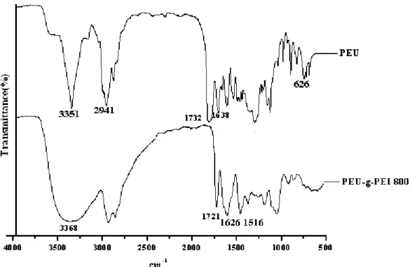
polymers were confirmed by FTIR and NMR spectroscopy. Figure 1 shows the FT-IR spectra of the
synthesized PEU and PEU-g-PEI800. The peaks at 1732 cm -1 (C=O, stretching, urethane), 1638
cm -1 (C=O, stretching, amide), 1560 cm -1 (N-H, bending, amide), and 3351 cm -1 (N-H, stretching,
urethane) represent the absorptions of urethane and amide groups in PEU, as shown at the top of
Figure 1. The peaks at 1721 cm -1 (C=O, stretching, urethane), 1626 cm -1 (C=O, stretching, amide),
1516 cm -1 (N-H, bending, amide), and 3368 cm -1 (N-H, stretching, urethane) represent the absorptions
induced from PEU-g-PEI800, as shown at the bottom of Figure 1. The chemical shifts of
characterized protons and carbons of PEU and PEU-g-PEI800 are shown in the materials and
methods section. The reaction between OH group in PEU and NCO group in ethyl isocyanatoacetate
was monitored by the disappearance of the characteristic peak of – CH(OH)- (4.06ppm) in PEOH and
10
the appearance of the peak, -CH(OCO-)- (4.36ppm), in PEU. However, the content of PEI segment in PEU-g-PEI800 could not be estimated from peak intensities of the relative proton signals at 2.30-3.40ppm (-CH 2 CH 2 -) of the PEI segment because of the overlap problem. Then element analysis was used to determine the PEI content in PEU-g-PEI800. The N element content in PEI(N PEI %: 32.71%), PEU(N PEU %: 8.88%), and PEU-g-PEI800(N PEU-g-PEI800 %: 26.26% ) are available, the PEI content (PEI%) in PEU-g-PEI800 could be calculated and its value (experimental value) was 72.93 % (theoretical value: 71.80%). From elemental data, PEU-g-PEI800 was completely synthesized by using the amionlysis reaction of the PEU and PEI800. In addition, the GPC data of PEU-g-PEI800 and PEU show that the weight-averaged molecular weights were 33100 and 22400 with polydispersities of 1.13 and 1.11, respectively, relative to polystyrene standards in THF.
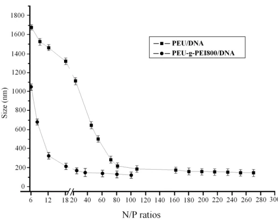
3.2. Size and Zeta-Potential analysis of polymer/DNA complexes
Figures 2 and 3 show the size and Zeta-potential of PEU/DNA and PEU-g-PEI800/DNA complexes
at various mass ratios, determined using dynamic light scattering (DLS) and electrophoretic mobility
at 25C with Zeta-Potential analysis. The size of complexes decreased with as increase of the mass
content of the polymers until the N/P ratio of PEU/DNA reached 120/1 and that of
PEU-g-PEI800/DNA reached 20/1. The results show that the average diameters (<200 nm at N/P
20/1) of PEU-g-PEI800/DNA and the average diameter (<200 nm at N/P ratio 120/1) of PEU/DNA
fall within the optimal range (40nm-200nm) required for cellular endocytosis [24]. The above data
demonstrates that PEU-g-PEI800 bearing short chain PEI800 in the side chain condenses DNA better
than PEU without the short chain PEI800.The Zeta-potential of the resulting complex changed from
negative charge to positive charge when the amounts of PEU-g-PEI800 and PEU increased. When
the N/P ratio of PEU-g-PEI800/DNA complexes was higher than 2/1, the surface of pCMV-β -gal
was fully occupied with the PEU-g-PEI800 molecules, forming positive charge complexes. When the
N/P ratio of PEU/DNA complexes was higher than 30/1, positive Zeta-potentials of the complexes
formed. Complexes with extra positive charges on their surfaces had better interaction with the target
11
cell membrane, resulting in an enhanced uptake.
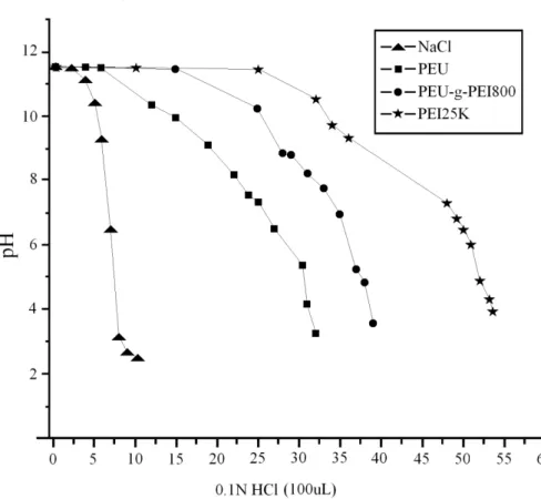
3.3. Buffering capacity of polymers
Titration studies were performed to determine the buffering capacities of the various polymers regarding a proton buffering effect within the endosomal / lysosomal compartments of the cell (Figure 4). All of the polycation solutions had a pH of 11.5 to 11.8 after the addition of 1.0 N NaOH.
The non-viral vector PEI showed a buffering capacity over a wide pH range, which is probably due to the high amount of amine functions present in the polymeric chain. The incorporation of low molecule weight PEI800 content changed the buffering region of the polycation. The data show the pH range of PEU-g-PEI800 to be between 11.8 and 5.5 and that of PEU to be between 11.8 and 6.4.
It was found that the polymers with short chain PEI800 content have a high buffering capacity.
3.4. Cytotoxicity of polymers/DNA
A critical element for the gene delivery system is cytotoxicity. Cell damage resulting from a cytotoxic delivery system is deleterious because cells must be capable of supporting translation and transcription. To determine the cytotoxicity of PEU-g-PEI800/DNA and PEU /DNA for comparison with that of PEI25k/DNA, we performed a XTT assay using the COS-7 cell line. Cells were incubated with increasing amounts of PEU-g-PEI800/DNA, PEU/DNA and PEI25k/DNA. Relative cell viabilities of PEU-g-PEI800/DNA, PEU/DNA, and PEI25k/DNA were shown in Figure 5. As can be seen, the cell viability of PEI25k/DNA complexes decreased rapidly with an increase in PEI25k concentration, whereas that of PEU-g-PEI800/DNA and PEU/DNA did not change much with an increase in PEU-g-PEI800 and PEU, respectively. The results show that PEU-g-PEI800/DNA and PEU/DNA exhibited substantially lower toxicity on COS-7 cells than did high molecule weight PEI25k/DNA. We infer that PEU-g-PEI800/DNA and PEU/DNA can provide better cytotoxicity profiles than these presently used for PEI25k/DNA due to the differences in the chemical structure, which reduces the high charge density of the polycation.
3.5. DNA gel retardation assay
The electrophoretic mobility behavior of free DNA, PEU-g-PEI800/DNA, and PEU/DNA is shown
12
in Figure 6 and 7. Increasing amounts of PEU-g-PEI800 and PEU led to the neutralization of DNA negative charges, as shown by gel retardation. The DNA mobility on agarose gel was influenced by the presence of PEU-g-PEI800 and PEU. With a low mass ratio of PEU-g-PEI800 (20/1), plasmid DNA was totally retained, as indicated in lane 5 of Figure 6. Plasmid DNA was partially retained by the presence of PEU at a mass ratio of 100/1 (lane 3 of Figure 7) and totally retained at a mass ratio of 150/1 (lane 4 of Figure 7). These results suggest that DNA was fully completed with PEU-g-PEI800 and PEU to form complexes, showing that amine groups of PEU-g-PEI800 and PEU with positive charges could interact with the charge of negative phosphate groups of DNA strands to form complexes.
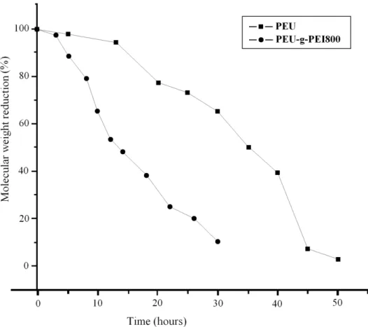
3.6. In vitro hydrolysis of polymers
Figure 8 shows the degradation profiles of polymers which were monitored at 37C at buffer pH values of 7.4 in order to approximate the environments within endosmal vesicles. The PEU-g-PEI800 generally degraded faster than that of PEU pH 7.4. The degradation studies indicate that the half-life of PEU-g-PEI800 and PEU in the HEPES buffer were 14 and 35 hours at pH 7.4, respectively. These results show that PEU-g-PEI800 exhibited higher hydrolytic degradation rate than did PEU at pH7.4.
3.7. Cellular delivery of plasmid DNA via PEU and PEU-g-PEI800 vectors
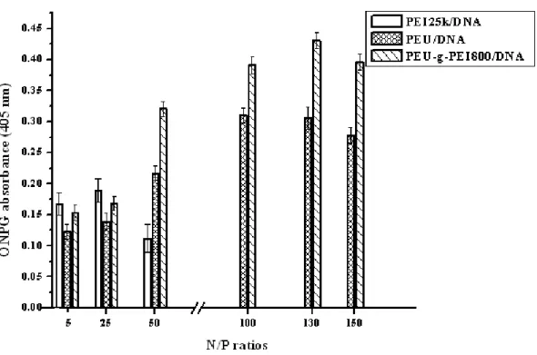
The transfection efficiency is expressed by the amount of β-galactosidase (equivalent to ONPG absorbance) measured in COS-7. The effect of N/P ratios of PEU/DNA, PEU-g-PEI800/DNA, and PEI25k/DNA complexes on transfection in COS-7 cells are shown in Fig. 9. As can be seen, PEU-g-PEI800 containing short chain PEI800 had the greater transfection efficiency than PEU without short chain PEI800. The transfection efficiencies of PEU/DNA and PEU-g-PEI800/DNA complexes increased with an increase in N/P ratios, whereas that of PEI25k/DNA complexes did not observe. The best relative transfection efficiency of PEU-g-PEI800/DNA complexes was reached at a N/P ratio 135/1, which was higher than that of PEI25k/DNA and PEU/DNA complexes. This
discrepancy may be due to the difference in proton sponage effect and size distribution of relative
13
complex. These results demonstrated that the introduction of short chain PEI800 into the side chain of poly(ester-co-urethane) has a significant effect on the size distribution and transfection ability of relative complex.
3.8. AFM (atomic force microscopy) images of PEU/DNA and PEU-g-PEI800 /DNA nano-particles
Figure 10 (a and b) shows the atomic force microscopy (AFM) images of PEU/DNA and PEU-g-PEI800/DNA complexes at a N/P ratio of 100/1 and 20/1, respectively. At a N/P ratio of PEU/DNA (100/1) and PEU-g-PEI800 (20/1), nearly all complexes condensed to a roughly spherical shape with a average particle size of 189 and 148 nm, respectively. The discretely spherical images verify the very efficient DNA condensation obtained using PEU and PEU-g-PEI800. The AFM images of complexes were captured after the complexes were coated on mica and dried with compressed air. The observed dimensions, therefore, are slightly different from the ones determined in aqueous media using DLS.
4. Conclusion
Poly(ester-co-urethane)(PEU-g-PEI800) bearing short chain PEI800 with low cytotoxicity, high biodegradability, and high transfection efficiency was synthesized and then characterized using FT-IR, NMR, and GPC in this study. PEU-g-PEI800 and DNA had a strong electrostatic interaction to self-assemble nano-particles. The positive charges in the backbone and side chain of PEU-g-PEI800 condense DNA to form DNA-polycation complexes. The transfection efficiency of PEU-g-PEI800 into COS-7 cells was better than that of the PEU without short chain PEI800, which resulted in numerous amine groups of PEU-g-PEI800 with short chain PEI800 in the side chain.
Biodegradable PEU-g-PEI800 could potentially be used in a non-viral gene delivery system.
Acknowledgement
The authors acknowledge with gratitude financial support from the National Science Council of the Republic of China through Grant No. NSC96-2221-E-041-018-MY3.
References
14
[1] Thomas CE, Ehrhardt A, Kay MA. Progress and problems with the use of viral vectors for gene therapy. Nat Rev Genet 2003;4:346-358.
[2] Pfeifer A, Verma IM. Gene therapy: promises and problems. Annu Rev Genomics Hum Genet 2001;2:177-211.
[3] Anderson WF. Human gene therapy. Nature 1998;392:25-30.
[4] Behr JP. The proton sponge: a trick to enter cells the viruses did not exploit. Chimia 1997;51:34-36.
[5] Boussif O, Lezoualch F, Zanta MA, Mergny MD, Scherman D, Demeneix B, et al. A versatile vector for gene and oligonucleotide transfer into cells in culture and in-vivo- polyethylenimine.
Proc Natl Acad Sci USA 1995;92:7297-7301.
[6] Haensler J, Szoka FCJ. Polyamidoamine cascade polymers mediate efficient transfection of cells in culture. Bioconjug Chem 1993;4:372-379.
[7] Godbey WT, Wu KK, Mikos AG. Poly(ethylenimine) and its role in gene delivery. J Controlled Release 1999;60:149-160.
[8] Cherng JY, van deWetering P, Talsma H, Crommelin DJA, Hennink WE. Effect of size and serum proteins on transfection efficiency of poly(2-dimethylamino)-ethyl methacrylate)-plasmid
nanoparticles. Pharm Res 1996;13:1038-1042.
[9] van deWetering P, Cherng JY, Talsma H, Hennink WE. Relation between transfection efficiency and cytotoxicity of poly(2-(dimethylamino)ethyl methacrylate)/plasmid complexes. J. Controlled
Release 1997;49:59-69.
[10] Lim Y, Kim C, Kim K, Kim SW, Park J. Development of a safe gene delivery system using biodegradable polymer, poly( α -(4-aminobutyl)-L-glycolic acid). J Am Chem Soc 2000;122:6524-6525.
[11] Putnam D, Langer R. Poly(4-hydroxy-L-proline ester): low temperature polycondensation and
15
plasmid DNA complexation. Macromolecules 1999;32: 3658-3662.
[12] Lynn DM, Langer R. Degradable poly(β-amino ester): synthesis, characterization, and self-assembly with plasmid DNA. J Am Chem Soc 2000; 122: 10761-10768.
[13] Akinc A, Lynn DM, Anderson DG, Langer R. Parallel synthesis and biophysical characterization of a degradable polymer library for gene delivery. J Am Chem Soc 2003;125:5316-5323.
[14] Zugates GT, Anderson GD, Little SR, Lawhorn IEB, Langer R. Synthesis of po l y( β-amino ester)s with thiol-reactive side chains for DNA delivery. J Am Chem Soc
2006;128:12726-12734.
[15] Choi MK, Arote R, Kim SY, Chung SJ, Shim CK, Cho CS, et al. Transfection of primary
human nasal epithelial cells using a biodegradable poly (ester amine) based on polycaprolactone and polyethylenimine as a gene carrier. J Drug Targ 2007;15:684-690.
[16] Wang J, Mao H, Leong KW. A novel biodegradable gene carrier based on phosphoester. J Am Chem Soc 2001;123:9480-9481.
[17] Shau MD, Tseng SJ, Yang TF, Cherng JY, Chin WJ. Effect of molecular weight on the
transfection efficiency of novel polyurethane as a biodegradable gene vector. J Biomed Mat Res Part A 2006;77A:736-746.
[18] Hung WC, Shau MD, Kao HC, Shih MF, Cherng JY. The synthesis of cationic polyurethanes to study the effect of amines and structures on their DNA transfection potential. J Controlled Release 2009;133:68-76.
[19] Lamba M, Woodhouse K, Cooper S. Polyurethane in biomedical applications. Boca Raton, FL:
CRC press; 1998.
[20] Zhang JY, Beckman EJ, Piesco NP, Agarwal S. A new peptide-based urethane polymer:
synthesis, biodegradation, and potential to support cell growth in vitro. Biomaterials 2000;21:1247-1258.
[21] Zhang S, Xu Y, Wang B, Qiao W, Liu D, Li Z. Cationic compounds used in lipoplexes and polyplexes for gene delivery. J. Controlled Release 2004;100:165-180.
[22] Lim K, Chae CB. A simple assay for DNA transfection by incubation of the cells in culture
16
dishes with substrates for beta-galactosidase. Biotech 1989;7:576-579.
[23] Crystal RG. Transfer of genes to humans: early lessons and obstacles to success. Science 1995;270:404-410.
[24] Kabanov AV, Kabanov VA. DNA complexes with polycations for the delivery of genetic
material into cells. Bioconjug Chem 1995;6:7-20.
17
Scheme 1. Synthesis of PEU and PEU-g-PEI800.
N
H
2CH
2NH
24
N
H2 CH2 OH 6 C
H2 C C O CH2CH CH2 CH3
O
OH
NH CH2 NH CH2CH CH2O C C CH2 CH3 O
OH 4
C
H2 C C O CH2 HC CH2 CH3
O O
CH2CH C O CH2CH CH2 CH3
O
OH
NH CH2 NH CH2 CH CH2O C CH CH2 CH3 O
OH
OH CH2 4 N
6 n
C
H3 CH2 O C CH2 N C O O
Glycidyl methacrylate 1,4-Diaminobutan
EOD
PEOH
6-Amino-1-hexanol
+
Ethyl isocyanatoacetate
CH2CH C O CH2CH CH2 CH3
O
O
NH CH2 NH CH2 CH CH2O C CH CH2 CH3 O
O
O CH2 N
CH3 CH2 O C CH2 NH C
O O
CH3 CH2 O C CH2 NH C
O O
CH3 CH2 O C CH2 NH C
O O 4
6 n
CH2CH C O CH2CH CH2 CH3
O
O
NH CH2 NH CH2 CH CH2O C CH CH2 CH3 O
O
O CH2 N
PEI C CH2 NH C
O O
PEI C CH2 NH C
O O
PEI C CH2 NH C
O O 4
6 n