行政院國家科學委員會專題研究計畫 成果報告
EGFR 的表現及其對血管新生的調控在胸腺瘤之研究
計畫類別: 個別型計畫
計畫編號: NSC91-2320-B-006-090-
執行期間: 91 年 08 月 01 日至 92 年 07 月 31 日 執行單位: 國立成功大學醫學系病理學科
計畫主持人: 陳芬芬
報告類型: 精簡報告
處理方式: 本計畫可公開查詢
中 華 民 國 92 年 10 月 13 日
Clinical Evaluation of HER-2/neu Protein in Malignant Pleural Effusion Associated Lung Adenocarcinoma and as a Tumor Marker
in Pleural Effusion Diagnosis
1Trei-Lien Hung, Fen-Fen Chen, Wu-Wei Lai, Ai-Li Hsiao, Wen-Tsung Huang, Helen H.W. Chen, and Wu-Chou Su
2Department of Pathology [T-L H, F-F. C.], Surgery [W-W. L.], Internal Medicine [W-T. H., W-C. S.], Radiotherapy [H. H. W. C], Institute of Microimmunology [I-L.
H.], College of Medicine, National Cheng Kung University, Tainan, Taiwan 704
1 Supported by NCKUH grant 88-02 and NSC-89-2314-B-006-141.
2 To whom requests for reprints should be addressed, at Wu-Chou Su, M.D., Division of Hematology/Oncology, Department of Internal Medicine, National Cheng Kung University Hospital, College of Medicine, National Cheng Kung University, 138, Sheng-Li Road, Tainan 704, Taiwan, R.O.C.
Tel: +886-6-2353535 ext 5401 Fax: +886-6-2766175
E-Mail: [email protected]
Running title: HER-2/neu in lung adenocarcinoma and pleural effusion Key words: CYFRA 21-1, tuberculosis, empyema, parapneumonic effusion,
heart failure.
ABSTRAT
Lung adenocarcinoma presented as malignant pleural effusion (MPE) is common in Taiwan. Microscopically, the involved pleurae were infiltrated by numerous tumor foci, which suggests the cancer cells are highly invasive. Overexpression of HER-2/neu has been related to proliferation, anti-apoptosis and high invasiveness of various cancer cells, we therefore were interested to study the role of HER-2/neu in MPE associated adenocarcinoma cell lung cancer (ADCLC). The expression of HER-2/neu in pleural effusion was measured by enzyme linked immunosorbent assay (ELISA) and the expression on tumor cells was evaluated by immunohistochemical staining. The mean value of HER-2/neu in pleural effusion of patients with ADCLC and other non-malignant lung diseases was 9.9 and 2.7 ng/ml, respectively. The difference is statistically significant (p<0.001). Compared with CYFRA 21-1, the performance of HER-2/neu as a tumor marker in pleural effusion diagnosis was better.
Overexpression of HER-2/neu in tumor tissues was found in 70% (23/32) patients of MPE associated ADCLC, 30% (13/43) patients of stage I/II non-small cell lung cancer (NSCLC), and 44% (14/32) patients of stage III NSCLC. The incidence of HER-2/neu overexpression in tumor tissues of patients with MPE associated ADCLC is significantly higher than that of stage I-III NSCLC without MPE. The findings indicate that HER-2/neu is important in the pathogenesis of MPE associated ADCLC
and is a potential tumor marker for pleural effusion diagnosis.
INTRODUCTION
Approximately 15% of lung cancer patients have a pleural effusion at the time of initial diagnosis and 50% develop pleural effusion later in their courses (1, 2). Patients with malignant pleural effusion have short life expectancy and are difficult to manage clinically (3, 4). Malignant pleural effusion commonly accompanies adenocarcinoma (5, 6). Since 1952, adenocarcinoma has become the most common cell type of lung cancers in Taiwan (7), and, subsequently, patients with MPE3 associated ADCLC have become common in Taiwan. Microscopically, we found that involved pleural surfaces were diffusely infiltrated by cancer nests. The observation suggested the neoplastic cells are highly invasive.
HER-2/neu, also referred to as c-erbB-2, is a member of epidermoid growth factor receptor (EGFR) family and has intrinsic tyrosine kinase activities (8).
Amplification and/or overexpression of HER-2/neu gene are found in approximately 30% of human breast carcinoma (9) and in a significant fraction of many other types of malignancies, including lung adenocarcinoma (10). The abnormal expression of HER-2/neu in primary tumors from human NSCLC has been correlated with poor clinical outcome (11,12). One of the underlying mechanisms is overexpression of HER-2/neu enhances metastasis-related properties(invasion, angiogenesis, increased survival)of cancer cells (13). The other is that overexpression of HER-2/neu confers
cancer cells’ resistance to various cancer therapies (14). Since the poor prognosis of patients with MPE associated ADCLC is related to the high invasiveness and drug resistance of tumor cells, we therefore were interested in studying the role of
HER-2/neu in this special disease category. First, we had collected pleural effusions from patients with ADCLC, pulmonary TB, empyema, pneumonia and CHF. We then analyzed expression of HER-2/Neu and CYFRA 21-1 proteins in the pleural fluids by ELISA. CYFRA 21-1 is a cytokeratin 19 fragment. Cytokeratin 19 is released into the bloodstream and other biological fluids upon cell death. There are many studies indicating that CYFRA 21-1 is a useful tumor marker in the management of patients with lung cancer and especially with NSCLC (15,16). By comparison with CYFRA 21-1, we evaluated the performance of HER-2/neu as tumor marker for pleural effusion diagnosis. We also studied the expression of HER-2/neu protein on neoplastic cells of NSCLC with or without MPE by IHC stains. The correlation between levels of HER-2/neu in pleural effusion and intensities of staining on tumor cells from patients with MPE associated ADCLC was analyzed, thereafter. The results suggest a strong contribution of HER-2/neu to the pathogenesis of MPE associated ADCLC. Furthermore, HER-2/neu in pleural effusion is a potential tumor marker for differential diagnosis and indicator of tumor biology.
MATERIAL and METHODS
Patients and Sample Processing
A total of 145 pleural effusion samples were collected from patients with various disease categories. Of them, 50 had ADCLC induced MPE, 20 TB pleural effusions, 20 empyema, 20 PPE, and 35 benign pleural effusions induced by CHF. Each
ADCLC associated malignant pleural effusion was either cytology or a pleural biopsy positive. The diagnosis of TB was based on positive culture from pleural effusion or pleural biopsy. All empyema effusions had positive culture for bacteriae. PPE indicates non-empyemic effusion accompanying pneumonia. Effusions of CHF were collected from patients with documented heart failure but without neoplastic or other major disease.
Effusions were collected in sterile tubes and centrifuged immediately at 4℃. Cell free supernatants were collected and aliquots were stored frozen at -70℃ until use.
Immunoassay for CYFRA 21-1
Levels of CYFRA 21-1 in pleural effusions were determined using
electrochemiluminescence immunoassay (ECLIA) on the Roche Elecsys 2010 immunoassay analyzers. The immunoassay of CYFRA 21-1 used a sandwich technology- the sample, a biotinylated monoclonal cytokeratin 19-specific antibody
and a monoclonal cytokeratin 19-specific antibody labeled with a ruthenium complex (Tris 2,2-bipyridyl ruthenium) reacted to form a sandwich complex. After addition of streptavidin –coated microparticles, the complex became bound to the solid phase via interaction of biotin and streptavidin. The reaction mixture was then aspirated into the measuring cell where the microparticles were magnetically captured onto the surface of the electrode. Application of a voltage to the electrode induced chemiluminescent emission, which was measured by a photomultiplier.
Immunoassay for HER-2/neu
Pleural effusion samples were assayed for HER-2/neu using the ELISA kit
manufactured by Oncogene Research Products (Cambridge, MA). The HER-2/Neu Rapid Format ELISA is a “sandwich “enzyme immunoassay employing two mouse monoclonal antibody. A capture antibody, specific for the human c-erbB2/c-neu protein immobilized onto the surface of microtiter wells was incubated with pleural effusion specimen. Another horseradish peroxidase –conjugated streptavidin detector antibody recognize the HER-2/neu protein and bind to the capture antibody. After washing steps, adding TMB (tetra-methylbenzidine) color reagent and stop reagent, then, the color reaction product was quantified by comparing the absorbance between unknown sample and the standards (known concentration provided by lyophilized) by constructed standard curve. Finally, the amount of HER-2/neu protein in the sample was determined.
Immunohistochemical staining for HER2/neu
The expression of HER-2/neu protein in tumor tissue was examined
immunohistochemically. Paraffin-embedded tumor sections were obtained from 43 patients with stageⅠ/Ⅱ NSCLC, 32 patients with stage Ⅲ NSCLC without pleural effusion , and 33 patients with MPE associated ADCLC. The 4μM-thick paraffin sections were de-waxed in xylene followed by rehydration in a decreasing ethanol series and washed in water. These samples were then incubated for 20 min with 0.3%
hydrogen peroxide (H2O2) in methanol at room temperature to quench endogenous peroxidase activity. Antigen retrieval was performed in an antigen-retrieval (BioGenex, San Ramon, CA, diluted 1:10) heated in a microwave oven at 700W (100%) for 10 min divided into two 5-min cycles. After washing with phosphate buffered saline (PH7.4), the samples were immersed in normal goat serum for 30 min to block non-specific protein binding. Tissue sections were then incubated with c-neu (Ab-3) OP 15 monoclonal antibody (1:100; Oncogene Research Product) at 4℃
overnight (17). Bound antibody was detected with biotinylated goat anti-rabbit IgG secondary antibody and streptavidin- peroxidase complex (MultiLink Supersensitive 500 Detection System, BioGenex), using diaminobenzidine tetrahydrochloride as the substrate. Sections were counterstained with Mayer’s hematoxylin. Incubation of some sections with non-immunized rabbit anti-IgG or without primary antibody did
not yield any immunoreactivity. A semi-quantitative scaling system was applied for evaluation of intensity of staining: Grade 0= no stained tumor cells; Grade1= a faint/barely perceptible cyto-membrane staining; Grade 2= a weak to moderate cyto-membrane staining; Grade 3= a strong cyto-membrane staining. Overepression of HER-2/Neu was considered in sections with a grade 2 staining in more than 30% of tumor cells or a grade 3 staining in more than 10% of tumor cells in this study (18).
Statistical Analysis
Differences between two independent groups were determined by the
Mann-Whitney U test. Differences between more than two groups were determined by means of one-way ANOVA-Bonferroni multiple-comparisons. ROC curves were calculated by logistic regression, considering the malignant /nonmalignant condition as dependent variable and the different tumor markers as independent variables.
Sensitivity and specificity patterns were studied by CDA (19). The two-sided Fisher exact test was used to assess associations between categorical variables. Significance testing of correlations was performed with the Spearman rank correlation analysis.
RESULTS
Expression of HER-2/neu and CYFRA 21-1 in Pleural Effusion
The median and range of HER-2/neu and CYFRA 21-1 in five disease categories are summarized in Table 1. Individual and mean levels of HER-2/neu and CYFRA 21-1 in the five subgroups are shown in Figure 1 and Figure 2, respectively. By one-way ANOVA-Bonferroni multiple-comparisons, the pleural levels of HER-2/neu and CYFRA 21-1 in ADCLC subgroup are significantly higher than those in other four subgroups. We then combined the 4 benign disease subgroups into a
nonmalignant group. Figure 3 depicts the individual and mean values of HER-2/neu in malignant and nonmalignant groups and Figure 4 depicts the individual and mean values of CYFRA 21-1 in malignant and nonmalignant groups. For both markers, the distribution of the values appeared significantly higher in malignant group than in nonmalignant group (both p values were <0.0001).
Comparison of CYFRA 21-1 and HER2/Neu as a diagnostic marker
The CDA curves (Figure 5, 6) indicate the maximum sensitivity and specificity that could be expected from pleural fluid was 84% for HER-2/neu assay, and 65% for CYFRA 21-1 assay. When 95% specificity is chosen, the cutoff point for HER-2/neu and CYFRA 21-1 was 5.5 ng/ml and 60 ng/ml, respectively; the sensitivity for
HER-2/neu and CYFRA 21-1 to detect malignant effusion induced by ADCLC was 72% and 40%, respectively. The ROC curves for HER-2/neu and CYFRA 21-1 are shown in Figure 7. As depicted, CYFRA 21-1 curve had worse curve characteristics than HER-2/neu. The area under curve of ROC, in fact, was 0.84 for HER-2/neu verse 0.65 for CYFRA 21-1. The above data indicated that the performance of HER-2/neu as tumor marker of pleural effusion diagnosis is better than that of CYFRA 21-2.
Expression of HER-2/neu by Immunohistochemistry
The expression of HER-2/neu in tumor tissues was studied in 108 patients with NSCLC, comprising 43 patients with stageⅠ/Ⅱ NSCLC, 32 patients with stage Ⅲ NSCLC but without MPE, and 33 patients with MPE associated ADCLC. Positive HER-2/neu expression (Figure 8) was observed in 13 patients (30%) of stageⅠ/Ⅱ NSCLC group, 14 patients (44%) of stageⅢ NSCLC group, and 23 patients (70%) of MPE associated ADCLC group, respectively. The differences between stage Ⅰ/Ⅱ NSCLC group vs. MPE associated ADCLC group (P=0.0011) and stage Ⅲ NSCLC group vs. MPE ADCLC group (p=0.046) are statistically significant. However, there is no statistical difference (p=0.336) between stage Ⅰ/Ⅱ and Ⅲ NSCLC groups.
Correlation of HER-2/neu expression between tumor tissue and pleural effusion
There were 32 patients with MPE associated ADCLC, whose tumor tissues and pleural effusions had been collected simultaneously. The expression of HER-2/neu by grading system in tumor tissues was compared with concentration of HER-2/neu in
paired pleural effusion samples (Figure 9). The correlation between tumor-tissue expression and pleural fluid values of HER-2/neu was highly significant (r = 0.90; P<
0.0001).
DISCUSSION
Membranous staining of HER-2/neu has been demonstrated in 2% to 40% of NSCLC cases (11,12). Although only membranous expression of HER-2/neu was considered functionally important, several studies from lung cancers have shown that cytoplasmic HER-2/neu staining, especially in adenocarcinoma, is associated with a poor prognosis. The cytoplasmic expression has been found in 10% to 60% of NSCLC cases (20-23). The enormous variations in frequencies and patterns of HER-2/neu expression in NSCLC might be due to different assay methods and scoring systems. In order to have a good correlation between IHC and ELISA tests, we used c-neu (Ab-3) OP15 monoclonal antibody for IHC staining and c-erbB2/c-neu RAPID FORMAT kit ( Cat # QIA10) for ELISA. Both were purchased from
Oncogene Research Products. Since membranous and cytoplasmic expressions of HER-2/neu have been related to prognosis of patients with NSCLC, we interpreted either membranous or cytoplasmic staining as positive in of IHC experiments.
Overexpression of HER-2/neu has frequently been linked to a poor prognosis for patients with NSCLC (12, 20-23), other studies found opposite results (24, 25), however. In our studies, cyto-membranous staining of HER-2/neu was found in 30%
of stage I/II NSCLC cases, 44% of stage III cases, and 70% of stage IIIB/IV MPE associated ADCLC cases. The correlation of HER-2/neu overexpression with
advanced stage in NSCLC from the current study is thus supporting a linkage between HER-2/neu expression and poor prognosis. The high percentage (70%) of HER-2/neu staining on tumor tissue from patients with MPE associated ADCLC may be due to cells those overexpressed HER-2/neu in a tumor are more likely to invade pleurae than others, or because that ADCLC with HER-2/neu overexpression tends to have pleural invasion and develop MPE. Although we could obtain tumor tissues from involved pleural via thoracoscopy-guided biopsy, we have difficulties in obtaining the tumor tissues from the primary lung tumors due to either surgical resection is not indicated for patients with MPE associated NSCLC or bronchoscopic biopsy is hardly reaching the peripherally located primary tumor. Therefore we don't have a definite answer for the question yet. A recent study reported by Simon et al. (26) found discordance in HER-2/neu status between the primary rumor and involved axillary lymph node metastases is small (5%), suggests the assumption that
HER-2/neu-overexpressed ADCLC tends to involve pleurae may be the case.
HER-2/neu protein is a product of oncogene. Its extracellular domain is solubilized and may shed into the culture medium of tumor cell lines and sera of advanced breast carcinoma patients (27). In this study, we investigated expression of HER-2/neu in the pleural fluids from patients with MPE associated ADCLC,
pulmonary TB, empyema, pneumonia and CHF. The pleural levels of HER-2/neu
were significantly higher in malignant group than in nonmalignant group. The 72%
sensitivity of HER-2/neu for diagnosis of MPE is very close to the 70% positive cyto-membrane staining of HER-2/neu on pleural tumor cells. Furthermore, the correlation between grades of HER-2/neu expression on pleural tumor cells and concentrations of HER-2/neu protein in pleural fluids was high and significant (r = 0.9, p < 0.0001). The finding was in consistent with the result from Osaki et al. (28) that serum levels of HER-2/neu in patients with tissue overexpression were higher than in patients without overexpression. On the contrary, Ardizzoni et al. didn’t find a good correlation between circulating HER-2/neu levels and tissue expression, but they found high pretreatment levels of serum HER-2/neu are associated with an adverse prognostic impact on survival in patients with locally advanced or metastatic NSCLC (29). The discrepancy may be caused by different assay systems been used or
different patient groups been studied. The strong correlation between pleural HER-2/neu levels and tumor tissue expression in the current study indicates
HER-2/neu in pleural effusion is shed from tumor cells and measurement of pleural HER2/Neu may substitute the direct IHC staining on tumor cells.
Malignancy is one of the main etiologies in causing pleural effusion. However, cytological examination of pleural fluid fails to detect neoplastic cells in about
40-50% of malignant effusions (30), and a blind pleural needle biopsy adds very little
to negative cytology (31), therefore, several investigators have tried to improve diagnostic performances by measuring tumor markers in pleural fluids. Tumor markers been frequently studied include carcinoembryonic antigen (CEA), cancer antigen 125(CA 125), carbohydrate antigens 15-3, 19-9 and 72-4(CA 15-3, CA19-9 and 72-4), cytokeratin 19 fragments (CYFRA 21-1), neuron-specific enolase (NSE) and squamous cell carcinoma antigen (SCC) (32,33). Among them, CYFRA 21-1 is one of the most reliable tumor markers in pleural effusion diagnosis (34,35). When compared with CYFRA 21-1, we found HER-2/neu had higher sensitivity to detect malignancies and had larger area under the ROC curve. In addition to its good performance in plural effusion diagnosis, HER-2/neu is also an indicator for
aggressive tumor biology. Therefore, HER-2/neu has a potential to be an ideal tumor marker in pleural effusion diagnosis and a good biomarker of MPE-inducing cancers.
The findings in lung cancer are consistent with experience from a breast cancer study by Sugano et al., who have shown serum HER-2/neu is more sensitive than the conventional tumor markers, such as CEA and CA 15-3, in predicting tumor relapse (36). In this study, we have only focused on MPE associated ADCLC. We didn’t investigate positive rate of HER-2/neu elevation in MPE from other malignancies.
Since the rates of HER-2/neu overexpression vary widely among different malignancies, it’ll be difficulty in interpretation of data when putting all cancers
together. Though the frequencies of HER-2/neu elevation in pleural fluids caused by other malignancies await further studies, a malignant origin should be highly
suspected in patients with pleural effusion, in which HER-2/neu level is higher than 5.5 ng/ml. An invasive diagnostic procedure, such as thoracoscopy-guided biopsy, has to be provided to the patient.
In conclusion, HER-2/neu may play an important role in the pathogenesis of MPE associated ADCLC and HER-2/neu in pleural fluid can be used as an ideal tumor marker for differential diagnosis and prediction of tumor biology.
FOOTNOTES
3 The abbreviations used are: MPE, malignant pleural effusion; ADCLC,
adenocarcinoma cell lung cancer; NSCLC, non-small cell lung cancer; TB,
tuberculosis; CHF, congestive heart failure; ELISA, enzyme linked immunosorbent assay; IHC, immunohistochemical; PPE, parapneumonic effusion; ROC, receiver operating characteristic; CDA, cumulative distribution analysis.
TABLES
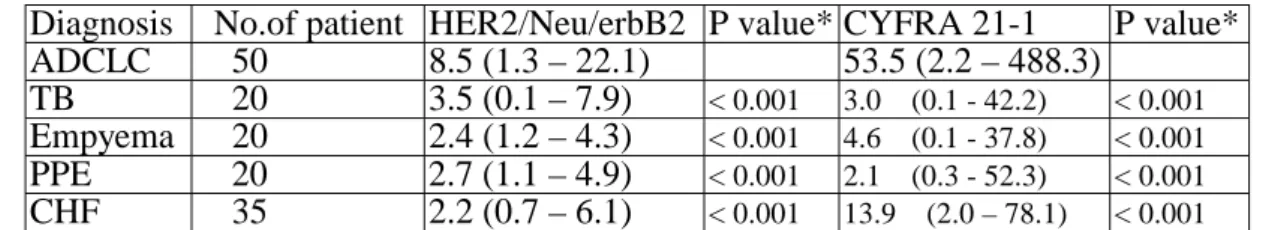
Table 1. Pleural fluid level of HER-2/neu and Cyfra 21-1 in various groups
# Diagnosis No.of patient HER2/Neu/erbB2 P value* CYFRA 21-1 P value*ADCLC 50 8.5 (1.3 – 22.1) 53.5 (2.2 – 488.3) TB 20 3.5 (0.1 – 7.9) < 0.001 3.0 (0.1 - 42.2) < 0.001
Empyema 20 2.4 (1.2 – 4.3) < 0.001 4.6 (0.1 - 37.8) < 0.001
PPE 20 2.7 (1.1 – 4.9) < 0.001 2.1 (0.3 - 52.3) < 0.001
CHF 35 2.2 (0.7 – 6.1) < 0.001 13.9 (2.0 – 78.1) < 0.001
# data are expressed as the median, minimum and maximum values.
* p value between ADCLC and other disease groups by ANOVA test
FIGURE LEGENDS
Figure 1. Scatterplot of individual and mean pleural fluid levels of HER-2/neu in patients of diverse etiologies. Thick black line: mean; ★: indicates the difference of mean values between indicated and ADCLC groups is statistically significant by Bonferroni test.
Figure 2. Scatterplot of individual and mean pleural fluid levels of CYFRA 21-1 in patients of diverse etiologies. Thick black line: mean; * : indicates the difference of mean values between indicated and ADCLC groups is statistically significant by Bonferroni test.
Figure 3. Scatterplot of individual and mean pleural fluid levels of HER-2/neu in patients of malignant and non-malignant groups. Thick black line: mean. The p value is < 0.0001.
Figure 4. Scatterplot of individual and mean pleural fluid levels of CYFRA 21-1 in patients of malignant and non-malignant groups. Thick black line: mean. The p value is < 0.0001.
Figure 5. CDA curves of HER-2/neu in patients with malignant and non-malignant pleural effusions.
Figure 6. CDA curves of CYFRA 21-1 in patients with malignant and non-malignant pleural effusions.
Figure 7. ROC curves of HER-2/neu and CYFRA 21-1 for distinguishing malignant and non-malignant pleural effusions. The areas under the curves for HER-2/neu and CYFRE 21-1 are 0.84 and 0.65, respectively.
Figure 8. Expression of HER-2/neu in MPE associated ADCLC by IHC staining. (A) Section from a pleural metastatic tumor of ADCLC showing strong (3+) membranous HER-2/neu reactivity. (B) Section from a pleural metastatic tumor of ADCLC
showing strong (3+) cytoplasmic HER-2/neu reactivity.
Figure 9. Correlation between pleural fluid HER-2/neu concentrations and IHC scoring (0 to 3+) on tumor tissues of the same patients (n=34). The correlation coefficient (r) = 0.90, and p < 0.0001.
REFERENCES:
1. Memon, A. N., and Zawadzki, G. A. Pleural effusions. Cur. Prob. Cancer 5:3-30, 1981.
2. Anderson, C. B., Philpott, G. W., and Ferguson, T. B. The treatment of malignant pleural effusions. Cancer 33:916-922, 1974.
3. Sugiura, S., Ando, Y., Minami, H., Ando, M., Sakai, S., and Shimokata, K.
Prognostic value of pleural effusion in patients with non-small cell lung cancer.
Clin. Cancer Res. 3:47-50, 1997.
4. Naito, T., Satoh, H., Ishikawa, H., Yamashita, Y. T., Kamma, H., Takahashi, H., Ohtsuka, M., and Hasegawa, S. Pleural effusion as a significant prognostic factor in non-small cell lung cancer. Anticancer Res. 17:4743-4746, 1997.
5. Hoffman, P. C., Mauer, A. M., and Vokes, E. E. Lung cancer. Lancet 355:479-485, 2000.
6. Chang, S. C., Lee, L. N., Kuo, S. H., Luh, K. T., and Wu S. C. [Survival of patients with malignant pleural effusion]. [Chinese]. Taiwan I Hsueh Hui Tsa Chih – J. Form. Med. Assoc. 86:672-678, 1987.
7. Yang, S. P., Luh, K. T., Kuo, S. H., and Lin, C. C. Chronological observation of epidemiological characteristics of lung cancer in Taiwan with etiological
consideration--a 30-year consecutive study. Jap. J. Clin. Oncol. 14:7-19, 1984.
8. Hynes, N. E., and Stern, D. F. The biology of erbB-2/neu/HER-2 and its role in cancer. Bioch. Bioph. Acta 1198: 165-184, 1994.
9. Slamon, D. J., Godolphin, W., Jones, L. A., Holt, J. A., Wong, S. G., Keith, D.
E., Levin, W. J., Stuart, S. G., Udove, J., and Ullrich, A. Studies of the HER-2/neu proto-oncogene in human breast and ovarian cancer. Science 244:707-12, 1989.
10. Weiner, D. B., Nordberg, J., Robinson, R., Nowell, P. C., Gazdar, A., Greene, M. I., Williams, W. V., Cohen, J. A., and Kern J. A. Expression of the neu gene-encoded protein (P185neu) in human non-small cell carcinomas of the lung. Cancer Res. 50:421-425, 1990.
11. Kern, J. A., Schwartz, D. A., Nordberg, J.E., Weiner, D. B., Greene, M. I., Torney, L., Robinson, R. A. p185neu expression in human lung
adenocarcinomas predicts shortened survival. Cancer Res. 50:5184-5187, 1990.
12. Pastorino, U., Andreola, S., Tagliabue, E., Pezzella, F., Incarbone, M., Sozzi, G., Buyse, M., Menard, S., Pierotti, M., and Rilke, F. Immunocytochemical markers in stage I lung cancer: relevance to prognosis. J. Clin. Oncol.15:2858-2865, 1997.
13. Yu, D., Wang, S. S., Dulski, K. M., Tsai, C. M., Nicolson, G. L., and Hung, M.
C. c-erbB-2/neu overexpression enhances metastatic potential of human lung cancer cells by induction of metastasis-associated properties. Cancer Res.
54:3260-3266, 1994.
14. Tsai, C. M., Chang, K. T., Perng, R. P., Mitsudomi, T., Chen, M. H., Kadoyama, C., and Gazdar A. F. Correlation of intrinsic chemoresistance of non-small-cell lung cancer cell lines with HER-2/neu gene expression but not with ras gene mutations. J. National Cancer Inst. 85:897-901, 1993.
15. Wieskopf, B., Demangeat, C., Purohit, A., Stenger, R., Gries, P., Kreisman, H., and Quoix, E. Cyfra 21-1 as a biologic marker of non-small cell lung cancer.
Evaluatio of sensitivity, specificity, and prognostic role. Chest 108:163-169, 1995.
16. Takada, M., Masuda, N., Matsuura, E., Kusunoki, Y., Matui, K., Nakagawa, K., Yana, T., Tuyuguchi, I., Oohata, I., and Fukuoka, M. Measurement of
cytokeratin 19 fragments as a marker of lung cancer by CYFRA 21-1 enzyme immunoassay. Br. J. Cancer 71:160-165, 1995.
17. Shi, D., He, G., Cao, S., Pan, W., Zhang, H. Z., Yu, D., and Hung, M. C.
Overexpression of the c-erbB-2/neu-encoded p185 protein in primary lung cancer. Molecular Carcinogenesis. 5:213-218, 1992.
18. Giatromanolaki, A., Gorgoulis, V., Chetty, R., Koukourakis, M. I., Whitehouse,
R., Kittas, C., Veslemes, M., Gatter, K. C., and Iordanoglou, I. C-erbB-2 oncoprotein expression in operable non-small cell lung cancer. Anticancer Res.
16:987-993, 1996.
19. Krouwer, J. S. Cumulative distribution analysis graphs--an alternative to ROC curves. Clin. Chemistry 33:2305-2306, 1987.
20. Weiner, D. B., Nordberg, J., Robinson, R., Nowell, P. C., Gazdar, A., Greene, M. I., Williams, W. V., Cohen, J. A., and Kern, J. A. Expression of the neu gene-encoded protein (P185neu) in human non-small cell carcinomas of the lung. Cancer Res. 50:421-425, 1990.
21. Tateishi, M., Ishida, T., Mitsudomi, T., Kaneko, S., and Sugimachi, K.
Prognostic value of c-erbB-2 protein expression in human lung adenocarcinoma and squamous cell carcinoma. Eur. J. Cancer 27:1372-1375, 1991.
22. Harpole, D. H., Herndon, J. E., Wolfe, W. G., Iglehart, J. D., and Marks, J. R. A prognostic model of recurrence and death in stage I non-small cell lung cancer utilizing presentation, histopathology, and oncoprotei expression. Cancer Res.
55:51-56, 1995.
23. Fontanini, G., De Laurentiis, M., Vignati, S., Chine, S., Lucchi, M., Silvestri, V., Mussi, A., De Placido, S., Tortora, G., Bianco, A. R., Gullick, W., Angeletti, C.
A., Bevilacqua, G., and Ciardiello, F. Evaluation of epidermal growth
factor-related growth factors and receptors and of neoangiogenesis in
completely resected stage I-IIIA non-small-cell lung cancer: amphiregulin and microvessel count are independent prognostic indicators of survival. Clin.
Cancer Res. 4:241-249, 1998.
24. Moldvay, J., Scheid, P., Wild, P., Nabil, K., Siat, J., Borrelly, J., Marie, B., Farre, G., Labib, T., Pottier, G., Sesboue, R., Bronner, C., Vignaud, J. M., Martinet, Y., and Martinet, N. Predictive survival markers in patients with surgically resected non-small cell lung carcinoma. Clin. Cancer Res. 6:1125-1134, 2000.
25. Pfeiffer, P., Clausen, P. P., Andersen, K., and Rose, C. Lack of prognostic significance of epidermal growth factor receptor and the oncoprotein
p185HER-2 in patients with systemically untreated non-small-cell lung cancer:
an immunohistochemical study on cryosections. Br. J. Cancer 74:86-91, 1996.
26. Simon, R., Nocito, A., Hubscher, T., Bucher, C., Torhorst, J., Schraml, P., Bubendorf, L., Mihatsch, M. M., Moch, H., Wilber, K., Schotzau, A., Kononen, J., and Sauter, G. Patterns of her-2/neu amplification and overexpression in primary and metastatic breast cancer. J. National Cancer Inst. 93:1141-1146, 2001.
27. Pupa, S. M., Menard, S. Morelli, D., Pozzi, B., De Palo, G., and Colnaghi, M. I.
The extracellular domain of the c-erbB-2 oncoprotein is released from tumor
cells by proteolytic cleavage. Oncogene 8:2917-2923,1993.
28. Osaki, T., Mitsudomi, T., Oyama, T., Nakanishi, R., and Yasumoto, K. Serum level and tissue expression of c-erbB-2 protein in lung adenocarcinoma.
Chest 108:157-162,1995.
29. Ardizzoni, A., Cafferata, M. A., Paganuzzi, M., Filiberti, R., Marroni, P., Neri, M., Fontana, V., Nicolo, G., Perdelli, L., Stampino, C. G., Rosso, R., and Puntoni, R. Study of pretreatment serum levels of Her-2/neu oncoprotein as a prognostic and predictive factor in patients with advanced nonsmall cell lung carcinoma. Cancer 92: 1896-1904, 2001.
30. Marel, M., Stastny, B., Melinova, L., Svandova, E., and Light, R. W. Diagnosis of pleural effusions. Experience with clinical studies, 1986 to 1990.
Chest 107:1598-1603,1995.
31. Prakash, B. S., and Reiman, H. M. Comparison of needle biopsy with cytologic analysis for the evalustion of pleural effusion: analysis of 414 cases. Mayo Clin.
Proc. 60:158-164, 1985.
32. Ferrer, J., Villarino, M. A., Encabo, G., Felip, E., Bermejo, B., Vila, S., and Orriols R. Diagnostic utility of CYFRA 21-1, carcinoembryonic antigen, CA 125, neuron specific enolase, and squamous cell antigen level determinations in the serum and pleural fluid of patients with pleural effusions. Cancer
86:1488-1495,1999.
33. Miedouge, M., Rouzaud, P., Salama, G., Pujazon, M. C., Vincent, C., Mauduyt, M. A., Reyre, J., Carles, P., and Serre G. Evaluation of seven tumour markers in pleural fluid for the diagnosis of malignant effusions. Br. J.
Cancer 81:1059-1065, 1999.
34. Satoh, H., Sumi, M., Yagyu, H., Ishikawa, H., Suyama, T., Naitoh, T., Saitoh, T., and Hasegawa, S. Clinical evaluation of CYFRA 21-1 in malignant pleural fluids. Oncology 52:211-214, 1995.
35. Toumbis, M., Rasidakis, A., Passalidou, E., Kalomenidis, J., Alchanatis, M., Orphanidou, D., and Jordanoglou J. Evaluation of CYFRA 21-1 in malignant and benign pleural effusions. Anticancer Res. 16:2101-2104, 1996.
36. Sugano, K., Ushiama, M., Fukutomi, T., Tsuda, H., Kitoh, T., and Ohkura, H.
Combined measurement of the c-erbB-2 protein in breast carcinoma tissues and sera is useful as a sensitive tumor marker for monitoring tumor relapse. Int. J.
Cancer 89:329-336, 2000.