A novel one-step reverse transcription loop-mediated isothermal
1amplification method for detection of infectious bursal disease virus
2Meng-Shiou Lee1ψ, Yi-Chiu Lin2ψ, Guan-Hua Lai3, Su-Yaun Lai4, Hsi-Jien Chen5, 3
Min-Ying Wang 2 4
1School of Chinese Medicine Resources, China Medical University, Taichung, Taiwan 5
2Graduate Institute of Biotechnology, College of Agricultural Resources, National 6
Chung Hsing University, Taichung, Taiwan 7
3 Institute of Biochemistry, College of Life Science, National Chung Hsing University, 8
Taichung, Taiwan 9
4
Dept. of Food Science, Central Taiwan University of Science and Technology, 10
Taichung, Taiwan 11
5Dept. of Safety, Health and Environmental Engineering, MingChi University of 12
Technology, Taipei, Taiwan. 13
ψThese authors contributed equally to this work. 14
Correspondence
15
Meng-Shiou Lee 16
School of Chinese Medicine Resources, China Medical University, 17
No. 91 Hseuh-shih Road, Taichung, Taiwan 40402. 18
E-mail: [email protected]; 19
Telephone: +886-4-2205-3366#5208; 1 Fax: +886-4-22078083. 2 3 Min-Ying Wang, 4
Graduate Institute of Biotechnology, College of Agricultural Resources, National 5
Chung Hsing University, 6
No. 250 Kuo Kuang Road,Taichung, Taiwan 40402 7 E-mail: [email protected]; 8 Telephone: +886-4-2284-0328#762; 9 Fax: +886-4-285-6697. 10 11
Abstract
1
Infectious bursal disease virus (IBDV) is an economically important viral 2
pathogen that affects young chickens worldwide and causes high mortality and 3
immunosuppression. An very fast, sensitive and specific reverse transcription 4
loop-mediated isothermal amplification (RT-LAMP) assay was developed involving a 5
single tube and a one step reaction for detecting IBDV infection. A set of four specific 6
designed RT-LAMP primers were used for the amplification of specific cDNA from 7
the IBDV genome, specifically the VP2 gene. Amplicons were successfully amplified 8
using Bst DNA polymerase under isothermal condition, and were verified by digestion 9
with the restriction enzyme BamH I. The amplified LAMP products were detected by 10
DNA electrophoresis and by direct observation using the naked eye in the presence of 11
SYBR Green I. The sensitivity of the RT-LAMP was determined to be 0.01 fg of 12
IBDV viral RNA. This RT-LAMP assay for IBDV is more sensitive than the 13
conventional RT-PCR assay, which has a detection limit of 1 ng. The specificity of the 14
LAMP assay was also assessed and it was found that it could precisely discriminate 15
between positive and negative test samples. This newly established LAMP assay, 16
combined with reverse transcription (RT) into RT-LAMP, is a practical diagnostic tool 17
because IBDV infected and uninfected clinical samples collected from an 18
experimental farm could be discriminated. Full verification of a sample’s IBDV status 19
was obtained within 40 min of extracting the viral RNA, which could be then directly 1
added to the RT-LAMP reaction mixture. 2
3
Keywords: infectious bursal disease virus (IBDV), reverse transcription
4
loop-mediated isothermal amplification (RT-LAMP), rapid detection, VP2, diagnosis 5
6
Introduction
7
Loop-mediated isothermal amplification (LAMP) is a novel method that able to 8
rapidly amplify a specific nucleic acid with high specificity under isothermal 9
conditions using four to six specifically designed primers [1]. This amplification 10
occurs by auto cycling strand displacement DNA synthesis, which is catalyzed by Bst 11
DNA polymerase. The LAMP reaction process has no denaturation step, which is 12
different from conventional PCR, and DNA amplification occurs by means of the 13
strand displacement activity of the Bst DNA polymerase [1,2]. So far, the LAMP 14
method has been applied to the detection of various microbes and pathogens in the 15
environmental, food and clinical samples including protozoa, bacteria, and viruses 16
[3-10]. Moreover, several uses of LAMP for pathogen detection have been 17
commercialized into LAMP kits [11]. When detecting the RNA genome of a pathogen 18
such as a RNA virus, LAMP has been merged with reverse transcription (RT) into 19
RT-LAMP to allow nucleic acid amplification [12]. For example, for medical 1
purposes, RT-LAMP has been employed in minimal residual disease (MRD) 2
monitoring of the WT1 mRNA expression level [13]. 3
Infectious bursal disease virus (IBDV) is an important veterinary pathogen 4
that infects young chickens. The virus belongs to the genus birnavirus of the family 5
Birnaviridae and contains two segments of double-stranded RNA genome, which are 6
designated A and B [14]. Epidemiological studies have shown that almost all 2 to 7
8-weeks-old chicks are susceptible to IBDV infection. IBDV infects the lymphoid 8
cells in the bursa of Fabricius (BF). Young chickens show various clinical signs 9
including whitish and watery diarrhea, anorexia, depression, trembling, ruffled 10
feathers and severe prostration on infection with IBDV [15]. General speaking, the 11
mortality rate may ranged from 1% to 50% across various outbreaks. In broilers, 12
infection may result in up to 50% morbidity, but mortality is seldom greater than 3% 13
in flocks aged 3-6 weeks. In addition to mortality, IBDV also often causes 14
immunosuppression in the chickens due to destruction of lymphoid tissue. This makes 15
these birds more susceptible to other pathogens, which can result in various secondary 16
infections [15]. At present, there are several conventional methods that can be used to 17
diagnose the IBDV pathogen including agar gel precipitation (AGP), virus 18
neutralization (VN), enzyme-linked immunosorbent assay (ELISA), dot blot 19
hybridization assay, indirect immunofluorescence (IIF) and electron microscopy 1
[16-21]. In addition, RT-PCR (reverse transcriptase polymerase chain reaction), 2
real-time RT-PCR, RT-PCR-RE (analysis of PCR product by restriction endonuclease) 3
and RT-PCR-RFLP (analysis of PCR product by restriction fragment length 4
polymorphism) have also been commonly used as molecular diagnostic methods to 5
detect IBDV [22-26] Among the assays mentioned above, RT-PCR is the most 6
convenient to apply because it allows the testing of a large number of samples. 7
However, the traditional RT-PCR method requires a relatively well-equipped 8
laboratory, needs well-trained staff and involves multiple reaction steps in order to 9
amplify the nucleic acid. Furthermore, the sensitivity of the method can be affected by 10
the fact that template RNA is limited. Therefore, there is a need to develop an 11
alternative assay that is more efficient, has greater sensitivity and has better specificity 12
that will be more economical when used for the diagnosis of IBDV infection. Recently, 13
the use of RT-LAMP for detection of the IBDV RNA genome has been reported; this 14
used M-MLV RTase (Moloney Murine Leukemia Virus Reverse Transciptase) and Bst 15
DNA polymerase in a two-stage reaction that needed at least 70 min [27]. This 16
two-step reaction is not very efficient and involved tedious processing. Nonetheless, it 17
had a sensitivity limit of 4.78 fg, which is about 100 times better than RT-PCR 18
detection of IBDV. 19
In this study, we described an improved RT-LAMP assay method to detect the 1
IBDV genome. This approach is characterized by a high sensitivity, a high specificity 2
and a very rapid procedure and is catalyzed by AMV RTase (Avian Myelobastosis 3
virus reverse transcriptase) and Bst DNA polymerase; the reaction involves one-step 4
process in a single tube. This newly established RT-LAMP should be a very valuable 5
and applicable tool for the detection of IBDV infection in the future. 6
7
Materials and methods
8
Virus strain, IBDV-infected bursa tissue and the plasmid used
9
Infectious bursal disease virus P3009 (Genebank Accession No. AF109154) was 10
provided by Professor Min-Ying Wang of National ChungHsin University (Taichung, 11
Taiwan). To propagate the virus, it was inoculated into specific pathogen free (SPF) 12
chickens that were one week old. The IBDV infected bursa of Fabricius from these 13
chickens were collected. This was done at 3-days post-infection; this involved 14
removing the individual bursa of the sacrificed chickens and homogenizing them 15
individually in phosphate-buffered saline. The tissue homogenate was then collected 16
and stored at -80oC until required. The recombinant plasmid, pFastBac-VP2, which 17
harbors the VP2 gene of P3009, has been used previously [28]. In this study, this 18
plasmid was used to produce VP2 cDNA as a template for testing the LAMP assay. 19
The VP2 cDNA was amplified by PCR using the previously described primers, P4F 1
and 1323R [28]. 2
3
RNA extraction from the chicken bursa specimens
4
Total viral RNA genomes were extracted from 100 μl of homogenized bursa 5
tissue infected with infectious bursal disease virus using the AxyprepTMBody Fluid 6
Viral DNA/ RNA Miniprep Kit (Axygen, USA) according to the manufacturer’s 7
protocol. The RNA was eluted from the column in a final volume of 50 μl of elution 8
buffer and stored at -20oC until required. In addition, 21 clinical samples were 9
obtained from an experimental broiler farm, 6 of these were infected with IBDV and 10
15 were uninfected; these were also extracted as described above. 11
12
Design of the primers for the RT-LAMP assay
13
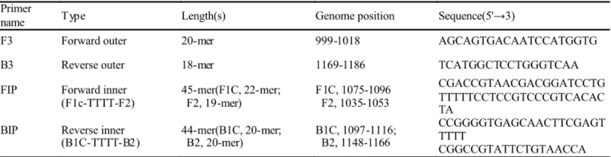
Four specific RT-LAMP primers (F3, B3, FIP, and BIP) for infectious bursal 14
virus detection were designed based on the sequence of the VP2 gene of infectious 15
bursal virus P3009 (Genebank Accession No. AF109154) using Primer Explorer V3 16
software (http://primerexplorer.jp; Eiken Chemical Co. Ltd, Japan). F3 and B3 were 17
the outer RT-LAMP primers, while FIP and BIP were inner RT-LAMP primers. FIP 18
consisted of 19 nucleotides of F2 and a 22nucleotides of F1c, which were linked by a 19
TTTT spacer. BIP consisted of 20 nucleotides of B2 and 20 nucleotides of B1c, which 1
were also linked by a TTTT spacer. The schematic positions on the VP2 gene are 2
showed in Figure 1. These primers were used in LAMP assay using VP2 3
complementary DNA as template as well as the viral assay itself. The sequences of the 4
primers are shown in Table 1. 5
6
LAMP/ RT-LAMP reaction conditions
7
The LAMP reaction was carried out in a total volume of 25μl. A reaction mixture 8
containing 12.5μl of 2×LAMP reaction buffer [20 mM Tri-HCl (pH8.8), 10 mM KCl, 9
10 mM (NH4)2SO4, 8 mM MgSO4and 0.2% Tween 20], 8U of Bst DNA polymerase 10
(NEW England Biolabs, Germany), 10μM of each F3 and B3 primers, 10μM of each 11
FIP and BIP primers, and 10μM of 2M betaine was used. After that, 2μl of the target 12
DNA was added to each LAMP reaction. The mixtures were incubated at 60oC for 40 13
min in a heating block (PCR Mechanics). For the RT-LAMP reaction, reverse 14
transcription and LAMP were carried out in single tube using a one step reaction that 15
was catalyzed by 10U (AMV) enhanced avian reverse transcriptase (Invitrogen, USA) 16
and 8U of Bst DNA polymerase at 60oC for 40 min. 17
18
Detection of the LAMP/RT-LAMP product
An aliquot of 1μl of LAMP product was subjected to DNA electrophoresis on a 1
1.6 % agarose gel and visualized under ultraviolet light after ethidium bromide 2
staining. Alternatively, the LAMP products were also detected using naked eye 3
observation of the color change during the LAMP reaction. SYBR Green I reagent 4
(Invitrogen, USA) was added to the LAMP reaction mixture before the reaction was 5
started. Finally, based on the target sequence, which contains a BamH I restriction 6
enzyme site, restriction digestion with BamH I was carried out for 1 hr at 37oC in a 7
water bath to test the amplification specificity of the target sequence. 8
9
Sensitivity and specificity of the LAMP assay
10
The sensitivity of the LAMP assay was evaluated using different amounts of 11
complementary DNA from the IBDV VP2 gene as template in a LAMP reaction. The 12
DNA consisted of the VP2 gene of IBDV, which had been amplified from the plasmid 13
pFastBac-VP2 [28]. The specificity of the LAMP assay for IBDV detection was tested 14
using the VP2 gene DNA of chicken anemia virus (CAV) and the H6 gene of avian 15
influenza virus H6N1 and the results were compared to those of the VP2 gene of 16
IBDV. These two additional virus templates had been created in our laboratory 17
previously. 18
RT-PCR for IBDV detection
1
Reverse transcription PCR (RT-PCR) was performed in one-tube contained 2
reverse transcriptase and Taq DNA polymerase. The RT-PCR reaction mixture was 3
carried out in a 25 l total volume that contained 1x ThermoPol Reaction Buffer (20 4
mM Tris-HCl, 10 mM KCl, 10 mM (NH4)2SO4, 2 mM MgSO4, 0.1% Triton X-100, 5
pH8.8 at 25℃), 0.4 mM of dNTPs, 5 pmol each of F3 and B3, 4U Taq DNA 6
polymerase (New England BioLabs), 25 U Reverse-iT™ RTase Blend (ABgene) and 7
5 l RNA template; the mixture was made up to 25 l by DEPC-treated water. The RT 8
conditions was 45oC for 30 min, and the reaction program of following PCR was 9
94oC for 2 min at pre-denaturation step, then followed by 35 cycles of 94oC for 20 sec, 10
55oC for 20 sec and 72oC for 20 sec, and a final extension cycle at 72oC for 10 min. 11
12
Western blotting analysis
13
Bursa samples containing IBDV VP2 protein were resolved on polyacrylamide 14
slab gels, and then were incubated in Tris-glycine running buffer (48 mM Tris-HCl, 15
39 mM glycine, 0.0375% SDS). The resulting gels containing the specific proteins 16
were transferred to a polyvinylidene difluoride (PVDF) membrane in a wet 17
electroblotter cassette using 100V for 60 min. Blots were then treated with blocking 18
buffer (5% skim milk in PBS) for 30 min and with 2000-fold PBS diluted primary 19
rabbit antibodies against IBDV VP2 protein for 2 hrs. The membrane was washed 1
three times in PBS-T buffer (1X phosphate-buffered saline + 0.5% Tween 20) and 2
incubated with an alkaline phosphatase-conjugated goat anti-rabbit antibody (ABcam) 3
for 2 hrs. Membranes were again washed three times and then finally incubated with 4
NBT and BCIP for color developing. 5
6
Results
7
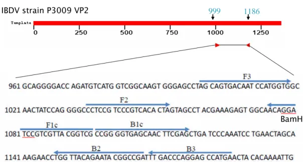
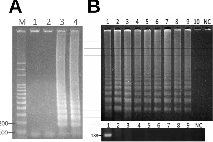
Establishing the LAMP assay for detection of the IBDV complementary genome
8
To establish the reverse transcription LAMP (RT-LAMP) method for IBDV 9
detection, primer explorer software was used to design the primers F3, B3, FIP and 10
BIP. These were then used to examine the amplification efficiency-using LAMP. 11
Using IBDV complementary VP2 cDNA as template, the LAMP reaction was carried 12
out using Bst DNA polymerase at 65oC for 1 hr. As illustrated in lane 3 of figure 2A, 13
visually distinct DNA ladder-like fragments could be seen on the ethidium 14
bromide-stained agarose gel after DNA electrophoresis. In contrast, LAMP reaction 15
mixtures without Bst DNA polymerase or target DNA template did not have LAMP 16
products present after the assay had been carried out. These results indicate that these 17
specifically designed LAMP primers are suitable for amplifying the target nucleic acid, 18
and are able to discriminate between the positive and negative test samples. To further 19
identify the presence of amplified LAMP products, the LAMP products were digested 1
with BamH I. As shown in lane 4 of figure 2A, BamH I digestion of the LAMP 2
products resulted in 108 bp and 80 bp DNA fragments. Theoretically, the LAMP 3
amplicon consists of a 188 bp length of DNA that is generated by the outer primers F3 4
and B3. The target sequence of BamH I is present within this DNA region. Therefore, 5
these results clearly demonstrated that the amplified LAMP products did indeed 6
originated from the IBDV VP2 gene. In addition to DNA electrophoresis, the LAMP 7
products were also detected by having SYBR Green I present. Tube 3 of figure 2B 8
shows that the LAMP products were stained and could be detected as strong 9
fluorescence under UV excitation. In contrast, the negative control showed only slight 10
background fluorescence. In addition, the reaction time and temperature for LAMP 11
were also determined in this study. The optimized reaction time and temperature were 12
determined over a range from 20-40 min and from 60-63oC as illustrated in figure 2C. 13
The LAMP product’s DNA ladder-like fragments were easily produced and detected 14
under all of these reaction conditions and optimized reaction conditions were 15
identified as . 16
17
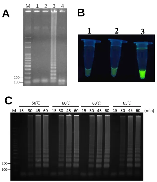
Sensitivity and specificity of the LAMP assay
18
To determine the sensitivity of the LAMP assay for detecting IBDV genome, 19
complementary DNA encoding the IBDV VP2 gene was serially diluted 10-fold and 1
used as template DNA. VP2 gene amounts ranging from 1 ng to 1 fg were used in the 2
LAMP reaction. After performing the LAMP reaction, LAMP products were detected 3
over the range of DNA from 1 ng to 10 fg; this is shown in figure 3A. There has no 4
LAMP products detected by DNA electrophoresis when less than 10 fg of template 5
DNA was added to the LAMP assay. Therefore, the sensitivity of LAMP when 6
detecting complementary DNA from the IBDV genome was determined to be 10 fg. 7
This is more sensitive than conventional PCR amplification of the IBDV VP2 gene. 8
Figure 3B shows that at least 1 pg of VP2 gene is required to amplify DNA by PCR in 9
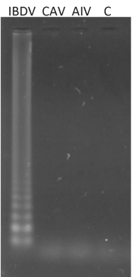
order to detect IBDV infection. Next, the specificity of the LAMP assay was 10
evaluated using the H6 gene of avian influenza virus (AIV) and the VP2 gene of CAV; 11
the results are shown in figure 4. No LAMP products were generated in these 12
reactions and only reaction mixtures containing IBDV VP2 gene cDNA gave a 13
positive signal by LAMP assay. These results suggest that this newly established 14
LAMP assay is highly specific and sensitive when detecting the IBDV complementary 15
genome. 16
17
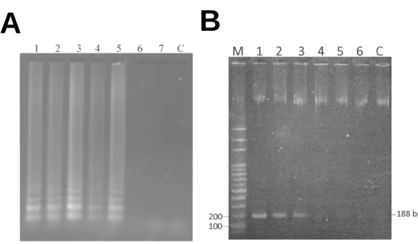
Detection of the IBDV genome by RT-LAMP
18
Having established that the LAMP assay can be used to detect the IBDV 19
complementary genome, it was necessary to establish whether the approach could be 1
expanded to detect the double-stranded RNA viral genome of IBDV. To do this, 2
reverse transcription (RT) is required to form complementary DNA from the viral 3
RNA genome before performing the LAMP assay. Thus, the LAMP assay was 4
combined into a RT-LAMP reaction by adding a reverse transcription step for cDNA 5
synthesis. As illustrated in lane 3 of figure 5A, the RT-LAMP reaction was performed 6
in a single tube and using one step. The results showed that a typical LAMP pattern 7
could be observed by DNA agarose gel electrophoresis using extracted viral RNA 8
from IBDV-infected bursa tissue that was added to the RT-LAMP reaction. In this 9
case, the negative sample (mock-infection) gave no LAMP product. Moreover, 10
IBDV-RT-LAMP did not amplify the RNA extracted from bursa tissue not infected 11
with IBDV (lane 2 of figure 5). Furthermore, it also did not amplify RNA extracted 12
from AIV or CAV infected liver tissue (data not shown). In terms of the amplification 13
limit, when the RT-PCR was performed using a 10 fold dilution series of viral RNA 14
from 1 ng to 0.001 fg, the IBDV viral RNA could be detected in all samples, which is 15
a RT-LAMP sensitivity of 0.01 fg of viral RNA. The results are shown in figure 5B. 16
17
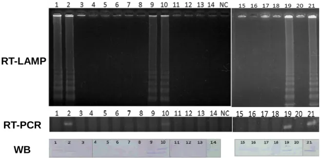
Application of the RT-LAMP assay as a diagnostic technique
18
Based on the above results, we evaluated the method for use as a practical tool 19
using 21 clinical bursa specimens from chickens. As shown in Figure 6, the genomic 1
viral RNAs were extracted from the bursa of six IBDV infected and fifteen IBDV 2
non-infected broilers; these were then analyzed by one-step RT-LAMP. The results 3
showed that six out of six of the clinical samples were positive when RT-LAMP 4
analysis was used (upper panel of figure 6). The IBDV non-infected samples were all 5
negative by RT-LAMP assay (lower panel of figure 6). These results completely agree 6
with the clinical testing by conventional Western blot assay. 7
8
Discussion
9
Infectious bursal disease virus (IBDV) causes an acute and highly contagious 10
viral disease. Once chickens are infected with this virus, it may result in high 11
mortality and immunosuppression. Therefore, IBDV is an economically important 12
viral pathogen that needs to be controlled within the poultry industry worldwide. 13
However, a simple, sensitive, specific and rapid assay is needed to detect IBDV 14
infection. In the past, several methods have been developed to diagnosis IBDV 15
infection. These methods include TEM observation, serological methods such as AGP 16
and ELISA and molecular diagnosis tools such as reverse transcription PCR 17
(RT-PCR). Among these methods, nucleic acid-based amplification techniques, such 18
as RT-PCR, are more rapid, sensitive and specific than the other approaches to IBDV 19
detection. However, even using real time RT-PCR, the detection limit can be as high 1
as 100 pg when SYBR Green I was used in the reaction [29]. In addition, there are a 2
number of shortcomings to these diagnosis methods, namely they are time-consuming, 3
tedious, have a low amplification efficiency, and need the use of a thermal cycler. In 4
this study, we have successfully developed a one-step RT-LAMP method for 5
detecting the IBDV RNA genome. The one-step approach involves combining the 6
LAMP method with AMV reverse transcriptase. In our study, the one-step RT-LAMP 7
method enable specific detection of IBDV infection in broilers using the optimized 8
condition of 60oC and a reaction time as short as 40 min. The assay can be performed 9
using avian reverse transcriptase and Bst DNA polymerase at the same time in a water 10
bath tank under isothermal conditions. The initial reverse transcription reaction uses 11
AMV RTase rather than M-MLV RTase because of the former enzyme’s broad range 12
of reaction temperatures, namely 42oC to 60oC. This allows the reaction to be carried 13
out with both enzymes, AMV RTase and Bst DNA polymerase, at the same time. The 14
simplicity of this approach together with its speed will allow this assay to be applied 15
to the detection of IBDV during infection screening or routine evaluation of 16
immunization efficacy. For the one-step reaction, the temperature chosen was 60oC 17
and therefore there is no necessity to pre-heat as is needed in conventional RT-PCR. 18
Overall, RT-LAMP is easier to carry out and save time compared to RT-PCR. 19
For RT-LAMP assay, the VP2 gene in segment A of the IBDV genome was 1
chosen and the designed primer set is able to recognize specifically a highly 2
conserved region of VP2. The RT-LAMP product gave the characteristic ladder-like 3
pattern by gel electrophoresis; this is specific because the reaction produces a mixture 4
of stem-loop structured DNA products with various lengths. The identity of the 5
amplified products was clearly confirmed by BamH I restriction enzyme digestion. To 6
save time when detecting the RT-LAMP products, SYBR Green I is the most 7
common approach for field operations.. When SYBR Green I is exposed to UV light, 8
it is possible to determine directly whether the RT-LAMP assay is positive or negative 9
by the naked eye. 10
The sensitivity of one-step RT-LAMP was established as 0.01 fg of viral RNA 11
using specifically designed RT-LAMP primers. This contrasts with a much poorer 12
sensitivity using RT-PCR of at least 1 ng of viral RNA. Thus, our results show that 13
the sensitivity of the RT-LAMP assay for IBDV detection is about 108 fold higher 14
than the RT-PCR assay. Based on the molecular weight of the IBDV genome at 4.7 15
MDa [30], this means that the detection limits for the RT-LAMP and RT-PCR assays 16
are approximately 1 and 108 copies of the IBDV genome, respectively. In addition, 17
there was complete agreement of the RT-LAMP results with the Western blot assay 18
results, which indicates that the one-step RT-LAMP method is both highly sensitive 19
and accurate. In order to test specificity, the VP2 gene of chicken anemia virus (CAV) 1
and hemagglutinin gene of H6 avian influenza virus (H6-AIV) were used. The results 2
showed that the RT-LAMP assay was specific to IBDV compared to these other two 3
viruses. IBDV is the sole member of the genus birnavirus, which is part of the 4
birnaviridae family. This genus also contains pancreatic necrosis virus (IPNV). 5
However, there is low sequence similarity between IBDV and IPNV and therefore 6
there is little likelihood of cross-reactivity between these two viruses. Based on the 7
specificity results, we suggest that the RT-LAMP assay will make a very good clinical 8
test for IBDV. 9
One of major problems with RT-PCR is that the assay needs an accurately 10
controlled thermocycling system to repeatedly perform the three temperatures shifts 11
needed to amplify using reverse transcriptase and Taq DNA polymerase over 2-3 hrs. 12
The one-step RT-LAMP needs no such expensive system, only a temperature 13
controlled waterbath or heating block. Conventional RT-PCR is currently used as the 14
“golden standard method” for IBDV detection, but the need for both the 15
thermocycling system and an elaborate method of detecting the product after 16
amplification is a disadvantage. Product detection using the RT-LAMP method is 17
easier than with RT-PCR and can be done by either using a fluorescence staining 18
reagent such as SYBR Green I, as was done in this study, or even by just observing 19
the turbidity [11]. Therefore, using RT-LAMP, a well-equipped laboratory, 1
well-trained staff, and multiple reaction steps are not needed, which makes this assay 2
more suitable to large-scale field investigations. 3
Xu et al. also used a RT-LAMP method to detect IBDV [27]. This method needed 4
1.5 hours and a two-step heating process to allow detection of the virus. Compared to 5
this earlier study, the sensitivity of our assay is 100 fold greater, as well as being 6
much faster and a one-step process. The primers used in Xu et al. targeted the VP3 7
gene of IBDV, while our assay targets the VP2 gene. In addition, in order to optimize 8
the primers, we focused on low free energy criteria. We suggested that the low free 9
energy of the primers used in our assay allowed us to achieve higher sensitivity. 10
Overall, the assay developed in this study is a sensitive, time saving and easier 11
one-step process for the detection of IBDV than the approach described in Xu et al. 12
Consequently, this one step RT-LAMP method can be easily applied to clinical 13
samples when performing IBDV diagnosis during epidemiological investigations and 14
to the assessment of immunization efficacy after vaccination. 15
In conclusion, the IBDV diagnostic method using one-step RT-LAMP assay 16
described here has the potential to become a valuable diagnostic system and could be 17
made into a simple-to-use diagnostic kit. This is the first report to demonstrate the 18
application of an improved and one-step RT-LAMP assay to the field diagnosis of 19
IBDV. In the future, the RT-LAMP assay for IBDV detection could be developed into 1
a portable mini-laboratory system for the on-site epidemiological detection of 2
infectious bursal disease virus infection among farm-bred young chickens. 3
4
Acknowledgements
5
This work has supported by a grant from the National Science Council (NSC 6
95-2313-B-039-004-, NSC96-2313-B-276-001-MY3) of Taiwan. 7
References
1
[1] Notomi T, Okayama H, Masubuchi H, Yonekawa T, Watanabe K, Amino N, Hase 2
T. Loop-mediated isothermal amplification of DNA. Nucleic Acids Res 2000; 28: 3
E63. 4
[2] Nagamine K, Watanabe K, Ohtsuka K, Hase T, Notomi T. Loop-mediated 5
isothermal amplification reaction using a nondenatured template. Clin Chem 2001; 6
47: 1742-3. 7
[3] Yeh HY, Shoemaker CA, Klesius PH. Sensitive and rapid detection of 8
Flavobacterium columnare in channel catfish Ictalurus punctatus by a
9
loop-mediated isothermal amplification method. J Appl Microbiol 2006; 100: 10
919-25. 11
[4] Salih DA, Liu Z, Bakheit MA, Ali AM, Hussein AM EI, Unger H, Viljoen G, 12
Seitzer U, Ahmed JS. Development and evaluation of a loop-mediated isothermal 13
amplification method for diagnosis of tropical theileriosis. Transbound Emerg Dis 14
2008; 55: 238-243. 15
[5] Sakuma T, Kurosaki Y, Fujinami Y, Takizawa T, Yasuda J. Rapid and simple 16
detection of Clostridium botulinum types A and B by loop-mediated isothermal 17
amplification. J Appl Microbiol 2009; 106:1252-9. 18
[6] Ohtsuki R, Kawamoto K, Kato Y, Shah MM, Ezaki T, Makino SI. Rapid detection 1
of Brucella spp. by the loop-mediated isothermal amplification method. J Appl 2
Microbiol 2008; 104:1815-23. 3
[7] Shivappa RB, Savan R, Kono T, Sakai M, Emmenegger E, Kurath G, Levine JF. 4
Detection of spring viraemia of carp virus (SVCV) by loop-mediated isothermal 5
amplification (LAMP) in koi carp, Cyprinus carpio L. J Fish Dis 2008; 31: 6
249-58. 7
[8] Liu Z, Teng Y, Xie X, Li H, Lv J, Gao L, Tian F, Jiang Y, Chu Z, Xie C, Liu H. 8
Development and evaluation of a one-step loop-mediated isothermal amplification 9
for detection of spring viraemia of carp virus. J Appl Microbiol 2008; 105: 10
1220-6. 11
[9]Yamazaki W, Taguchi M, Ishibashi M, Nukina M, Misawa N, Inoue K. 12
Development of a loop-mediated isothermal amplification assay for sensitive and 13
rapid detection Camylobacter fetus. Vet Microbiol 2009; 14
Doi:10.1016/j.vetmic.2008.11.018 15
[10] Huang CH, Lai GH, Lee MS, Lin WS, Lien YY, Hsueh SC, Kao JY, Chang WT, 16
Lu TC, Lin WN, Chen HJ, Lee MS. Development and evaluation of a 17
loop-mediated isothermal amplification assay for rapid detection of chicken 18
[11] Mori Y, Notomi T. Loop-mediated isothermal amplification (LAMP): a rapid, 1
accurate, and cost-effective diagnostic method for infectious diseases. J Infect 2
Chemother 2009; 15: 62-69. 3
[12] Soliman H, Midtlyng PJ, El-Matbouli M. Sensitive and rapid detection of 4
infectious pancreatic necrosis virus by reverse transcription loop mediated 5
isothermal amplification. J Virol Methods 2009; 158: 77-83. 6
[13] Morishita S, Tani H, Kurata S, Nakamura K, Tsuneda S, Sekiguchi Y, Noda N. 7
Real-time reverse transcription loop-mediated isothermal amplification for rapid 8
and simple quantification of WT1 mRNA. Clin Biochem 2009; 42: 515-20. 9
[14] Azad A A, Barrett SA, Fahey KJ. The characterization and molecular cloning of 10
the double-stranded RNA genome of an Australian strain of infectious bursal 11
disease virus. Virology 1985; 143: 35-44. 12
[15] Lasher HN, Davis VS. History of infectious bursal disease in the U.S.A.--the first 13
two decades. Avian Dis 1997; 41: 11-9. 14
[16]
Hirai K, Shimakura S, Hirose M. Immunodiffusion reaction to avian infectious 15bursal virus. Avian Dis 1972; 16: 961-64. 16
[17] Weisman J, Hitchner SB. Virus-neutralization versus agar-gel precipitin tests for 17
detecting serological response to infectious bursal disease virus. Avian Dis 1978; 18
[18] Marquardt WW, Johnson RB, Odenwald WF, Schlotthober B A. An indirect 1
enzyme-linked immunosorbent assay (ELISA) for measuring antibodies in 2
chickens infected with infectious bursal disease virus. Avian Dis 1980; 24: 3
375-385. 4
[19] Wang MY, Hu HL, Suen SY, Chiu FY, Shien JH, Lai SY. Development of an 5
enzyme-linked immunosorbent assay for detecting infectious bursal disease virus 6
(IBDV) infection based on the VP3 structural protein. Vet Microbiol 2008; 131: 7
229-36. 8
[20] Henderson KS, Jackwood DJ. Comparison of the dot blot hybridization assay with 9
antigen detection assays for the diagnosis of infectious bursal disease virus 10
infections. Avian Dis 1990; 34: 744-748. 11
[21] Harkness JW, Alexander DJ, Pattison M, Scott AC. Infectious bursal disease 12
agent: morphology by negative stain electron microscopy. Arch Virol 1975; 48: 13
63-73. 14
[22] Lee LH, Ting LJ, Shien JH, Shieh HK. Single-tube, noninterrupted reverse 15
transcription-PCR for detection of infectious bursal disease virus. J Clin Microbiol 16
1994; 32: 1268-1272. 17
[23] Tham KM, Young LW, Moon CD. Detection of infectious bursal disease virus by 18
reverse transcription-polymerase chain reaction amplification of the virus segment 19
A gene. J Virol Methods 1995; 53: 201-212. 1
[24] Moody A, Sellers S, Bumstead N. Measuring infectious bursal disease virus RNA 2
in blood by multiplex real-time quantitative RT-PCR. J Virol Methods 2000; 85: 3
55-64. 4
[25] Jackwood DJ, Nielsen CK. Detection of infectious bursal disease viruses in 5
commercially reared chickens using the reverse transcriptase/polymerase chain 6
reaction-restriction endonuclease assay. Avian Dis 1997; 41: 137-143. 7
[26] Jackwood DJ, Sommer SE. Restriction fragment length polymorphisms in the 8
VP2 gene of infectious bursal disease viruses. Avian Dis 1997; 41: 627-637. 9
[27] Xu J, Zhang Z, Yin Y, Cui S, Xu S, Guo Y, Li J, Wang J, Liu X, Han, L. 10
Development of reverse transcription loop-mediated isothermal amplification for 11
detection of infectiou bursal disease virus. J Virol Methods 2009; 12
doi:10.1016/j.jviromet.2009.07.010. 13
[28] Wang MY, Kuo YY, Lee MS, Doong, SR, Ho JY, Lee LH. Self-Assembly of the 14
Infectious Bursal Disease Virus Capsid Protein, rVP2, Expressed in Insect Cells 15
and Purification of Immunogenic Chimeric rVP2H Particles by Immobilized 16
Metal-Ion Affinity Chromatography. Biotechnol Bioeng 2000; 67: 104-111. 17
[29] Ling KL, Omar AR, Bejo MH, Ideris A, Tan SW. Development of SYBR green I 18
based one-step real-time RT-PCR assay for the detection and differentiation of 19
Methods 2009; dio:10.1016/j.jviromet.2009.06.023. 1
[30] Dobos P, Hill BJ, Hallett R, Kells DT, Becht H, and Teninges D. Biophysical and 2
biochemical characterization of five animal viruses with bisegmented 3
double-stranded RNA genomes. J Virol 1979; 32:593-605. 4
Legends
1 2
Figure 1. Schematic presentation of the primers used in the RT-LAMP assay for
3
IBDV detection. The partial nucleotide sequence of the VP2 cDNA of IBDV, the six
4
target regions for primers annealing, and the four specifically designed RT-LAMP 5
primers used in the assay are indicated. The underlined sequence is the BamH I 6
restriction enzyme site. 7
8
Figure 2. Analysis of the LAMP products using agarose gel electrophoresis and
9
SYBR Green I staining. (A) The LAMP products were detected by agarose gel
10
electrophoresis and the products was also digested with BamH I. LAMP in some cases 11
as described in Materials and Methods. Lane M, the 100 bp DNA ladder as size 12
marker. Lane 1, LAMP reaction mixture without Bst DNA polymerase; Lane 2, 13
LAMP reaction mixture without target DNA template; Lane 3, complete LAMP 14
reaction mixture; Lane 4, LAMP products after digestion with BamH I. (B) 15
Visualization of LAMP products by staining with SYBR Green I and using UV 16
excitation. Tubes 1-3 are the LAMP products showed in lanes 1 to 3 of (A). (C) 17
LAMP was performed for different reaction times and at various temperatures. Lane C, 18
LAMP reaction without target DNA as a negative control. 19
Figure 3. Sensitivity of the LAMP assay (A) and compare with PCR (B) for
1
detecting the cDNA of IBDV VP2 gene. The PCR was carried out using the outer
2
LAMP primers, F3 and B3. A 188 bp length fragment of the IBDV VP2 DNA was 3
obtained after the PCR reaction. Lane 1-7, 100 pg, 10, pg, 1 pg, 100 fg, 10 fg, 1 fg, 4
0.1 fg of VP2 cDNA were added to the LAMP reactions; Lane M, 100 bp DNA ladder; 5
Lane C, negative control. 6
7
Figure 4. Specificity of the LAMP assay for the detection of the VP2 gene of
8
IBDV. The LAMP reaction was performed at 65oC for 1 hr. Specific viral 9
complementary DNA from the three viruses tested was prepared as described in the 10
Materials and Methods. IBDV, VP2 gene of infectious bursal disease virus added as 11
the template. CAV, VP2 gene of chicken anemia virus added as the template. AIV, 12
H6 gene of avian influenza virus added as the template. Lane C, negative control. 13
14
Figure 5. The one-step RT-LAMP assay for detecting IBDV-infected bursa tissue
15
from chickens. The RT-LAMP primers were used with genomic RNA for the
16
diagnosis of IBDV in a one step RT-LAMP reaction (A). Lane M, 100 bp DNA ladder 17
marker; lane 1, RT-LAMP reaction mixture containing no template; lane 2, RT-LAMP 18
reaction mixture containing no reverse transcriptase; lane 3, extracted IBDV viral 19
to the RT-LAMP reaction. (B) One-step RT-LAMP and RT-PCR were performed after 1
adding various amounts of viral RNA. Upper panel is the RT-LAMP pattern, lower 2
panel is the RT-PCR pattern. Lane 1-10 were 1 ng, 100 pg, 10 pg, 1 pg, 100 fg, 10 fg, 3
1 fg, 0.1 fg, 0.01 fg, 0.001 fg of IBDV total RNA, respectively. Lane NC, negative 4
control. 5
6
Figure 6. Detection of IBDV-RNA from experimental chickens by RT-LAMP. 1
7
ng of viral genomic RNA extracted from the bursa was used for each RT-LAMP and 8
RT-PCR assay. The RT-LAMP and RT-PCR patterns were analyzed using 1.6% 9
agarose electrophoresis gel. The numbers on each of the lanes represent the different 10
clinical collected bursa samples from either IBDV infected or non-infected chickens. 11
These samples were also analyzed by Western blotting using antibody against the 12
IBDV VP2 protein. Lane NC, negative control for the RT-LAMP and RT-PCR assays. 13
WB, Western blotting assays. 14
Table 1. Details of oligonucleotide prime rs used for one -step RT-LAMP to amplify VP2 ge ne of IBDV. Primer
name Type Length(s) Genome position Sequence(5'→3)
F3 Forward outer 20-mer 999-1018 AGCAGTGACAATCCATGGTG B3 Reverse outer 18-mer 1169-1186 TCATGGCTCCTGGGTCAA FIP Forward inner
(F1c-TTTT-F2) 45-mer(F1C, 22-mer; F2, 19-mer) F1C, 1075-1096 F2, 1035-1053 CGACCGTAACGACGGATCCTG TTTTTCCTCCGTCCCGTCACAC TA
BIP Reverse inner
(B1C-TTTT-B2) 44-mer(B1C, 20-mer;B2, 20-mer) B1C, 1097-1116;B2, 1148-1166
CCGGGGTGAGCAACTTCGAGT TTTT
IBDV strain P3009 VP2 999 1186
BamHI