國立交通大學
生物科技學系
碩士論文
開發奈米鑽石連結紫杉醇作為藥物運送與癌症治療
Development of nanodiamond-conjugated paclitaxel for drug
delivery and cancer therapy
研究生: 王繼慶
指導教授: 趙瑞益
開發奈米鑽石連結紫杉醇作為藥物運送與癌症治療
Development of nanodiamond-conjugated paclitaxel for drug
delivery and cancer therapy
研究生: 王繼慶 Student: Chi-Ching Wang
指導教授: 趙瑞益 Advisor: Prof. Jui-I Chao
國 立 交 通 大 學
生物科技學系
碩 士 論 文
A Thesis
Submitted to Department of Biological Science and Technology College of Biological Science and Technology
National Chiao Tung University in partial Fulfillment of the Requirements
for the Degree of Master
In
Biological Science and Technology
July 2010
Hsinchu, Taiwan, Republic of China
致 謝
兩年的碩士研究生活,在一轉眼間就過了,而這篇論文的完成,要感謝許多 幫助過我的人,首先要感謝我的指導教授趙瑞益教授,感謝老師這兩年來悉心的 指導與關照,在撰寫論文期間,謝謝老師不辭辛勞的幫我校正與定稿。在研究所 的日子哩,實驗室就像是第二個家,實驗室的夥伴們都是幫助我成長的人。我要 感謝惠芳學姐和光凱學長,每當我在實驗遇到困難時,總是不厭其煩的替我找出 問題,也耐心的幫助我解決口試及生活上的問題,讓我覺得能夠認識學長姐你 們,是我研究所中最重要的收穫。已畢業的真宜學姐和瑞良學長,感謝你們在我 碩一時的幫助及照顧。還有厚巽、靜宜、勝壹、同屆的夥伴們,互相的體諒與幫 助完成這兩年的學業。實驗室的學弟妹們,芷漪、淳如、佩萱、毓婷、昱耀、旻 翰,有你們的陪伴,讓我的研究所生活更加精采。最後,我要感謝我的家人,謝 謝爸爸、媽媽對我的付出,給我莫大的支持,弟弟總是鼓勵與幫助我解決生活上 雜事。最後我要感謝曾經幫助過我的師長與朋友們,謝謝你們。Table of contents
page Chinese Abstract………...Ⅲ English Abstract………...Ⅳ List of figures………...Ⅴ Abbreviations………...Ⅶ 1. Introduction………12. Materials and methods………...5
2.1 Reagents and antibodies……….………..5
2.2 Cell lines………….………..5
2.3 Cell culture……….………..6
2.4 Synthesis of ND-conjugated paclitaxel……….………...6
2.5 Cytotoxicity assay……….………...9
2.6 Fluorescence intensity of ND-paclitaxel in cells by flow cytometer……...9
2.7 Confocal microscopy……….……….10
2.8 Cell cycle analysis……….……….11
2.9 Mitotic index analysis………..……...………...11
2.10 Apoptotic nuclear counting……….………...12
2.11 Annexin V- PI staining……….………..12
2.12 Time-lapse observation of apoptosis induction……….…13
2.13 Western blot analysis ...……….…13
3. Results………...……….…..15
3.1 ND-paclitaxel reduces cell viability in lung carcinoma cells …………....15
3.2 Uptake ability of ND-paclitaxel in lung carcinoma cells ………..15
3.3 ND-paclitaxel blocks microtubules to induce abnormal mitotic cells…...16
3.4 ND-paclitaxel induces the cell cycle arrest and apoptosis in lung carcinoma cells …….……….…..16
3.5 ND-paclitaxel inhibits tumorigenesis of human lung tumor xenograft in SCID mice……….……….…...18
3.6 ND-paclitaxel induces cytotoxicity in various human cancer cells...…....18
3.7 Uptake ability of ND-paclitaxel in human colon carcinoma cells…...…..19
3.8 ND-paclitaxel increases sub-G1 and G2/M fractions in human colon carcinoma cells ……….….19
3.9 ND-paclitaxel induces apoptosis formation in human colon carcinoma cells……….…………....19
4. Discussion ……...……….……..….21
5. Conclusion………...24
中文摘要
利用奈米物質攜帶抗癌藥物,提供一個嶄新的機會作為癌症的治療。在本篇 研究中,我們利用一種由碳所組成的奈米材料,稱作奈米鑽石,以共價鍵的方式 連結紫杉醇,作為藥物的運送和癌症的治療。透過一連串化學修飾的合成方法, 將紫杉醇鍵結到奈米鑽石的表面。在濃度 0.1-50 μg/mL 的奈米鑽石-紫杉醇處理 A549 人類肺癌細胞 48 小時之後,明顯降低癌細胞的存活率,然而單獨處理奈米 鑽石或是經強鹼處理過後的奈米鑽石-紫杉醇,並不會誘發 A549 肺癌細胞的死 亡。由共軛焦顯微鏡觀察,我們發現奈米鑽石-紫杉醇會進入 A549 肺癌細胞,並 且位在細胞質及微小管。奈米鑽石-紫杉醇仍然具有紫杉醇的抗癌活性,會造成 細胞的有絲分裂停止和細胞凋亡,並且抑制 CDC2、磷酸化 CDC2 及 cyclin B1 蛋白的表達。此外,在異體移植人類肺癌細胞到先天免疫不全的老鼠之動物實驗 中,奈米鑽石-紫杉醇會抑制老鼠體內腫瘤的形成。再者,我們也發現奈米鑽石-紫杉醇在其他種類的人類癌細胞,包括大腸癌細胞(ROK 和 HCT116)和膀胱癌細 胞(BFTC 905)也會誘發細胞毒性和細胞凋亡,並且造成 caspase-3 蛋白的活化及 PARP 蛋白被切割。綜合以上結果,我們已經開發出一種功能性之共價鍵結奈米 鑽石-紫杉醇,具有促使有絲分裂停止、誘發細胞凋亡及抑制腫瘤形成的抗癌活 性。Abstract
Nanoparticle-conjugated anticancer drugs provide novel opportunities for cancer therapy. In this study, we evaluated nanodiamond (ND), a carbon nanomaterial, to covalently bind paclitaxel for drug delivery and cancer therapy. Paclitaxel was bound to the ND’s surface through a succession of chemical modification. Treatment with 0.1-50 μg/mL ND-paclitaxel for 48h significantly reduced the cell viability in A549 human lung cancer cells. However, ND alone or denatured ND-paclitaxel (after treatment with strong alkaline solution, 1M NaOH) did not induce the damage effects on A549 cells. The ND-paclitaxel was taken into cell and located in the microtubules and cytoplasm of A549 cells observed by confocal microscopy. Moreover, ND-paclitaxel still reserves the anticancer activity of paclitaxel. ND-paclitaxel was attributed both mitotic blockage and apoptotic induction in cancer cells. The protein levels of CDC2, phosphorylated CDC2, and cyclin B1 were decreased by treatment with ND-paclitaxel. Besides, ND-paclitaxel inhibited the tumorigenesis of xenograft human lung tumor in SCID mice. Moreover, we also found ND-paclitaxel was significantly induced cytotoxicity and apoptosis in other cancer cells including colon cancer cells (RKO and HCT116) and bladder cancer cells (BFTC 905). ND-paclitaxel induced the caspase-3 activation and the protein cleavage of PARP. In summary, we have developed a functional covalent ND-paclitaxel, which still preserves its anticancer activities on the mitotic blockage, apoptosis induction and anti-tumorigenesis.
List of figures
page Scheme 1. Chemical synthesis of ND-paclitaxel……….32Figure 1. Effect of ND-paclitaxel on the cell viability in human lung carcinoma
Cells………33
Figure 2. Uptake ability of ND-paclitaxel in human lung carcinoma cells……..34
Figure 3. Effect of ND-paclitaxel on the blockage of microtubule and chromosome
segregation in human lung carcinoma cells………...35
Figure 4. Location and distribution of ND-paclitaxel in human lung carcinoma
cells……….36
Figure 5. Effect of ND-paclitaxel on the cell cycle progression in human lung
carcinoma cells………...37
Figure 6. The effect of ND-paclitaxel on the protein levels of phospho-CDC2 and total
CDC2 in human lung carcinoma cells………38
Figure 7. Effect of ND-paclitaxel on the percentages of mitotic index and apoptosis in
human lung carcinoma cells………...39
Figure 8. The effect of pretreated ND and ND-paclitaxel on tumor growth in A549
xenograft SCID mice model………...40
Figure 9. Effect of ND-paclitaxel on the cell viability in various human cancer
Figure 10. Uptake ability of ND-paclitaxel in RKO cells………42
Figure 11. Effect of ND-paclitaxel on cell cycle progression in RKO cells……43
Figure 12. Effect of ND-paclitaxel on the protein levels of phospho-CDC2, total
CDC2 and cyclin B1 in RKO cells...………44
Figure 13. Effect of ND-paclitaxel for apoptosis in the RKO cells………45
Figure 14. Effect of ND-paclitaxel on the active of caspase-3 and cleavage of
poly(ADP-ribose) polymerase (PARP) in RKO cells………...46
Figure 15. Time-lapse observation of apoptotic formation following treatment with
Abbreviations ND nanadiamond
PARP anti-poly(ADP-ribose) polymerase
DMSO dimethyl sulfoxide
PI propidium iodide
PBS phosphate buffered saline
SDS sodium dodecyl sulfate
CDKs cyclin-dependent kinases
CDC2 cell division control protein 2
FBS fetal bovine serum
Caspase-3 cysteine-aspartic acid protease
MTT 3-(4,5-dimethyl-thiazol-2-yl) 2,5-diphenyl tetrazolium bromide
PTX paclitaxel
THF tetrahydrofuran
FITC fluorescein isothiocyanate
1. Introduction
Cancers have been become the leading reason of death in the world. In Taiwan,
lung cancer has become the first mortality among all cancer patients (Department of
health, Executive Yuan, 2009). Although treatments of cancers include surgery,
radiation, and chemotherapy, patients in late stage diseases are usually managed
primarily with chemotherapy. However, poor intracellular uptake, limited
circulation stability, and normal cell damages reduce the abilities of
chemotherapeutic drugs. Therefore, development of novel strategies for cancer
therapy is highly desired. Nanoparticles have been evaluated for biomedical
applications in recent years. Moreover, nanoparticles can be developed for
biomedical application such as cancer detection and drug delivery (Kang et al., 2010;
Liu et al., 2008).
Nanoparticles refer to are the substance of investigation size at range 1-100 nm
at one dimension (McNeil, 2005). Nanomaterials as drug delivery systems facilitate
approach for cancer therapy (Alexis et al., 2010). Nanoparticles can improve cancer
therapeutics by conjugated with drugs and biological molecules (Akerman et al.,
2002; Gao et al., 2004; Michalet et al., 2005; Tada et al., 2007). For example,
quantum dots, which exhibit varying colors of fluorescence, have been applied for
(Akerman et al., 2002; Gao et al., 2004; Tada et al., 2007). Nanoliposomal carrying
cancer drugs have been successfully used in cancer therapy, demonstrating benefits
of prolonged tissue residence and reduced toxicity (Chau et al., 2006; Koshkina et
al., 2003; Noble et al., 2006). Utilization liposome-encapsulated doxorubicin have
been successfully used in cancer therapy, demonstrating doxorubicin displayed less
cardiac toxicity (Batist et al., 2001). Besides, carbon nanotube has been used as a
carrier for cancer drug delivery that effectively inhibited tumor growth in mice (Liu
et al., 2008).
It has been concerned on the issue of toxic potential of nanomaterials (Nel et
al., 2006; Service, 2004), although it is intensively developed for biomedical
applications. A non-toxic and biocompatible nanomaterial is desired for clinical
applications. Nanodiamond (ND) is a carbon derivative nanomaterial, which has
been evaluated for biomedical applications in recent years. It has been shown that
ND do not induce cytotoxicity in a variety of cells including lung (Liu et al., 2007;
Liu et al., 2009), neuronal (Schrand et al., 2007), renal (Lechleitner et al., 2008; Yu
et al., 2005), and cervical cells (Chang et al., 2008). Moreover, ND particles did not
alter cell division and differentiation (Liu et al., 2009). In addition, the intravenous
injection of ND particles into mice did not significantly induce symptoms of
non-cytotoxicity and biocompatibility, although ND should be to further evaluation
before clinical use.
The surface of ND particles has a unique platform for conjugation of chemicals and bio-molecules after functional modifications (Yang et al., 2002; Chao et al.,
2007; Ushizawa et al., 2002; Huang et al., 2004; Cheng et al., 2007; Krueger et al.,
2008). ND is suitable for bioconjugation either chemically (covalently or
noncovalently) or physically (adsorption) (Cheng et al., 2005; Krueger, 2008).
The modified ND’s surfaces have been shown to conjugate with DNA (Yang et al.,
2002; Ushizawa et al., 2002), lysozyme (Chao et al., 2007), cytochrome c (Huang et
al., 2004), growth hormone (Cheng et al., 2007), biotin (Krueger et al., 2008),
alpha-bungarotoxin (Liu et al., 2008), and insulin (Shimkunas et al., 2009), and
folate (Zhang et al., 2009). Lysozyme and alpha-bungarotoxin proteins can be
absorbed on the surface of carboxylated ND via non-covalent bonding that still
preserve the biological activities of these proteins (Chao et al., 2007; Liu et al.,
2008). The hydrogel of ND with chemotherapeutic drugs such as doxorubicin was
developed for drug delivery by non-covalent adsorption (Huang et al.,2007). In
addition, ND has been covalently linked to folate for targeting cancer cells (Zhang
et al., 2009).
anticancer activities of these conjugates are poorly understood. Paclitaxel is the one
of most widely used chemotherapeutic drugs in the clinic for the treatment of
advanced solid carcinomas (Wang et al., 2009). Paclitaxel disturbs microtubule
dynamics and impairs the transition of cells in mitosis, and leading to cell death
(Wang et al., 2010).
In this study, we create a novel covalent bonded ND-paclitaxel for drug
delivery and cancer therapy. ND-paclitaxel can be taken into human lung and colon
carcinoma cells. More importantly, ND-paclitaxel displays anticancer activities by
inducing mitotic arrest, apoptosis and anti-tumorigenesis. Accordingly, we create a
functional covalent conjugation of ND and paclitaxel, which provides the possible
2. Materials and methods
2.1 Reagents and antibodies
Paclitaxel was purchased from Tokyo Chemical Industry Co. (Ltd. Japan).
Powdered ND particles with diameters of 3-5 nm were purchased from
Nanostructured and Amorphous Materials Inc. (Houston, TX).
3-(4,5-dimethyl-thiazol-2-yl) 2,5-diphenyl tetrazolium bromide (MTT), Hoechst
33258, and the Cy3-labeled mouse anti--tubulin (c-4585) were purchased from
Sigma Chemical Co. (St Louis, Mo.). Anti-CDC2, anti-phospho-CDC2 (tyrsion-15,
threonine-14, and threonine-161), and anti-poly(ADP-ribose) polymerase (PARP)
antibodies were purchased from Cell Signaling Technology Inc. (Beverly, MA).
Anti-caspase-3 antibody was purchased from BioVision (BioVision, Inc., USA).
Anti-cyclin B1 (Ab-2) antibody was purchased from Oncogene Sciences
(Cambridge, MA). Anti-actin (I-19) antibody was purchased from Santa Cruz
Biotechnology, Inc. (Santa Cruz, CA).
2.2 Cell lines
A549 cell line (ATCC number: CCL-185) was derived from the lung
adenocarcinoma. RKO (ATCC number: CRL-2677) was a colon carcinoma cell line.
HCT116 (ATCC number: CCL-247) was a colon carcinoma cell line. BFTC 905
transitional cell carcinoma.
2.3 Cell culture
A549 and BFTC 905 cells were maintained in RPMI-1640 medium (Invitrogen
Co., Carlsbad, CA). RKO cells were cultured in DMEM medium (Invitrogen).
HCT116 cells were maintained in McCoy’s 5A medium. The complete media were
supplemented with 10% FBS, 100 units/ml penicillin, and 100 g/ml streptomycin. These cells were cultured at 37 ℃ and 5% CO2 in a humidified incubator (Thermo,
Forma Scientific, Inc., Marietta, OH).
2.4 Synthesis of ND-conjugated paclitaxel
ND-paclitaxel was synthesized and kindly provided by our collaborator Dr.
Chinpiao Chen (Department of Chemistry, National Dong-Hwa University,
Hualien). The chemical synthetic procedure for the conjugation of ND and
paclitaxel was been shown in Scheme 1. Initial chemical treatment of ND powders
(1) by carboxylation was carried out according to standard procedure. The ND
particles were stirred in a 3:1 (v/v) mixture of concentrated HCl and HNO3 at room
temperature for three days, then diluted with deionized H2O and separated by
centrifugation at 900 rpm. After centrifugation, the pellets were extensively rinsed
with deionized H2O three times. Thereafter, ND particles were heated in 0.1 M
90 ºC for 2 h. The resulting carboxylated-ND (2) was dried under vacuum for 24 h.
A mixture of ND-(CO2H)x and THF was sonicated under argon for 5 min. After this
time, LiAlH4 was added, and the system was refluxed for 24 h. The reaction mixture
was cooled to room temperature and quenched with deionized H2O. The supernatant
liquid was removed by centrifugation at 900 rpm, and the residue was rinsed with
deionized H2O three times. The residue was then heated in 6 M NaOH at 90 ºC
overnight. The reaction mixture was cooled to room temperature, washed, and
treated with 0.1 M HCl as described earlier. The repeatedly-washed ND-(CH2OH)n
(3) was dried under vacuum at 50 oC. To the ND-(CH2ONa)n mixture generated
from (3) and NaH in THF was added 6-(chloro-hexyloxy)-tetrahydropyran, and the
mixture was stirred at 45 ºC for 24 h. The reaction mixture was cooled to room
temperature, washed with THF and water as before, and finally dried under a
vacuum. A suspension of ND-(CH2O(CH2)6OTHP)x (4) in MeOH/H2O (3:1) was
sonicated for 5 min; p-TsOH was then added until the solution became acidic, after
which it was stirred at room temperature overnight. The reaction mixture was
worked-up as described earlier to generate (5) as a dry powder. Triethylamine was
added to a slurry of ND-(CH2O(CH2)6OH)x (5) in THF at 0 ºC, and the mixture was
stirred under argon for 30 min. Methanesulfonyl chloride was then added dropwise,
overnight. Deionized H2O was added, and the centrifuged residue was washed
repeatedly with THF, water, and finally dried under a vacuum to yield
ND-(CH2O(CH2)6OMs)x (6). ND-(CH2O(CH2)6OMs)x (6) was suspended in DMF
and sonicated under argon for 5 min; NH4OH was then added and the mixture was
again sonicated for 3 min. It was then stirred at 70 ºC for 12 h. The reaction mixture
was cooled to room temperature and rinsed with THF three times and deionized
H2O twice. Separation by centrifugation at 900 rpm yielded ND-(CH2O(CH2)6NH2)x
(7), which was then dried under a vacuum. Paclitaxel-2′-succinate (8) was prepared
according to the known procedure (Zakharian et al., 2005). EEDQ was added to a
solution of paclitaxel-2'-succinate (8) in dry CH2Cl2 and stirred for 30 min at room
temperature. To this was added an ultrasonicated suspension of (7) and Et3N in
CH2Cl2. The resulting mixture was sonicated for an additional 5 min and stirred at
room temperature for 3 h. Paclitaxel-conjugated ND (9) was separated by
centrifugation at 900 rpm and then rinsed three times with CH2Cl2, three times with
THF, and twice with deionized H2O; the system was then separated by
centrifugation at 900 rpm. The resulting pellet was transferred to a round flask using
a small amount of deionized H2O and dried under a vacuum to give
paclitaxel-conjugated ND (9) as a dry grey powder. The basic hydrolysis of (9) was
and stirring overnight at room temperature. The Paclitaxel-hydrolyzed-ND (10) thus
obtained was separated by centrifugation at 900 rpm, rinsed with THF three times
and deionized H2O twice, and dried under a vacuum.
2.5 Cytotoxicity assay
In all experiments, the powders of ND or ND-paclitaxel were prepared with
sterilized distillated water in laminar flow. To avoid aggregation, the samples were
ultrasonicated for 20 min at room temperature before use. The MTT systematic
name is 3-(4,5-dimethyl-thiazol-2-yl) 2,5-diphenyl tetrazolium bromide, that is
agent of offer cell toxicity test. The surviving cells were converted MTT to
formazan that generates a blue-purple colour. Briefly, the cells were plated in
96-well plates at a density of 1 × 10⁴ cells/well for 16–20 h. Then the cells were
treated with ND or ND-paclitaxel in complete medium for 48 h. Subsequently, the
medium was replaced and the cells were incubated with 0.5 mg/ml of MTT in
complete medium for 4 h. After 4 h discard supernatant MTT then replaced DMSO.
The intensity was measured at 565 nm using a plate reader. The cell viability was
calculated by dividing the absorbance of the treated cells by that of the untreated
cells.
2.6 Fluorescence intensity of ND-paclitaxel in cells by flow cytometer
complete medium for 16-20 h. After treatment with or without ND-paclitaxel, the
cells were collected and fixed with ice-cold 70% ethanol overnight at -20°C. The
samples were analyzed by flow cytometer. A minimum of ten-thousand cells were
analyzed. The fluorescence from the ND-paclitaxel was excited with wavelength
488 nm and the emission was collected in the green light signal range. The
fluorescence intensity was quantified by a CellQuest software (BD Biosciences, San
Jose, Cal.).
2.7 Confocal microscopy
The cells were cultured on coverslips and kept in a 35-mm Petri dish for 16-20
h before treatment. After treatment with or without ND, ND-paclitaxel, or paclitaxel,
the cells were washed with isotonic PBS (pH 7.4) and then were fixed with 4%
paraformaldehyde solution in PBS for 1 h at 37ºC. Thereafter, the coverslips were
washed three times with PBS, and non-specific binding sites were blocked with PBS
that contained 10% FBS and 0.25% Triton X-100 for 1 h. The nuclei and -tubulin
were stained with Hoechst 33258 and the Cy3-labeled anti--tubulin, respectively.
At the end of staining, the samples were examined under a confocal laser scanning
microscope (Leica TCS SP2, Mannheim, Baden-Württemberg). The fluorescence
images were displayed using the frames stored in the computer, and the images were
2.8 Cell cycle analysis
To investigate the effect of ND and ND-paclitaxel on the cell cycle progression.
The cells were plated at a density of 7× 10⁵ cells per 60-mm Petri dish in complete
medium for 16-20 h. After drug treatment, the cells were collected and fixed with
ice-cold 70% ethanol overnight at -20 ºC. Thereafter, the cell pellets were treated
with 4 g/mL propidium iodide solution containing 1% Triton X-100 and 100
g/mL RNase for 30 min. To avoid cell aggregation, the cell solutions were filtered through a nylon mesh membrane. A minimum of ten thousand cells in each samples
were analyzed by CellQuest software in flow cytometer (BD Biosciences, San Jose,
Cal.). The percentage of cell cycle phases was quantified by ModFit LT software
(Ver. 2.0 and Ver. 3.2, Becton-Dickinson)
2.9 Mitotic index analysis
To determine whether G2 or M phases increased by ND-paclitaxel, the cells
were analyzed by mitotic index. The mitotic cells showed round-up morphology,
compact chromosomes, spindle formation, and contained a complete cell membrane
but did not produce the cell membrane blebbing or the formation of apoptotic bodies.
The adherent cells were cultured on coverslips in a 60-mm Petri dish for 16-20 h
before treatment. After treatment, the cells were carefully and gently washed with
paraformaldehyde solution in PBS for one hour at 37 ºC. The -tubulin was stained
with the Cy3-labeled mouse anti--tubulin (1:50) for 30 min at 37 ºC. Finally, the
nuclei were stained with 2.5 g/mL Hoechst 33258 for 30 min. Mitotic index
indicated the percentage of mitotic cell number/total counted cells that was counted
under a fluorescence microscope (Leica TCS SP2, Mannheim, Baden-Württemberg)
in each treatment.
2.10 Apoptotic nuclear counting
After treatment with or without ND-paclitaxel, the cells were carefully and
gently washed with isotonic PBS (pH 7.4), and fixed with 4% paraformaldehyde
solution in PBS for one hour at 37 ºC. The -tubulin was stained with the
Cy3-labeled mouse anti--tubulin (1:50) for 30 min at 37 ºC. The nuclei were
stained with 2.5 g/mL Hoechst 33258 for 30 min. The number of apoptotic nuclei
was counted under a fluorescence microscope (Leica TCS SP2, Mannheim,
Baden-Württemberg). The cell morphology of apoptosis was confirmed by the
observation of nuclear fragmentation, cell membrane blebbing, and cytoskeleton
disruption. The apoptotic percentage (the apoptotic cell number/total counted cells ×
100%) was counted under a fluorescence microscope (Leica TCS SP2) in each
treatment.
Theapoptosis induction by ND-paclitaxel was analyzed by Annexin
V-propidium iodide (PI) assays. The cells were plated in P60 dish at a density of 7 ×
105 cells/well for 16–20 h. Then the cells were treated with or without
ND-paclitaxel in complete medium for 48 h. Then the cells were collected by
centrifugation at 1500 rpm. Thereafter, the cells were resuspended in 500 μl of 1X
binding buffer and then add 5 μl of Annexin V-FITC and 50 μg/mL PI. Finally, the
samples were incubated at room temperature for 5 min in the dark and analyzed by
flow cytometer. The cells shows Annexin V+/PI- indicated at the early stage
apoptosis. The Annexin V+/PI+ indicated the late stage apoptosis. The percentage of
Annexin V-PI staining cells was quantified from a minimum of 10,000 cells by
CellQuest software (BD Biosciences, San Jose, Cal.).
2.12 Time-lapse observation of apoptosis induction
The cells were plated in a 35-nm Petri dish for 16-20 h, then treatment with 1 μg/mL ND-paclitaxel. Immediately, using has the live cell imaging system microscope in long-duration observation for 24 h (OLYMPUS IX71, Japan). The
pictures were edited by DP manager software (Ver. 3.3.1, OLYMPUS)
2.13 Western blot analysis
At the end of treatment, the cells were lysed in the ice-cold whole cell extract
subjected to electrophoresis using sodium dodecyl sulfate-polyacrylamide gels.
After electrophoretic, the proteins transfer onto polyvinylidene fluoride membranes
(Millipore, Anaheim, Cal.). Then, they were sequentially hybridized with primary
antibody and followed with a horseradish peroxidase-conjugated secondary
antibody. Finally, the protein bands were visualized using the enhanced
chemiluminescence detection system (PerkinElmer Life Sciences, Waltham, MA).
2.14 Xenograft animal model
Lung carcinoma xenograft was developed in 4-6 week-old
CB17/Icr-Prkdcscid/Crl mice that were obtained from BioLASCO (BioLASCO Co.,
Ltd., Taipei, Taiwan). The A549 cells were treated with or vehicle, ND, and
ND-paclitaxel (10 μg/mL for 48 h). The A549 cells were collected, each mouse was
s.c. injection of 1×10⁶ cells in 100 μl PBS into the right flank area. The tumor sizes
in SCID mice were measured by a digital caliper every four days and calculated
tumor volume by the following formula: (length) × (width)2 × 0.5. Finally, the
visible lung tumors were separated from sacrificed xenograft mice.
2.15 Statistical analysis
Each experiment was repeated at least three times. The data were analyzed
using t-test, and a p value of < 0.05 was considered as statistically significant in the
3. Results
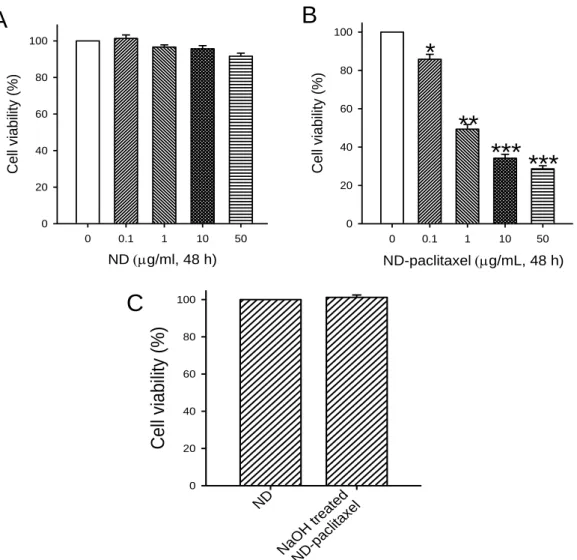
3.1 ND-paclitaxel reduces cell viability in lung carcinoma cells
The cell viability of ND, ND-paclitaxel, and NaOH-treated ND-paclitaxel on the
A549 human lung carcinoma cells were analyzed by MTT assay. Fig. 1A shows that
treatment with 0.1-50 g/mL ND particles for 48 h did not significantly induce the
cytotoxicity of A549 cells. However, ND-paclitaxel for A549 cells cytotoxicity, fig.
1B shows that treatment with ND-paclitaxel for 48 h significantly reduced the cell
viability and in a concentration-dependent manner. To further prove the biological
activity of paclitaxel on ND, ND-paclitaxel was treated with strong alkaline solution
(1 M NaOH), which caused dysfunction of paclitaxel. Indeed, denatured
ND-paclitaxel lost the activity to cause the lung cancer cell death (Fig. 1C).
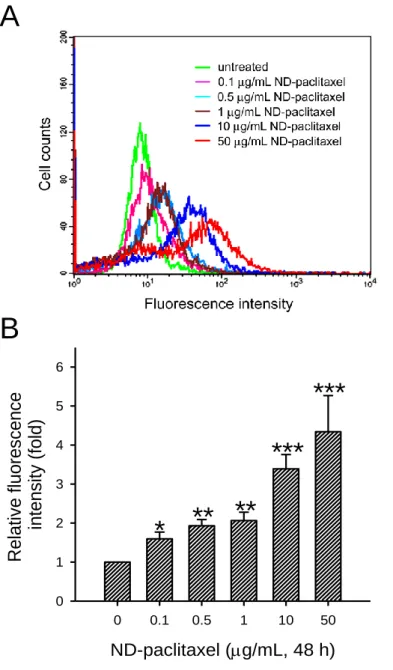
3.2 Uptake ability of ND-paclitaxel in lung carcinoma cells
To analyze the uptake ability of ND-paclitaxel, the cells were analyzed by flow
cytometer. Fig. 2A shows that treatment with ND-paclitaxel (0.1-50 μg/mL for 48 h)
increased the green fluorescence intensities that the spectra were shifted to right in
A549 cells. The quantified fluorescence intensities showed the uptake ability of
ND-paclitaxel via a concentration-dependent manner in A549 cells (Fig. 2B). The
fluorescence intensities were increased to 4-5-folds in A549 cells than untreated
3.3 ND-paclitaxel blocks microtubules to induce abnormal mitotic cells
To examine whether ND-paclitaxel disturbed microtubule dynamics, A549 cells
were treated with ND-paclitaxel and subjected to cytoskeleton and nuclear staining.
The green fluorescence from the ND particles was excited by a wavelength of 488
nm and the emission was collected in the range 510-530 nm, the red color was
indicated the location of microtubules, and blue color was represented the location
of nuclei in A549 cells. Treatment with ND-paclitaxel or paclitaxel markedly
increased the abnormal mitotic cell number (Fig. 3A, stars). Fig. 3B shows that
treatment with ND-paclitaxel or paclitaxel blocked spindle formation and
chromosomes segregation in A549 cells. The disturbance of chromosomes was
elicited by ND-paclitaxel or paclitaxel (Fig. 3B, arrows). In contrast, ND particles
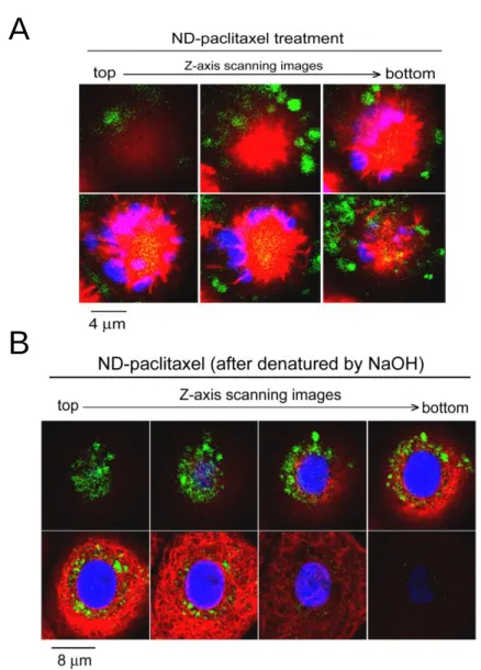
did not induce the aberrant chromosomes in the A549 cells. Fig. 4A shows that
ND-paclitaxel particles were located on the microtubules and blocked the spindle
formation. Nevertheless, NaOH-treated ND-paclitaxel particles did not block
microtubules and located in cytoplasm by dissection of confocal scanning of Z-axis
(Fig. 4B).
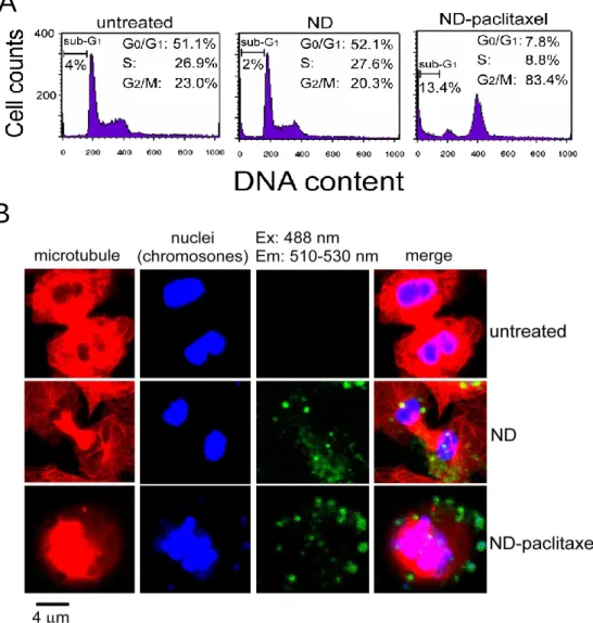
3.4 ND-paclitaxel induces the cell cycle arrest and apoptosis in lung carcinoma cells
To investigate the effect of ND-paclitaxel on the cell cycle progression, A549
index. Comparing with untreated and ND-treated samples, ND-paclitaxel
dramatically decreased the G1/G0 fractions and increased the G2/M fractions in
A549 cells (**p <0.01) (Fig. 5A). The average percentages of G2/M fractions were
elevated at 83.4% after treatment with ND-paclitaxel. In untreated and ND treated
samples, the cells were dividing into two daughter cells during cytokinesis; however,
ND-paclitaxel blocked cell division and arrested in the prophase of mitosis (Fig. 5B).
Furthermore, treatment with 0.1-50 g/mL ND-paclitaxel for 48 h decreased the
mitosis-regulated protein levels of CDC2 and phospho-CDC2 via a
concentration-dependent manner in A549 cells (Fig. 6). Actin protein was as an
internal control protein, which was not altered by ND-paclitaxel. Additionally, the
mitotic index was increased by treated with ND-paclitaxel but not in the untreated,
ND alone, and NaOH-treated ND-paclitaxel samples (Fig. 7A).
ND-paclitaxel also significantly increased the sub-G1 fractions (apoptosis
fractions) at the average value of 13.4% in A549 cells, but the sub-G1 fractions of
untreated or ND alone were at the basal level of 2-4% (p < 0.01) (Fig. 5A). Besides,
we have confirmed and counted the percentage of apoptotic nuclear number by
morphological changes under a fluorescence microscope. Consistently,
ND-paclitaxel significantly elevated ~12% apoptosis in A549 cells; in contrast, the
7B).
3.5 ND-paclitaxel inhibits tumorigenesis of human lung tumor xenograft in SCID
mice
The model of xenograft lung tumor in SCID mice was used to study the effect of
ND-paclitaxel on anti-tumorigenesis. The xenograft tumor was developed in
five-week-old SCID mice. After treatment with vehicle, ND, or ND-paclitaxel in
A549 cells, then cells were collected and mice received 1 × 10⁶ cells by
subcutaneously injected. The visible lung tumors that were separated from
sacrificed xenograft SCID mice (Fig. 8A). The tumors of mice were growth to
average 300–400 mm3 in control and ND groups after inoculation for 70 days.
Moreover, ND-paclitaxel dramatically reduced the tumor size at an average of ~25
mm3 (Fig. 8B). ND alone did not significantly alter the tumorigenesis of A549 cells
in mice during 70 days observation.
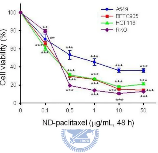
3.6 ND-paclitaxel induces cytotoxicity in various human cancer cells
To examine the effect of ND-paclitaxel in other human cancer cells, the
colorectal (RKO and HCT116) and bladder (BFTC905) cancer cells were exposed
to ND-paclitaxel and analyzed by MTT assay. Fig. 9 shows that treatment with
0.1-50 g/mL ND-paclitaxel for 48 h significantly reduced the cell viability in all
cell death in RKO cells than other cancer cell lines.
3.7 Uptake ability of ND-paclitaxel in colon carcinoma cells
The uptake ability of ND-paclitaxel in RKO cells was examined by flow
cytometer. Treatment with ND-paclitaxel (0.1-1 g/mL for 48 h) elevated the green
fluorescence intensities that the spectra were shifted to right in RKO cells (Fig.
10A). The quantified fluorescence intensities showed the uptake ability of
ND-paclitaxel (Fig. 10B). The fluorescence intensities were significantly increased
by treatment with 0.5-1 μg/mL ND-paclitaxel comparing to untreated cells (Fig.
10B).
3.8 ND-paclitaxel increases sub-G1 and G2/M fractions in colon carcinoma cells
We have examined the effect of ND-paclitaxel on the cell cycle progression and
sub-G1 formation in RKO cells. ND-paclitaxel dramatically decreased the G1/G0
fractions and increased the G2/M fractions in RKO cells (Fig. 11A and 11B).
ND-paclitaxel also significantly increased the sub-G1 fractions (apoptosis fractions) in
RKO cells (Fig. 11A and 11B). Moreover, ND-paclitaxel decreased the protein levels
of CDC2, phospho-CDC2 (Tyr-15 and Thr-161), and cyclin B1 in RKO cells (Fig. 12).
The protein level of phosphor-CDC2 (Thr-14) was slightly seduced by ND-paclitaxel.
Actin was as an internal control, which was not altered by ND-paclitaxel.
To further examine the effect of ND-paclitaxel apoptosis induction, Annexin
V-PI assay was analyzed by flow cytometry. Treatment with 0.5-1 μg/mL
ND-paclitaxel for 48 h increased the early and late apoptosis percentage in RKO
cells (Fig. 13A). Fig. 13B shows quantified results that treatment with
ND-paclitaxel significantly increased the apoptosis. Furthermore, the active forms
of caspase-3 were induced following treatment with 0.5-1 μg/mL ND-paclitaxel (Fig.
14). The protein cleavage of PARP, was significantly increased by ND-paclitaxel
treatment (Fig. 14). We also confirmed the apoptosis formation following
ND-paclitaxel by direct observation of time-lapse cell morphology alteration.
Treatment with 1 g/mL ND-paclitaxel induced apoptosis cells formation (Fig. 15
4. Discussion
Chemical drugs linked covalently with nanoparticles have been developed for
diagnostic and therapeutic applications in recent years. In this study, we created a
novel covalent linkage of ND-paclitaxel. ND-paclitaxel significantly induced the cell
death in a variety of cancer cell types. ND-paclitaxel can be taken into lung and colon
cancer cells in a concentration-dependent manner. More importantly, ND-paclitaxel
exerts its anticancer abilities by inducing mitotic arrest, apoptosis, and anti-tumorigenesis.
However, ND alone or denatured ND-paclitaxel did not induce the damage effects on
cancer cells. The covalent linkage of nanoparticles and drugs provides the advantage
for stabilization to avoid drug dissociation during delivery process. We suggest that
ND is a potential nanomaterial for drug delivery and cancer therapy. Recently, it has
been demonstrated that carbon nanotubes conjugated with paclitaxel exerted drug
delivery for tumor suppression in mice (Liu et al., 2008). Recently, Danhier et. al.
reported that paclitaxel-loaded PEGylated PLGA-based nanoparticles displayed
greater on the inhibition of tumorigenesis by compared with paclitaxel (Danhier et al.,
2009). Moreover, nanoliposomal delivering cancer drugs that have the benefits of
prolonged drug in tissue residence during cancer therapy (Chau et al., 2006;
Koshkima et al., 2003; Noble et al., 2006). Therefore, nanoparticles provide the
The toxicity of nanoparticles is a critical issue of concern for clinical
applications. Several studies showed that ND did not induce cytotoxicity in various
cell types (Liu et al., 2007; Liu et al., 2009; Schrand et al., 2007; Lechleither et al.,
2008; Yu et al., 2005; Chang et al., 2008). It is presented that ND particles are
non-cytotoxic during cellular division and differentiation (Liu et al., 2009). In the
present study, ND particles did not elicit the mitotic blockage and apoptosis in lung
cancer cells. Moreover, it is the first time demonstrating that ND particles did not
influence the tumorigenesis of human lung cancer cells in xenograft SCID mice.
However, ND-paclitaxel is effective to induce the mitotic blockage, apoptosis and
anti-tumorigenesis in cancer cells. Recently, it has been reported that no mice showed
any symptoms of abnormality after intravenous injection of ND particles (Yuan et al.,
2009). We propose that ND is a benign nanomaterial for drug delivery based on its
non-cytotoxicity and bio-compatibility.
It has been shown that ND is detectable by its fluorescence property but without
photobleaching (Chao et al., 2007; Liu et al., 2009; Yu et al., 2005). We have
examined the location and uptake ability of ND-paclitaxel in human carcinoma cells
by confocal microscope and flow cytometer. Using confocal microscopy, we observed
that ND-paclitaxel was taken into cancer cells and located in the microtubules and
clathrin-mediated endocytosis and macropinocytosis (Liu et al., 2009; Faklaris et al.,
2008). Interestingly, we found that ND-paclitaxel was uptake into cancer cells in a
concentration-dependent manner by flow cytometer analysis. The uptake ability of
ND-paclitaxel is correlated to the anticancer activity of paclitaxel on inducing cell
death and mitotic blockage in lung cancer cells. ND-paclitaxel has an ester bond
between ND and paclitaxel that can be hydrolyzed by esterases. The ester linkage of
ND-paclitaxel may be cleaved by esterases of cancer cells for releasing paclitaxel to
execute microtubule inhibition and apoptosis. As a whole, the visualization, uptake
ability, and paclitaxel released of ND-paclitaxel contain the advantages for cancer
drug delivery.
Paclitaxel induced apoptosis pathway through multiple mechanisms (Wang et al.,
2000). Moreover, paclitaxel initiated apoptosis has been associated with CDC2 and
other CDKs (Donaldson et al., 1994; Meikrantz et al., 1996). In this study, we found
that the protein levels of CDC2 and phosphorylated CDC2 (Tyr-15 and Thr161) were
decreased by treatment with ND-paclitaxel. We also observed that ND-paclitaxel
increased caspase-3 activation and the cleavage of PARP indicating ND-paclitaxel can
induce apoptosis pathway in cancer cells. Accordingly, ND-paclitaxel still reserves
5. Conclusion
In summary, we have provided a novel covalent linkage of ND and paclitaxel.
More importantly, the covalent bonding of ND-paclitaxel still preserves its anticancer
activities on the mitotic blockage, apoptosis induction and anti-tumorigenesis in human
lung and colon carcinoma cells. A functional ND-paclitaxel conjugate is potential for
6. Reference
Akerman, M.E., Chan, W.C., Laakkonen, P., Bhatia, S.N., and Ruoslahti, E. (2002).
Nanocrystal targeting in vivo. Proc Natl Acad Sci U S A 99, 12617-12621.
Alexis, F., Pridgen, E.M., Langer, R., and Farokhzad, O.C. (2010). Nanoparticle
technologies for cancer therapy. Handb Exp Pharmacol, 197, 55-86.
Batist, G., Ramakrishnan, G., Rao, C.S., Chandrasekharan, A., Gutheil, J., Guthrie, T.,
Shah, P., Khojasteh, A., Nair, M.K., Hoelzer, K., Tkaczuk, K., Park, Y.C., and
Lee, L.W. (2001). Reduced cardiotoxicity and preserved antitumor efficacy of
liposome-encapsulated doxorubicin and cyclophosphamide compared with
conventional doxorubicin and cyclophosphamide in a randomized, multicenter
trial of metastatic breast cancer. J Clin Oncol 19, 1444-1454.
Chang, I.P., Hwang, K.C., and Chiang, C.S. (2008). Preparation of fluorescent
magnetic nanodiamonds and cellular imaging. J Am Chem Soc 130,
15476-15481.
Chou, H.H., Wang, K.L., Chen, C.A., Wei, L.H., Lai, C.H., Hsieh, C.Y., Yang, Y.C.,
Twu, N.F., Chang, T.C., and Yen, M.S. (2006). Pegylated liposomal doxorubicin
(Lipo-Dox) for platinum-resistant or refractory epithelial ovarian carcinoma: a
Taiwanese gynecologic oncology group study with long-term follow-up.
Chao, J.I., Perevedentseva, E., Chung, P.H., Liu, K.K., Cheng, C.Y., Chang, C.C., and
Cheng, C.L. (2007). Nanometer-sized diamond particle as a probe for
biolabeling. Biophys J 93, 2199-2208.
Cheng, C.L., Chen, C.F., Shaio, W.C., Tsai, D.S., and Chen, K.H. (2005). The CH
stretching features on diamonds of different origins. Diamond Relat Mater 14,
1455–1462.
Cheng, C.Y., Perevedentseva, E., Tu, J.S., Chung, P.H., Chenga, C.L., Liu, K.K., Chao,
J.I., Chen, P.H., and Chang, C.C. (2007) Direct and in vitro observation of
growth hormone receptor molecules in A549 human lung epithelial cells by
nanodiamond labeling. Appl Phy Lett 90, 163903
Danhier, F., Lecouturier, N., Vroman, B., Jerome, C., Marchand-Brynaert, J., Feron,
O., and Preat, V. (2009). Paclitaxel-loaded PEGylated PLGA-based
nanoparticles: in vitro and in vivo evaluation. J Control Release 133, 11-7.
Department of health, Executive Yuan, Taiwan (2009). www.doh.gov.tw
Donaldson, K.L., Goolsby, G.L., Kiener, P.A., and Wahl, A.F. (1994). Activation of
p34cdc2 coincident with taxol-induced apoptosis. Cell Growth Differ 5,
1041-1050.
Faklaris, O., Garrot, D., Joshi, V., Druon, F., Boudou, J.P., Sauvage, T., Georges, P.,
diamond nanoparticles in cells and study of the internalization pathway. Small 4,
2236-9.
Gao, X., Cui, Y., Levenson, R.M., Chung, L.W., and Nie, S. (2004). In vivo cancer
targeting and imaging with semiconductor quantum dots. Nat Biotechnol 22,
969-976.
Huang, H., Pierstorff, E., Osawa, E., and Ho, D. (2007). Active nanodiamond
hydrogels for chemotherapeutic delivery. Nano Lett 7, 3305-3314.
Huang, L.C., and Chang, H.C. (2004). Adsorption and immobilization of cytochrome
c on nanodiamonds. Langmuir 20, 5879-5884.
Kang, J.H., Asami, Y., Murata, M., Kitazaki, H., Sadanaga, N., Tokunaga, E., Shiotani,
S., Okada, S., Maehara, Y., Niidome, T., Hashizume, M., Mori, T., and
Katayama, Y. (2010). Gold nanoparticle-based colorimetric assay for cancer
diagnosis. Biosens Bioelectron 25, 1869-1874.
Koshkina, N.V., Waldrep, J.C., and Knight, V. (2003). Camptothecins and lung cancer:
improved delivery systems by aerosol. Curr Cancer Drug Targets 3, 251-64.
Krueger, A. (2008). New Carbon Materials: Biological Applications of Functionalized
Nanodiamond Materials. Chem Eur J 14, 1382-1390
Krueger, A., Stegk, J., Liang, Y., Lu, L., and Jarre, G. (2008). Biotinylated
Langmuir 24, 4200-4.
Lechleitner, T., Klauser, F., Seppi, T., Lechner, J., Jennings, P., Perco, P., Mayer, B.,
Steinmuller-Nethl, D., Preiner, J., Hinterdorfer, P., Hermann, M., Bertel, E.,
Pfaller, K., and Pfaller, W. (2008). The surface properties of nanocrystalline
diamond and nanoparticulate diamond powder and their suitability as cell
growth support surfaces. Biomaterials 29, 4275-4284.
Liu, K.K., Cheng, C.L., Chang, C.C., and Chao, J.I. (2007). Biocompatible and
detectable carboxylated nanodiamond on human cell. Nanotechnology 18,
325102.
Liu, K.K., Wang, C.C., Cheng, C.L., and Chao, J.I. (2009). Endocytic carboxylated
nanodiamond for the labeling and tracking of cell division and differentiation in
cancer and stem cells. Biomaterials 30, 4249-4259.
Liu, Z., Chen, K., Davis, C., Sherlock, S., Cao, Q., Chen, X., and Dai, H. (2008).
Drug delivery with carbon nanotubes for in vivo cancer treatment. Cancer Res
68, 6652-6660.
Liu, K.K., Chen, M.F., Chen, P.Y., Lee, T.J.F., Cheng, C.L., Chang, C.C., Ho, Y.P., and
Chao, J.I. (2008). Alpha-bungarotoxin binding to target cell in a developing
visual system by carboxylated nanodiamond. Nanotechnology 19, 205102
Meikrantz, W., and Schlegel, R. (1996). Suppression of apoptosis by dominant
negative mutants of cyclin-dependent protein kinases. J Biol Chem 271,
10205-10209.
Michalet, X., Pinaud, F.F., Bentolila, L.A., Tsay, J.M., Doose, S., Li, J.J., Sundaresan,
G., Wu, A.M., Gambhir, S.S., and Weiss, S. (2005). Quantum dots for live cells,
in vivo imaging, and diagnostics. Science 307, 538-44.
Nel, A., Xia, T., Madler, L., and Li, N. (2006). Toxic potential of materials at the
nanolevel. Science 311, 622-627.
Noble, C.O., Krauze, M.T., Drummond, D.C., Yamashita, Y., Saito, R., Berger, M.S.,
Kirpotin, D.B., Bankiewicz, K.S., and Park, J.W. (2006). Novel nanoliposomal
CPT-11 infused by convection-enhanced delivery in intracranial tumors:
pharmacology and efficacy. Cancer Res 66, 2801-6.
Schrand, A.M., Huang, H., Carlson, C., Schlager, J.J., Omacr Sawa, E., Hussain, S.M.,
and Dai, L. (2007). Are Diamond Nanoparticles Cytotoxic? J Phys Chem B
Condens Matter Mater Surf Interfaces Biophys 111, 2-7.
Service, R.F. (2004). Nanotoxicology. Nanotechnology grows up. Science 304,
1732-1734.
Shimkunas, R.A., Robinson, E., Lam, R., Lu, S., Xu, X., Zhang, X.Q., Huang, H.,
pH-dependent protein delivery vehicles. Biomaterials 30, 5720-8.
Tada, H., Higuchi, H., Wanatabe, T.M., and Ohuchi, N. (2007). In vivo real-time
tracking of single quantum dots conjugated with monoclonal anti-HER2
antibody in tumors of mice. Cancer Res 67, 1138-1144.
Ushizawa, K., Sato, Y., Mitsumori, T., Machinami, T., Ueda, T., and Ando, T. (2002).
Covalent immobilization of DNA on diamond and its verification by diffuse
reflectance infrared spectroscopy. Chem Phys Lett 351 105-8.
Wang, T., Lv, J.H., Zhang, X.F., Li, C.J., Han, X., and Sun, Y.J. (2010). Tissue
inhibitor of metalloproteinase-1 protects MCF-7 breast cancer cells from
paclitaxel-induced apoptosis by decreasing the stability of cyclin B1. Int J
Cancer 126, 362-370.
Wang, T.H., Wang, H.S., and Soong, Y.K. (2000). Paclitaxel-induced cell death: where
the cell cycle and apoptosis come together. Cancer 88, 2619-2628.
Wang, X., Li, J., Wang, Y., Cho, K.J., Kim, G., Gjyrezi, A., Koenig, L., Giannakakou,
P., Shin, H.J., Tighiouart, M., Nie, S., Chen, Z., Shin, D.M. (2009). HFT-T, a
targeting nanoparticle, enhances specific delivery of paclitaxel to folate
receptor-positive tumors. ACS Nano 3, 3165-3174.
Yang, W., Auciello, O., Butler, J.E., Cai, W., Carlisle, J.A., Gerbi, J.E., Gruen, D.M.,
(2002). DNA-modified nanocrystalline diamond thin-films as stable,
biologically active substrates. Nat Mater 1, 253-257.
Yu, S.J., Kang, M.W., Chang, H.C., Chen, K.M., and Yu, Y.C. (2005). Bright
fluorescent nanodiamonds: no photobleaching and low cytotoxicity. J Am Chem
Soc 127, 17604-17605.
Yuan, Y., Chen, Y., Liu, J.H., Wang, H., and Liu, Y. (2009). Biodistribution and fate of
nanodiamonds in vivo. Diam Relat Mater 18, 95-100.
Zhang, B., Li, Y., Fang, C.Y., Chang, C.C., Chen, C.S., Chen, Y.Y., and Chang, H.C.
(2009). Receptor-mediated cellular uptake of folate-conjugated fluorescent
nanodiamonds: a combined ensemble and single-particle study. Small 5,
ND ND (CH2O(CH2)6-O-THP )x (CO2H)x ND ND (CH2O(CH2)6-OMs)x OH O N H O O ND O OH AcO O AcO O O Ph NH O O Ph OCOP h H OH O HO O ND O N H O O OH AcO O AcO O O Ph NH O O Ph OCOP h H OH O ND (CH2OH)x ND (CH2O(CH2)6-OH)x ND (CH2O(CH2)6-NH2)x x (1) (2) (3) (4) (5) (6) (7) (9): R = H (d-9): R = D a b c d e f g h R R R R R R R R R R R R (10): R = H (d-10): R = D (8): R = H (d-8): R = D
Scheme 1. Chemical synthesis of ND-paclitaxel. The chemical products are indicated by the numbers. The succession of chemical synthetic processes is the following: (a) HCl/HNO3 (3:1), rt, 3 d; 1M NaOH, 90 ºC, 2 h; 1M HCl, 90 ºC, 2 h; (b) LiAlH4, THF, reflux 24 h; 6M NaOH, 90 ºC, overnight; (c) NaH, THF, THP-O-(CH2)5CH2Cl; (d) p-TsOH, MeOH/H2O (3:1), rt, overnight; (e) MsCl, Et3N, THF, 0 ºC, 1 h, rt, overnight; (f) NH4OH(aq), reflux, 24 h; (g) 8 (paclitaxel-2’-succinate), EEDQ, Et3N, CH2Cl2, rt, 3 h; (h) 1M NaOH, rt, overnight.
Figure 1. Effect of ND-paclitaxel on the cell viability in human lung carcinoma cells. (A) A549 cells were treated with or without ND (0.1-50 μg/mL for 48 h). (B) A549 cells were treated with or without ND-paclitaxel (0.1-50 μg/mL for 48 h). At the end of treatment, the cell viability was measured by MTT assay. Results were obtained from four-twelve separate experiments and the bar represents mean ± S.E. *p < 0.05, **
p < 0.01, and ***p < 0.001 indicate significant difference between control and ND-paclitaxel treated samples. (C) A549 cells were treated with 50 μg/mL ND particles or NaOH-treated ND-paclitaxel for 48 h. The cell viability was measured by MTT assay. Results were obtained from eight experiments and the bar represents mean ± S.E. C el l via bi lity ( %) 0 20 40 60 80 100 ND NaOH t reated ND-pacli taxel ND-paclitaxel g/mL, 48 h) 0 0.1 1 10 50 Cell viabili ty (%) 0 20 40 60 80 100
**
*
***
***
ND g/ml, 48 h) 0 0.1 1 10 50 Cell viabili ty (%) 0 20 40 60 80 100C
A
B
Figure 2. Uptake ability of ND-paclitaxel in human lung carcinoma cells. (A) A549 cells were treated with 0-50 μg/mL ND-paclitaxel for 48 h. At the end of treatment, the cells were trypsinized and then subjected to flow cytometer. (B) The fluorescence intensities were quantified by a CellQuest software of flow cytometer. Results were obtained from four separate experiments and the bar represents the mean ± S.E. *p < 0.05, **p < 0.01, and ***p < 0.001 indicate significant difference between untreated and ND-paclitaxel samples.
ND-paclitaxel (g/mL, 48 h) 0 0.1 0.5 1 10 50 R e la tiv e flu o re sc e n ce in ten si ty (fo ld ) 0 1 2 3 4 5 6
*
** **
***
***
B
A
Figure 3. Effect of ND-paclitaxel on the blockage of microtubule and chromosome segregation in human lung carcinoma cells. (A) A549 cells were treated with ND (50
g/mL), ND-paclitaxel (50 g/mL), or paclitaxel (50 nM) for 24 h. The microtubule were stained with Cy3-labeled mouse anti--tubulin. The nuclei were stained with Hoechst 33258. Microtubules and nuclei were exhibited red and blue color, respectively. The green fluorescence from ND particles was excited by a wavelength of 488 nm and the emission was collected in the range 510-530 nm by using confocal microscope. The stars indicate that the cell morphology (mitotic round-up) was affected by paclitaxel or ND-paclitaxel compared to untreated or ND treated samples. (B) The pictures are amplified from (A). The arrows indicate the derangement of chromosomes after treatment with paclitaxel or ND-paclitaxel.
B
A
Figure 4. Location and distribution of ND-paclitaxel in human lung carcinoma cells. (A) An A549 cell from ND-paclitaxel (50 g/mL for 24 h) treatment was visualized by examining Z-axis scanning images using a confocal microscope. The distribution of ND-paclitaxel interacting with microtubules was observed by scanning in the vertical direction from top to bottom. Microtubule and nuclei exhibited red and blue color, respectively. The green color indicates the location of ND particles. The yellow color indicated that ND-paclitaxel particles were co-localized with microtubules. (B) An A549 cell from NaOH-treated ND-paclitaxel (50 g/mL for 24 h) treatment was visualized by examining Z-axis scanning images of a confocal microscope.
B
A
Figure 5. Effect of ND-paclitaxel on the cell cycle progression in human lung carcinoma cells. A549 cells were treated with 50 g/mL ND or ND-paclitaxel for 24 h. (A) At the end of treatment, the A549 cells were trypsinized and then subjected to flow cytometry analyses. These data of each cell cycle phases and sub-G1 fractions were represented the average values from three separate experiments. (B) The
-tubulin and nuclei were stained with the Cy3-labeled mouse anti--tubulin and Hoechst 33258, respectively. Microtubules and nuclei were exhibited red and blue color, respectively. The green fluorescence from ND particles was excited by a wavelength of 488 nm and the emission was collected in the range 510-530 nm by using confocal microscope. The untreated and ND-treated cells were separating to two daughter cells during cytokinesis. The ND-paclitaxel-treated cell was arrested in the prophase of mitosis.
B
A
Figure 6. The effect of ND-paclitaxel on the protein levels of phospho-CDC2 and total
CDC2 in human lung carcinoma cells. A549 cells were treated with or without
ND-paclitaxel (0.1-50 μg/mL for 48 h). The total protein extracts were subjected to
Western blot analysis by using anti- phospho-CDC2, anti-CDC2, and actin antibodies.
Representative Western blot results were shown from one of three separate
Figure 7. Effect of ND-paclitaxel on the percentages of mitotic index and apoptosis in human lung carcinoma cells. A549 cells were treated with 50 g/mL ND, ND-paclitaxel, or NaOH-treated ND-paclitaxel for 24 h. (A) After the end of treatment, the -tubulin and nuclei were stained with the Cy3-labeled mouse anti--tubulin and Hoechst 33258, respectively. Mitotic index (the percentage of mitotic cell number/total cell number) was counted under a fluorescence microscope. (B) The percentage of apoptosis was counted by apoptotic nuclei. Results were obtained from three separate experiments and the bar represents the mean ± S.E. **p < 0.01 and ***p < 0.001 indicate significant difference between the controls and
ND -pac litaxel Mit otic index ( %) 0 5 10 15 20 25 30 35 40 ND NaO H treated ND -pac litaxel untr eated
***
ND -pac litaxel Apopto sis ( %) 0 2 4 6 8 10 12 14 16 18 ND NaO H treated ND -pac litaxel untr eated**
B
A
Figure 8. The effect of pretreated ND and ND-paclitaxel on tumor growth in xenograft SCID mice model. (A) A549 cells were treated with ND or ND-paclitaxel (10 mg/mL for 48 h). Then the SCID mice were subcutaneously injected with 1×10⁶ cells. The visible lung tumors were separation from sacrificed xenograft mice after inoculation for 70 days. (B) The tumor volume in SCID mice were measured every four days until 70 days. The bar represents the mean ± S.E. **p < 0.01 indicates significant difference between the controls and ND-paclitaxel.
Days (after cancer cell inoculation)
0 5 10 15 20 25 30 35 40 45 50 55 60 65 70 Tu mo r vo lume (mm 3) 0 50 100 150 200 250 300 350 400 450 500 Vehicle (n=5) ND (n=5) ND-paclitaxel (n=5)
**
B
Tumors
A
Figure 9. Effect of ND-paclitaxel on the cell viability in various human cancer cells. A variety of cancer cell lines including lung (A549), colorectal (RKO and HCT116), and bladder (BFTC 905) cancer cells were treated with or without ND-paclitaxel (0.1–50 μg/mL for 48 h). The cell viability was measured by MTT assay. The results were from 4-8 separated experiments. The bar represents mean ± S.E. **p < 0.01, and ***p < 0.001 indicate significant difference between control and ND-paclitaxel treated sample.
Figure 10. Uptake ability of ND-paclitaxel in human colon cancer cells. (A) RKO cells were treated with 0-1 μg/mL ND-paclitaxel for 48 h. At the end of treatment, the cells were harvested and subjected to flow cytometer. (B) The fluorescence intensities of ND-paclitaxel in RKO cells were quantified by CellQuest software of flow cytometer. Results were obtained from three separate experiments and the bar represents the mean ± S.E. *p < 0.05 indicate significant difference between untreated
Cell co u n ts Fluorescence intensity untreated 0.1 μg/mL ND-paclitaxel 0.25 μg/mL ND-paclitaxel 0.5 μg/mL ND-paclitaxel 1 μg/mL ND-paclitaxel
B
A
A
B
Figure 11. Effect of ND-paclitaxel on cell cycle progression in human colon cancer
cells. (A) RKO cells were treated with or without ND-paclitaxel (0.1-1 g/mL for 48
h). At the end of treatment, the RKO cells were trypsinized and then subjected to flow
cytometry analyses. (B) The each cell cycle phases were quantified by ModFit LT
software of flow cytometer. Results were obtained from three experiments and the bar
represents mean ± S.E. *p < 0.05, **p < 0.01, and ***p < 0.001 indicate significant
difference between control and ND-paclitaxel treated samples.
0 0.1 0.25 0.5 1
RKO cells
Cell cou nt s DNA content ND-paclitaxel (μg/mL, 48 h) ND-paclitaxel (g/mL, 48 h) 0 0.1 0.25 0.5 1 Sub-G1 (%) 0 20 40 60 80 ** * *** ND-paclitaxel (g/mL, 48 h) 0 0.1 0.25 0.5 1 G1 p hase (%) 0 10 20 30 40 50 *** ND-paclitaxel (g/mL, 48 h) 0 0.1 0.25 0.5 1 S pha se (%) 0 10 20 30 40 * * ND-paclitaxel (g/mL, 48 h) 0 0.1 0.25 0.5 1 G2/M ph ase (%) 0 10 20 30 40 50 * ***Figure 12. Effect of ND-paclitaxel on the protein levels of phosphorylated CDC2, total CDC2 and cyclin B1 in human colon cancer cells. RKO cells were treated with or without ND-paclitaxel (0.1-1 μg/mL for 48 h), total protein extracts were prepared for Western blot analysis using specific anti-phospho-CDC2 (Thr-14, Tyr-15, and Thr-161), anti-CDC2, anti-cyclin B1 and actin antibodies. Western blot data were shown from one of three separate experiments with similar findings.
ND-paclitaxel
(μg/mL, 48 h)
phospho-
CDC2 (Tyr-15)
phospho-
CDC2 (Thr-14)
CDC2
cyclin B1
Actin
0
0.1
0.25
0.5
1
phospho-
CDC2 (
Thr-161)
ND-paclitaxel (g/mL, 48 h) 0 0.1 0.25 0.5 1 Percen tage of apo ptosis (%) 0 20 40 60
**
**
Figure 13. Effect of ND-paclitaxel for apoptosis in the RKO cells. (A) RKO cells were treated with or without 0.1-1 g/mL ND-paclitaxel for 48 h. At the end of treatment, the A549 cells were trypsinized and then the cells were analyzed by Annexin V-FITC assay. The percentages of the cell forms mentioned below were calculated by the CellQuest software (mean values are given). (B) The early and late apoptosis cells were quantified by CellQuest software of flow cytometer. Results were obtained from three experiments and the bar represents the mean ± S.E. **p < 0.01 indicate significant difference between untreated and ND-paclitaxel samples.B
A
P
I
0.6% 2.4% 95.2% 1.8% 1.1% 2.5% 94.6% 2% 2% 7.8% 84.8% 5.28% 2.4% 31.3% 39.3% 27.8% 3.2% 33.2% 33.7% 29.9%Annexin V-FITC
ND-paclitaxel (μg/mL, 48 h) 0 0.1 0.25 0.5 1Figure 14. Effect of ND-paclitaxel on the active of caspase-3 and cleavage of
poly(ADP-ribose) polymerase (PARP) in human colon cancer cells. Induce of
caspase-3 was activated and PARP cleavage by ND-paclitaxel at the indicated
concentrations after 48 h of treatment.
PARP
Actin
ND-paclitaxel
(μg/mL, 48 h)
cleaved PARP
0 0.1 0.25 0.5 1pro-caspase-3
active caspase-3
Figure 15. Time-lapse observation of apoptotic formation following treatment with
ND-paclitaxel. RKO cells were treated with ND-paclitaxel 1 g/mL. The cell were
immediately observed by live-cell imaging microscope for 0-24 h. The arrows
indicate the apoptotic cells by ND-paclitaxel treatment.
0 h 4 h 8 h 12 h 24 h
ND-paclitaxel 1 μg/mL