1
Parasitic Diseases as an Index of Environmental Changes in
Twentieth-Century Taiwan: A Preliminary Study
This paper was originally delivered at the Second International conference on Environmental History, held on November 8-10, 2006, organized by the Institute of Taiwan History, Academia Sinica.
Ts’ui-jung Liu*, Shi-yung Liu**, and Ya-wen Ku***
Introduction
In the early twentieth century, parasitic infection rate among Taiwanese was rather high. For instance, a health examination conducted during 1921-1929 with a sample of 77,011 males and females from 32 localities in 8 administrative areas found that parasitic infection rate was 78.3%. Among various parasites, the infection rate of Ascaris lumbricoides (abbr. Al) was 54.4%, Trichuris trichiura (Tt) 29.3%, and Ancylostoma duodenale (Ad) 14.4%. These rates were quite close to those found in
Japan around the same time.1 As for the cause of death, the statistics during
1906-1942 showed that in addition to malaria induced by plasmodium, two entries related to parasites were specified as “diseases caused by parasitic protozoa and helminth” and “other infectious and parasitic diseases”; and the figures of these two entries are either separated or combined. If combined together, the death rate was
lowest at 0.4% in 1909 and highest at 6.4% in 1941.2 As for malaria induced by
plasmodium, it was the most important cause of death among Taiwanese before 1911 and in 1915-1916; the death rate during 1906-1916 was around 10% and mostly
above 5% during 1917-1929, once fell to 3% in 1934 but returned to 5.35% in 1942.3
In 2000, according to the health statistics of the Department of Health, Executive Yuan, classified by international standard of the cause of death, the total number of death was 124,481 and those died of “infectious and parasitic diseases” were 3,016, or 2.4%. Under this major entry, details related to parasitic diseases were as follows: 1 amoebiasis, 1 malaria, 3 helminthiases, 1 other trematode infections, 1 ancylostomiasis and necatoriasis, and 1 other and unspecified helminthiases; adding
*Distinguished Research Fellow, Institute of Taiwan History, Academia Sinica.. **Assistant Research Fellow, Institute of Taiwan History, Academia Sinica.
***Post-doctoral Fellow, Research Center for Humanities and Social Sciences, Academia Sinica.
1
Health Division of the Bureau of Police Affairs, Taiwan Government-general (comp.), 1931, The
Tenth Report of Health Examination: The Second Part of Field Examination on Diseases (Taipei:
Taiwan Sotokufu), pp. 55-56. Also see Liu Ts’ui-jung and Liu Shi-yung, 1999, “Disease and Mortality in the History of Taiwan”, Taiwan Historical Research, 4:2, p. 114. For a list of parasites mentioned in this paper, see Appendix 1.
2
The Statistic Department of the Head Administrator’s Office in Taiwan (comp.), 1946, Summaries of
statistics in Taiwan during the past 51 years (Taipei: Head Administrator’s Office in Taiwan). The
percentages are calculated with statistics in Table 91.
3
2
up to 8 persons died of these causes. When 1 died of “other and unspecified infectious and parasitic diseases” and 204 died of “late effects of infectious and parasitic diseases” were added again, there were altogether 213 deaths and the rate was 0.17%.4 Although it is difficult to separate precisely the shares of infectious and parasitic diseases with existing statistics, the above rates at least suggest that the share of parasitic disease could be in decline during the twentieth century. In other words, changes in parasitic infections can be used as an index for investigating environmental change in Taiwan during the twentieth century.
Parasites in human body are classified into three main categories: protozoa, helminth (including trematoda, cestoidea, nematoda, gordiacea, acanthocephala, and hirudinea), as well as insect. Parasitic infections are related to food, residence, custom and migration of mankind. This is not only testified in individuals but also in groups. Geographical distribution of parasites relates to various factors. The way of life and the custom of human beings may confine living conditions for the host. From the perspective of parasites, those living with a complicated life cycle will have more limitations than those with a simple one. In the course of its growth, a parasite may require a change of host and needs to have other animals than humans to serve as its host, and thus limitations are even more obvious. In other words, in the case that a parasite requires a certain host or disseminator, it can only survive at places where there are this species. On the contrary, those do not need to change the host will survive according to human conditions and migrations. However, the growth of parasites is also related to the environment and climate. Perfect condition for a single host is not a sufficient condition for the survival of parasites. With a prerequisite of sufficient humidity, places and seasons with high temperature are more suitable for the growth of parasites. In the tropical area, parasites have more varieties and spread more widely, particularly in the summer. In addition to the environment, it is obvious that the survival of parasites is also affected by animals serving as hosts and disseminators.5
This paper aims to provide a review of literature in two periods: the Japanese colonial period (1895-1945) and the period after Taiwan restoration in 1945. The discussion will be focused on the infection of helminthes with some mentions of protozoa. Among protozoa, this paper will not particularly deal with malaria as there are already some studies in recent years.6
4
Department of Health, Executive Yuan, Health and National Insurance Annual Statistics Information Service, http://www.doh.gov.tw/statistic/data/生命統計/89/08.XLS, Table 8 and 8.3-8.5.
5
Yokogawa Sadamu and Morishita Kaoru, 1931, Human Parasitology, Vol. 1 (Tokyo: Tohodo), p. 5, pp. 66-70;Yokogawa Sadamu and Morishita Kaoru, 1943, The Newest Summary of Human
Parasitology (Tokyo: Tohodo, second edition), pp. 1-5. 6
For example see, Fann Yann-chiou, 1996, “Medicine and Colonial Expansion: Taiwan’s Malaria Research under Japanese Rule,” New History, 7:3, pp. 133-173. Ku Ya-wen, 2004, “Anti-parasite vs.
3 I. Japanese Colonial Period (1895-1945)
Reports and essays published in the Journal of the Formosan Medical Association (hereafter JFMA) demonstrate that during the Japanese colonial period many examinations of parasitic infections were conducted among patients, prisoners, soldiers, students of primary, middle and vocational schools, inhabitants in general, and aborigines. As the scale and representation of these examinations are different and may imply certain limitations, it is thus appropriate to illustrate these results in separate classifications.
1. Patients of Hospitals
The first report regarding parasitic infections among patients was presented in 1908 by Wang Chen-chien concerning the results of stool examinations conducted from January to December 1907 among 1,648 patients, of them 1,360 males and 288 females, at the Red Cross Taiwan Branch Hospital. The age of these patients ranged from under 1 year old to 65 years old. It was found that 151 males (or 11.1% of males examined) and 1 female suffered from ancylostomiasis. Of those males suffered from this disease, 134 (88.7%) were farmers.7
In 1912, Hou Chi-fa and Li Chin-fu presented another report with statistics compiled from the record of internal medicine of the Red Cross Taiwan Branch
Hospital in 1906-1911.8 Table 1 showed that during 1906-1911, the Red Cross
Taiwan Branch Hospital had 1,587 patients suffered from ancylostomiasis; they counted for 8.8% of the total number of patients in the internal medicine, and 19.4% for those suffered with nutrition deficiency diseases. During these 6 years, the highest infection rate of Ad was found in 1919, it was 19.7% among patients of the internal medicine and 32.1% among patients of nutrition deficiency diseases.
Of the 1,587 patients, there were 1,516 Taiwanese and 71 Japanese; and among Taiwanese, there were 1,140 (75.2%) farmers and 118 (7.8%) laborers. These patients came mostly from the Taipei area, including 506 from Paichieh Pao 擺接堡, 345 from Tachiana Pao 大加蚋堡, 282 from Wenshan Pao 文山堡, 120 from Hsingchih Pao 興直堡, 48 from Palifen Pao 八里坌堡, 54 from Chihlanyi Pao 芝蘭一堡, 51
Anti-mosquito-Anti-malaria Program in Colonial Taiwan,” Taiwan Historical Research, 11:2, pp. 185-222. Ku Ya-wen, 2005a, “The development and the epidemic of malaria in Taiwan during Japanese colonial period-the ‘bad environment’ built up,” Shakai keizai shigaku (Social Economic History), 70: 5, pp. 67-89. Ku Ya-wen, 2005b, The transition of malaria epidemic and its control
policy in Taiwan (Yokohama: Ph.D. dissertation of division of international social sciences,
Yokohama University, 2005/09). Liu Shi-yung, 2001, “GIS, Malaria and Highland Environment in Colonial Taiwan,” in Proceedings of the International Workshop on Colonial Medicine, (2001/10/25-26), Institute of Taiwan History, Academia Sinica.
7
Wang Chen-chien, 1908, “On Ancylostomiasis in Taiwan,” JFMA, No. 69, pp. 289-308.
8
Hou Chi-fa and Li Chin-fu, 1912, “Some statistics concerning Ancylostomiasis around Taipei,”
4
from Chihlaner Pao 芝蘭二堡, 9 from Chihlansan Pao 芝蘭三堡, 39 from Shihting Pao 石碇堡, 18 from Keelung Pao 基隆堡, and 7 from Chinpaoli Pao 金包里堡 (see Map 1).
Table 1: Infections of Ancylostoma duodenale (Ad) among Patients at the Red Cross Taiwan Branch Hospital, 1906-1911
Year Total No. Patients of Internal Medicine (1) No. Patients of Nutrition Deficiency Diseases (2)
No. Infected with Ad
Percentage 1 (3)/(1) Percentage 2 (3)/(2) Japanese Taiwanese Total
(3) 1906 2,912 1,085 2 61 63 2.16 5.81 1907 2,919 1,154 4 172 176 6.03 15.25 1908 3,709 1,166 4 259 263 7.09 22.56 1909 2,184 1,344 21 410 431 19.73 32.07 1910 2,583 1,499 17 301 318 12.31 21.21 1911 3,674 1,925 23 313 336 9.15 17.45 Total* 17,981 8,173 71 1,516 1,587 8.83 19.42 * Total is calculated with the figures listed in the table; the original figures showed the total of col. (1)
as 20,117 and col. (2) as 8,902;percentage 1 as 7.8% and percentage 2 as 17.6%, the Japanese as 72 and Taiwanese as 1,515。
Source: Hou Chi-fa and Li Chin-fu, 1912, pp. 358-359, Tables 1 and 2.
Map 1: Distribution of Patients Infected with Hookworm at the Red Cross Taiwan Branch Hospital, Taipei Area, 1906-1911
5
In addition, there were 92 from Taoyuan Ting 桃園廳, 6 from Hsinchu Ting 新 竹廳, 6 from Taichung Ting 臺中廳, 2 from Chiayi Ting 嘉義廳, 1 from Taitung Ting 臺東廳 and 1 from Yilan Ting 宜蘭廳. In general, there were more patients came from places nearby Taipei city and the number declined with the distance. However, because the majority of the patients suffered from ancylostomiasis were farmers and those living far away would not come to the hospital unless they were ill seriously, therefore, the above statistics might not reflect the true situation of geographical distribution of hookworm infection.9
Moreover, this report also pointed out that, together with ancylostomiasis, there were 609 patients suffered from ascariasis, 77 chronic splenomegalia, 48 gastrointestinal catarrh, 14 bronchial catarrh, 10 malaria, 7 enterobiasis, 4 beriberi, 3 pleurisy, 2 epilepsy, and 1 each paragonimiasis, cirrhosis of liver, tuberculosis, and nephritis. It was pointed out that as quite a number of patients suffered both from chronic splenomegalia and ancylostomiasis, there was a coexistence of malaria and ancylostomiasis in rural area of Taiwan.10
There are three reports concerning patients of Taichung 臺 中 Hospital established by the Taiwan Government-general. In 1915, Ooi conducted stool examinations with in-patients, of them 41 Taiwanese and 80 Japanese. The Taiwanese patients came from Taichung Ting and Nantou Ting; among them 31 (75.6%) were from Taichung, 2 each from Hulutun 葫蘆墩 and Nantou 南投, and 1 each from Changhua 彰化, Shalu 沙轆, Erlin 二林, Peitou 北斗, Lukang 鹿港, and Wushe
霧社. Their families were mostly belonging to middle class and above.11
In Ooi’s 1919 report, there were results of examinations with 120 Taiwanese in-patients and out-patients after 1916.12 Furuichi’s report in 1919 showed the result of examinations with in-patients, of them 130 Japanese and 44 Taiwanese, but it was not clear where did these Taiwanese patients come from.13
For the patients of Taipei Hospital, Yamasaki Shigeru presented a report in 1925 concerning the result of examinations from October 6, 1924 to October 5, 1925 with
555 Japanese out-patients.14 For the patients of Hualienkang 花蓮港 Hospital, Ooi
presented in 1927 the result of examinations conducted from November 1924 to
9
Hou Chi-fa and Li Chin-fu, 1912, pp. 361-363. It should be noted that this paper adopts the Wade- Giles system (with omission of phonetic symbols) for spelling place names in Taiwan to avoid making many differences with those currently in use.
10
Hou Chi-fa and Li Chin-fu, 1912, pp. 363-364.
11
Ooi Tsukasa, 1915, “An investigation of intestinal parasites in Middle Taiwan,” JFMA, No. 154, pp 816-825.
12
Ooi Tsukasa, 1919, “On the infection of Clonorchis sinensis among Taiwanese, with a supplement on the second host of Clonorchis sinensis,” JFMA, No. 195-196, pp. 107-117.
13
Furuichi Torakuma, 1919, “Some experiences about intestinal parasitosis,” JFMA, No. 195-196, pp. 117-131.
14
Yamazaki Shigeru, 1925, “An investigation of human intestinal parasites in Taipei area with an emphasis on Clonorchis sinensis,” JFMA, No. 249, pp. 1129-1135.
6
November 1926 with 220 out-patients. Ooi identified these patients as inhabitants around Hualienkang; it was possible that most of them were Taiwanese.15
Table 2 shows parasitic infections among patients of different hospitals. In 1915 and 1919, the highest infection rate among in-patients of Taichung Hospital was with Tt, Taiwanese had 92.7% and 70.5% and Japanese had 82.5% and 71.5% respectively. The infection rates of Al among Taiwanese were 65.9% and 38.6% and among Japanese 60.0% and 54.6% respectively. The infection rates of Ad among Taiwanese were 43.9% and 43.2% and among Japanese 35.0% and 40.0% respectively. The infection rates of Necator americanus (Na) among Taiwanese were 12.2% and 6.8% and among Japanese 15.0% and 6.2% respectively. The two species of hookworm together made the infection rates of 56.1% and 50.0% for Taiwanese and 50.0% and 46.2% for Japanese respectively.
Table 2: Parasitic Infections among Patients in Hospitals, 1915-1929 ( % in parenthesis) Hospital Taichung Taichung Taichung Taipei Hualienkang
Year 1915 1916 1919 1924-25 1924-26
Patients Taiwanese Japanese Taiwanese Taiwanese Japanese Japanese Inhabitants
No. Examined 41 80 120 44 130 555 230 No. Infected -- -- 118 (98.3) 38 (86.4) 120 (92.3) 394 (71.0) 216 (93.9) Al (Ascaris lumbricoides) 27 (65.9) 48 (60.0) 88 (73.0) 17 (38.6) 70 (53.8) 194 (35.0) 97 (42.7) Tt (Trichuris trichiura0 38 (92.7) 66 (82.5) 106 (90.0) 31 (70.5) 93 (71.5) 259 (46.7) 115 (50.0) Ev (Enterobius vermicularis) 0 3 (3.8) 3 (2.4) 1 ( 2.3) 6 (4.6) 16 ( 2.9) 2 (0.9) Ad (Ancylostoma duodenale) 18 (43.9) 28 (35.0) 67 (56.0) 19 (43.2) 52 (40.0) 72 (13.0)* 56 (24.3)* Na (Necator americanus) 5 (12.2) 12 (15.0) 29 (24.0) 3 (6.8) 8 (6.2) Ss (Strongyloides stercoralis) 0 2 (2.5) 6 (5.0) 0 0 5 ( 0.9) 0 To (Trichostrongy- lus orientalis) 2 (4.9) 8 (10.0) 6 (5.0) 2 (4.5) 5 (3.8) 7 ( 1.3) 4 (1.7) Heterophyes 1 (2.4) 16 (20.0) 2 (1.6) 2 (4.5) 10 (7.7) 39 ( 7.0) 2 (0.9) Cs (Clonorchis sinensis) 3 (7.3) 32 (40.0) 6 (5.0) 4 (9.1) 38 (29.2) 59 (10.6) 7 (3.0) Tae (Cestoidea, Taenia sp.) 0 2 (2.5) 0 0 1 (0.8) 0 2 (0.9) Unspecified 0 1 (1.3) 0 0 0 4 ( 0.7) 0 *A combination of Anchylostoma duodenale (Ad) and Necator americanuse (Na).
Source: Ooi Tsukasa, 1915, pp. 820-821, Tables 2 and 3. Ooi Tsukasa, 1919, p. 109, Table 3. Furuichi Torakuma, 1919, p. 119, Table 3 (Errors of percentages in the original table had been corrected). Yamazaki Shigeru, 1925, pp. 1130-1131, Table 1. Ooi Tsukasa, 1927, p. 230, Table 2.
Regarding the high infection rate of hookworm, Ooi pointed out that the
15
Ooi Tsukasa, 1927, “Some statistical observations on the parasitic infections among residents of colonist villages in eastern Taiwan with a particular emphasis on Ancylostoma duodenale and
7
infection was usually through skin and the majority among Taiwanese patients was farmers and laborers, it was easy for them to become infected because they used to work barefoot.16 Furuichi also noted with a similar observation.17 It should be noted that the infection rate of hookworm among patients at Taichung Hospital can be a cross reference with the estimate of James Maxwell (1836-1921). Maxwell considered the two species of hookworm together and estimated that nearly 40% of inhabitants in Taiwan harbored the parasite. He also said that the ordinary channel of infection was through the unbroken skin of legs and feet, and depended almost entirely on the use of night soil as a fertilizer in agriculture. At places where vegetable and flower gardening were predominant the infestation rate was 90%.18
In addition, in 1915 the infection rate of Trichostrongylus orientalis (To) among Japanese was 10% and among Taiwanese 4.9%; but in 1919, the rate was 3.8% among Japanese and 4.5% among Taiwanese. The infection rate of Metagonimus & Heterophyes in 1915 was 20% among Japanese but only 2.4% among Taiwanese; and in 1919, Japanese had 7.7% and Taiwanese 4.5%. As for pinworm (Enterobius vermicularis, Ev), Strongyloides stercoralis (Ss) and tapeworm (Cestoidea, Tae), only a few Japanese and none Taiwanese patients were infected in 1915, but in 1919 one Taiwanese was infected with pinworm.
In 1916, the parasitic infection rate of Taiwanese in- and out-patients at Taichung Hospital was as high as 98.3%. Among various parasites, the highest infection rate was 90% counted for Tt, followed by 80% for Ad and 73% for Al. Moreover, the infection rates of Ss, To, and Clonorchis sinensis (Cs) were all 5%; the rate of pinworm (Ev) was 2.4% and that of Metagonimus & Heterophyes 1.6%.
It is notable that the infection rate of Cs was 40% among Japanese and 7.3% among Taiwanese in 1915, while in 1919 Japanese had 29.2% and Taiwanese had 9.1%. Regarding the infection rate of Cs, Ooi pointed out that the rate among Japanese was much higher than among Taiwanese, but the later had gradually been influenced by Japanese culture and there were more opportunities to eat Japanese food and thus got infected.19 Furuichi also noted that Taiwanese had gradually adopted the
habit of eating raw fish and this was related to the infection of this parasite.20
Moreover, Ooi discovered that among Taiwanese patients he examined in 1916, one came from Yuanlin 員林. In order to understand the fact thoroughly, Ooi asked the Police Office to assist investigating at every district. He summarized reports gathered from various districts and said, “After inquiring, it is known that at Yuanlin and Peitou 16 Ooi Tsukasa, 1915, p. 824. 17 Furuichi Torakuma, 1919, p. 121. 18
James Maxwell, 1929, The Disease of China, including Formosa and Korea, second edition (Shanghai: A. B. C. Press), pp. 180, 182.
19
Ooi Tsukasa, 1915, p. 825.
20
8
districts the inhabitants used to eat raw fish since the old days. The patient himself often prepared a dish called ‘yu-sheng’ with slices of raw ‘grass fish’ (Chinese ide, Ctenopharyngodon idellus) and thus the larva of the fluke could enter easily into human body. Besides, it is particularly notable that food of the Japanese style such as sliced raw fish (sashimi) was gradually popularized among Taiwanese and thus also increased the opportunity of infection.”21
Compared with Japanese patients at Taichung Hospital, those at Taipei Hospital were also infected mostly with Tt, less with Al and further less with Ad, but with lower rates at 46.5%, 35.0%, and 13.0% respectively. Also, the infection rate of Cs among Japanese patients at Taipei Hospital was 10.6%, lower than 40.0% and 29.2% at Taichung. Yamasaki pointed out that except for 5 among the 59 Japanese infected with Cs, these patients all resided in Taipei city at that time.22 As for patients at Hualienkang Hospital, Table 2 shows that the total infection rate was 93.9% and the infection rates of various parasites were somewhat lower than those at Taichung and Taipei Hospital.
Another report concerning patients of hospital was presented by Muto in 1941. This report showed the result of stool examinations with infants and children
hospitalized in Muto Children’s Hospital in 1938-1939.23 During these two years,
stool examinations were conducted with 1,434 patients aged 1-15, of them 990 were Japanese and 444 Taiwanese. Table 3 shows that the total infection rate among Japanese children was 13.7% and among Taiwanese children 35.1%. With specific parasite, the infection rate of Al Taiwanese was 88.5%, of Tt 31.4% and of Ad 1.3%, all higher than those among Japanese, 66.2%, 20.6% and 0.7% respectively. However, the infection rate of Ev among Japanese was 12.5% which was much higher than 1.9% accounted for Taiwanese. Moreover, one Japanese child, but no Taiwanese, was infected with Metagonimus yokogawai (My). Muto did not specified the species of parasites for those multiple infections, but it was clear that Japanese infected with 2 species accounted for 7.4%, while Taiwanese infected with 2 to 3 species accounted for 24.3%.
Muto also tried to illustrate the relation between living standard and parasitic infection. He found that among Japanese, the infection rate of children from the upper class family was 15.2%, from the middle class family 13.3% and from the lower class family 28.2%; and among Taiwanese, the rates were 37.1%, 25.7%, and 70.8% respectively; similarly, the lowest rate was found with the middle class family and the highest rate with the lower class family. From the point of view of location, Muto 21 Ooi Tsukasa, 1919, pp. 111-113. 22 Yamasaki Shigeru, 1925, p. 1132. 23
Muto Sangoro, 1941, “On the results of a faecal examination of patients in Muto Children’s Hospital during the last 2 Years,” JFMA, No. 432, pp. 490-500.
9
divided the area into three parts: Taipei city center, near center and outside the city. The infection rates of Japanese were 11.8%, 15.6% and 34.5% and those of Taiwanese were 26.7%, 33.9% and 47.5% respectively. In other words, the lowest rate was found in the city center and the highest rate outside the city.24 Moreover, Muto pointed out that because Taiwanese did not eat raw food, infections of roundworm and whipworm were due less from food directly, but rather from unclean hands due to contacting with polluted soil.25
Table 3: Parasitic Infection Rates among Patients of Muto Children’s Hospital, Taipei, 1938-1939 (% in parenthesis)
Examinee Japanese Taiwanese
Male Female Total Male Female Total
No. Examined 561 429 990 316 128 444 No. Infected 75(13.4) 61(14.2) 136(13.7) 112(35.4) 44(34.4) 156(35.1) Al (Ascaris lumbricoides) 52(69.3) 38(92.3) 90(66.2) 102(91.1) 36(81.8) 138(88.5) Tt (Trichuris trichiura) 14(18.7) 14(23.0) 28(20.6) 35(31.3) 14(31.8) 49(31.4) Ev (Enterobius vermicularis) 7(9.3) 10(16.4) 17(12.5) 3 (2.7) 0 3 (1.9) Ad (Ancylostoma duodenale) 1(1.3) 0 1(0.7) 0 2 (4.5) 2 (1.3) My (Metagonimus yokogawai) 0 1 (1.0) 1(0.7) 0 0 0 1 species 68(90.6) 52(85.2) 120(88.2) 85(75.9) 29(65.9) 114(73.1 2 species 4(5.3) 6 (9.8) 10 (7.4) 22(19.6) 12(27.3) 34(22.4) 3 species 0 0 0 2 (1.8) 1 (2.3) 3 (1.9)
Source: Muto Sangoro, 1941, pp. 491-492, Tables 1 and 2.
There are two reports concerning the infection of parasitic protozoa. The first one presented by Kodama Tarou of the Health Department of Central Research Institute in 1924 included both in- and out-patients, 27 Taiwanese and 27 Japanese, at Taipei
Hospital and Hakuai Hospital.26 The second one, by Namikawa Hiroshi of Taipei
Hospital, included 250 in-patients of internal medicine in 1931.27
As shown in Table 4, among patients of Taipei Hospital in 1924 and 1931, infection rates of Entamoeba histolytica (Eh) were 3.7% and 3.6% and those of Entamoeba coli (Ec) both 9.2%. But in 1931, the infection rates of Taiwanese (Eh 4.5%, Ec 11.3%) were higher than those of Japanese (Eh 3.1%, Ec 8.0%). In addition, the infection rate of Endolimax nana (En) was higher in 1931 but that of Iodamoeba bütschlii (Ib) was higher in 1924.
It should be noted that these two reports are relatively brief. There was no differentiation between Taiwanese and Japanese in the first report and in the second one, differentiations were only available in Eh and Ec. However, Kotama pointed out 24 Muto Sangoro, 1941, p. 494. 25 Muto Sangoro, 1941, p. 496. 26
Kodama Tarou, 1924, “On carriers of amoebic cysts in Taipei (a brief report),” JFMA, No. 236, pp. 393-395.
27
Namikawa Hiroshi, 1931, “On parasitic protozoa in human intestines in Taiwan,” JFMA, No. 321, p. 1496.
10
in his 1924 report that the two persons infected with Eh were both Taiwanese. One of them suffered from disease like dysentery in the summer in the past two to three years and a large number of cysts were discovered from the examination; the other person only found one cyst and it was not clear whether he suffered from amoebic dysentery before.28 Namikawa pointed out that Taiwanese had a higher rate of infections with the Entamoeba group (Eh and Ec), but Japanese had higher rates with the other 5 specises of protozoa.29
Table 4: The Infection of Protozoa among Patients of Taipei Hospital, 1924 and 1931 (% in parenthesis)
Year 1924* 1931
Examinee Total Total Taiwanese Japanese
Number examined 54 250 Eh (Entamoeba histolytica ) 2(3.7) (3.6) (4.5) (3.1) Ec (Entamoeba coli) 5(9.2) (9.2) (11.3) (8.0) En (Endolimax nana) 3(5.5) (8.0) Ib (Iodamoeba bütschlii) 2(3.7) (2.8) Df (Dientamoba fragilis) 0 (1.6) Gl (Giardia lamblia) 0 (1.2)
Tricho (Trichomonas hominis) 0 (0.4) *Including Hakuai Hospital.
Source: Kodama Tarou, 1924, pp. 393-395; Namikawa Hiroshi, 1931, p. 1496.
2. Prisoners
The parasitic infection rates of prisoners appeared first in Ooi’s report in 1915. In that report, Ooi examined 120 prisoners in Taichung Prison. These prisoners lived originally in the following places: 88 in Taichung Ting (including 42 Taichung, 1 Hulutun, 12 Changhua, 1 Yuanlin, 8 Shalu, 2 Erlin, 7 Peitou 北斗, 5 Lukang, 1 Tachia 大甲, and 6 in Tungshihchiao 東勢角, 15 in Nantou Ting (including 8 Nantou, 3 Tsaohsiehtun 草鞋墩, and 4 Pulishe 埔里社, 2 in Taipei, 4 in Hsinchu 新 竹, 7 in Taoyuan 桃園, 3 in Chiayi 嘉義, and 1 in Tainan 臺南. Most of them were laborers.30
Another report was presented by Oono Ryouya in 1918. There were two groups of prisoners under examination. The first group consisted of 300 prisoners (of which 4 females) captured during the event of rebels (probably the Silaian 西來庵 event) in the south in 1915 and they were transferred from Tainan to Taipei Prison. Most of these prisoners suffered with scabies and eczema, or had edemata on the face and limbs, and apparently had symptoms of nutrition deficiency. They were sent directly to the health department of the prison. The Taiwan Government-general’s Medical School conducted stool examinations with all of them in order to check the level of 28 Kodama Tarou, 1924, p. 395. 29 Namigawa Hiroshi, 1931, p. 1496. 30 Ooi Tsukasa, 1915, pp. 819-820, 823.
11
hookworm infection. These prisoners came mostly from hillside villages in Tainan Ting and Akou Ting (阿緱廳,today’s Pingtung). The youngest of them was 18 years old and the oldest 69. Among them 283 were farmers. The result of examinations showed that 271 (90.3% of the total) were infected with hookworm; and among them 255 were farmers (90.1% of the farmers). The second group was 916 ordinary prisoners at Taipei Prison; the stool examinations were conducted among them in 1916-1917.31
Table 5 lists the parasitic infection rates of prisoners. In 1915-1917, the infection rates of Al among Taiwanese at Taichung and Taipei Prisons were about the same, 65.8% and 66.4% respectively. These rates were higher than 45.4% found among Japanese at Taipei Prison. The infection rate of Tt among Taiwanese at Taichung Prison was 100%, but in Taipei Prison, the rates of Taiwanese and Japanese were rather close to each other, 66.1% and 67 % respectively. As for the infection rate of Ad, it was 73.3% for Taiwanese at Taichung Prison, higher than 59.3% for Taiwanese and 36.1% for Japanese at Taipei Prison.
Table 5: The Parasitic Infections of Prisoners in Taichung and Taipei Prisons 1915-1917 (% in parenthesis)
Place of Prison Taichung 1915
Taipei 1916-1917
Prisoner Taiwanese Taiwanese Japanese
Male Female Youngster* Total
No. Examined 120 684 50 85 819 97 No. Infected 120 (100) 627(91.7) 48(96.0) 83 (97.6) 758(92.6) 86(88.6) Al (Ascaris lumbricoides) 79 (65.8) 430(62.8) 42(84.0) 72 (84.7) 544(66.4) 44(45.4) Tt (Trichuris trichiura) 120 (100) 434(63.4) 39(78.0) 69(81.1) 542(66.1) 65(67.0) Ad (Ancylostoma duodenale) 79 (73.3) 403(58.9) 36(72.0) 47(55.3) 486(59.3) 35(36.1) Ad, Al and Tt 196(24.7) 25(50.0) 32(37.6) 253(30.9) 12(12.4) Ad and Al 68 ( 9.9) 6(12.0) 8( 9.4) 82(10.0) 4( 4.1) Ad and Tt 71(10.4) 4( 8.0) 5( 5.9) 80 ( 9.8) 9 ( 9.3) Al and Tt 109(15.9) 9(18.0) 28(32.9) 146(17.8) 21 (21.6) Ad only 68 ( 9.9) 1( 2.0) 2( 2.4) 71 ( 8.7) 10 (10.3) Al only 57 ( 8.3) 2( 4.0) 4( 4.7) 63 ( 7.7) 7( 7.2) Tt only 58 ( 8.5) 1( 2.0) 4( 4.7) 63 ( 7.7) 23(23.7) *Youngsters aged 14-18.
Source: Ooi Tsukasa, 1915, p. 820, Table 2; Oono Ryouya, 1918, pp. 112-113, data in three tables.
There was no statistics concerning single or multiple infections in Ooi’s report, but Oono’s report showed that both Taiwanese and Japanese prisoners at Taipei Prison had rather high rates of multiple infections. Those infected simultaneously with Ad, Al and Tt accounted for 30.9% among Taiwanese and 12.4% among Japanese. Those infected with Al and Tt accounted for 17.8% among Taiwanese and 21.6% among
31
Oono Ryouya, 1918, “On the infection of Ancylostoma duodenale among prisoners at Taipei Prison with an appendix of survey on Ascaris lumbricoides andTrichuris trichiura,” JFMA, No. 182-183,
12
Japanese. In the case of single infection, the infection rate of Tt among Japanese was 23.7%, much higher than 7.7% among Taiwanese. In addition, in Ooi’s report, there were infection rates of other parasites: To 3.3%, Ev 1.7%, Ss 1.7%, Cs 1.7%, and unspecified parasites 1.7%, but these specises were not included in Oono’s report.
As for the infection with protozoa among prisoners, there was a report by Namikawa Hiroshi in 1936 with examinations of 429 male prisoners in Tainan Prison, including 23 Japanese, 1 Korean, and 405 Taiwanese. These Taiwanese came mostly from Tainan and Kaohsiung 高雄 prefectures. Their ages were from 20 to 57; 77% of them aged 20-40 and 21% aged above 40. The results showed that the infection rates were in the order of Eh 18%, En 14.1%, Gl 9%, Ec 6.25%, Ib 5.6%, and Df and Tricho only 0.65%. Moreover, Namikawa pointed out that among 181 prisoners infected with protozoa, there were 29 double infections, 8 triple infections, and 2 quadruple infections, altogether 39 cases (22%) were multiple infections.32
3. Soldiers
There were two reports concerning parasitic infection of soldiers, the one by Ikeda Zenzou in 1918 and the other by Kan Yoshio in 1934. These two reports had different focal points and difficult to compare, here only their main points are mentioned for reference.
Ikeda Zenzou conducted stool examinations with 321 soldiers newly enlisted in December 1916 to Taichung military station and the focal point was Ancylostoma duodenale. The results showed that the total infection rate of Ad was 15.9% and the infection rate varied among seven regiments. The Tsu 津 regiment had no soldier infected while the Kumamoto 熊本 regiment had 26.3% with the rates of other 5 regiments in between, Hachidai 八代 21.9%, Tojo 都城 20.4%, Hamamatsu 濱松 19.6%, Gifu 岐阜 13.6% and Nagoya 名古屋 5.6%. In addition, this report also mentioned that the infection rate of Ev was 12.5%, of Tt 21.18% and of Al 65.73%. These 321 soldiers were recruited from Japan and those infected with hookworm
came mostly from Kyushu 九州 area.33
Kan Yoshio conducted examinations from April to November 1933 with 40 officers and soldiers at the Wireless Telegraph Station of Navy at Fengshan 鳳山 and the focal point was intestinal protozoa. These officers and soldiers were young men aged 20-30. The results found 5 persons (12.5%) were infected with Ec, 10 (25.0%) with En, 4 (10.0%) with Ib, and 2 (5.0%) with Gl.34
32
Namikawa Hiroshi, 1936, “On human intestinal protozoa in Tainan, Formosa (An observation on the prisoners in Tainan),” JFMA, No. 381, pp. 2817-2819.
33
Ikeda Zenzou, 1918, “On the infection of Ancylostoma duodenale among Japanese soldiers at military stations,” JFMA, No. 182-183, pp. 107-109.
34
Kan Yoshio, 1934, “The results of examinations on intestinal protozoa in southern Taiwan,” JFMA, No. 350, pp. 823-831.
13
4. Students
There were more reports concerning parasitic infections among students than the above three categories. In below the students will be discussed by classifying into elementary, middle, and vocational schools.
4.1 Elementary School Children
Under the Japanese colonial rule, elementary schools in Taiwan were divided into two groups: the one for Japanese children and the other for Taiwanese children.35 There were six reports concerning parasitic infection among school children; most of them dealt with Taiwanese but some of them also dealt with Japanese children.
Tanaka Seigo and Chen Tu-chin conducted stool examinations with both Japanese and Taiwanese school children in Yilan 宜蘭 in 1922. There were 303 Japanese (143 males and 160 females) and 1,296 Taiwanese (988 males and 308
females) received examinations.36 Ooi Tsukasa undertook examinations from
October to December 1925 with 168 Taiwanese school children at Hualienkang, and
from February to May 1926 with 242 Japanese children at Yoshino Village.37
Yokogawa Sadamu and Wakejima Osamu conducted examinations, from April 1929 to January 1931, with 1,820 Taiwanese children at 5 schools in Taipei prefecture, including 463 at Lungshan 龍山, 332 at Nankang 南港, 270 at Shihpai 石牌, 611 at
Peitou 北投, and 139 at Kuantu 關渡.38 Kan Yoshio undertook examinations in
1934 with 156 Taiwanese school children at Fengshan for the infection of protozoa.39
Narihara Norio et al. undertook examinations, from October 1936 to February 1937, with 971 Japanese children at Shou (Su壽) elementary school and 1,363 Taiwanese children at Laosung 老松 elementary school located at the same district in Taipei
city.40 In July-August, 1939, Loo Wan-teh conducted examinations at Shihpai
elementary school with 354 children for both helminth and protozoa; these children
took santonin for expelling roundworm a month before.41 Lastly, in 1943, Huang
35
For establishment of Taiwanese elementary school, see Pei-hsuen Hsu, 2005, Modern School of
Colonial Taiwan (Taipei: Yuanliu Publishing Company). 36
Tanaka Seigo and Chen Tu-chin, 1922, “The results of examinations on intestinal parasites among Japanese and Taiwanese elementary school children in Yilan,” JFMA, No. 225, pp. 641-642. Also see a report by Taiwan Nichi Nichi Shinho, 1922/06/11/3.
37
Ooi Tsukasa, 1927, pp. 225-226.
38
Yokogawa Sadamu and Wakejima Osamu, 1932, “A survey on parasitic infections among Taiwanese elementary school children especially with medical and biological observations of Ascaris
lumbricoides,” JFMA, No. 326, pp. 552-570;No. 327, pp. 654-687. 39
Kan Yoshio, 1934, pp. 823-824.
40
Narihara Norio, Yumoto Yoshika, Osaka Kiyoshi and Maeda Toshinori, 1938, “Intestinal Parasitic Infections of Japanese and Formosan Chinese Children in Taihoku (Taipei) City,” JFMA, No. 403, pp. 1581-1606.
41
Loo Wan-teh, 1940, “On the Results of a Facal Examination of Sekihai Elementary School Children in Hokuto District, Sitisei County, Taihoku Prefecture and the Efficacy of Santonin on Ascaridiasis,”
14
Teng-yun conducted examinations with 85 students of agricultural school at Wantan 萬 丹 and 3,610 children at 7 elementary schools in Tungkang 東 港 district, including 565 at Shepi 社皮, 682 at Wantan, 242 at Hsinchuangtsu 新庄子, 390 at Hsinyuan 新園, 474 at Wulung 烏龍, 917 at Tungkang, and 255 at Liuchiu 琉球. It should be noted that Huang’s statistics included the agricultural school students as there was no seprate figures for them.42
Some of the aforementioned reports concentrated either with helminth or with protozoa, but some included both. Here, the statistics of helminth are listed in Table 6 and those of protozoa in Table 7.
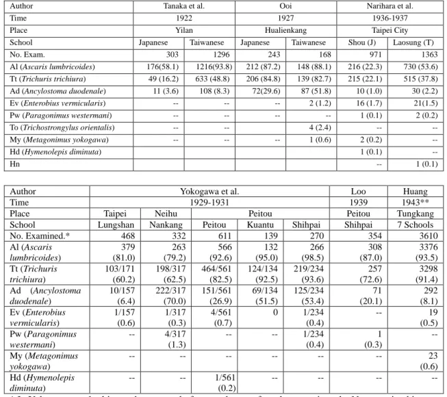
Table 6 shows that in the cases in which both Japanese and Taiwanese children were examined, the infection rates among Taiwanese school children were higher than those among Japanese. In the case of Al infection, in 1922 in Yilan, Taiwanese had 93.8% infected while Japanese had 58.1%; in 1927 in Hualienkang, Taiwanese had 88.1% infected while Japanese had 87.2%; in 1938 in Taipei City, Taiwanese had 53.6% infected while Japanese had 22.6%. In the case of Tt, Taiwanese had 48.8%, 82.7% and 37.8% in the three years (places), while Japanese had 16.2%, 84.8% and 22.1% respectively. In the case of hookworm, Taiwanese had 8.3%, 51.8% and 2.2%, while Japanese had 3.6%, 29.6% and 1.0% respectively. Besides, the Taiwanese children at Hualienkang had 2.4% infected with To. As for the infection rates of Ev, lung fluke (Paragonimus westermani, Pw), Metagonimus yokogawai (My), Hymenolepis diminuta (Hd), and Hymenolepis nana (Hn), both Taiwanese and Japanese had rather low infection rate, mostly below 1%.
As for the cases in which only Taiwanese children were examined, in 1929-1931, the infection rates of Al among children at 5 schools in Taipei area ranged from 79.2.% to 98.5%, while in 1939 the children at Shihpai had 80.7% and in 1943 the children at Tungkang had 93.5%, both within the range. It is notable that the Al infection rate of Shihpai school children reduced from 98.5% to 80.7% in about 10 years reflecting possibly the effect of taking expellant. In the case of Tt, the infection rates among children of 5 schools ranged from 60.2% to 93.2%, while the children at Shihpai had 72.6% and at Tungkang 91.4%, also within the range. However, the average infection rate of Ad among children of 5 schools in Taipei area was 41.1%, with the rate at each school varied as follows: Lungshan 6.4%, Peitou 26.9%, Kuantu 51.5%, shihpei 53.4% and Nankang 70.0%; except for Lungshan, these rates were much higher than 8.3% at Yilan and 8.1% at Tungkang. Comparatively, children of Laosung elementary school in Taipei City had only 2.2%. Yokogawa and Wakajima had specified the locations of these 5 elementary schools: Lungshan was located at a
42
Huang Teng-yun, 1943, “The results of stool examinations among elementary school children and agricultural school students in Tungkang district,” JFMA, No. 454, p. 88.
15
zone where the health condition and living standard were comparatively lower than other zones in Taipei City, Nankang, Shihpai and Kuantu all located at Chihsing (七星) district where people engaged mostly in agriculture. Thus, it was clear that the rate
was higher among those living in the rural area than those in the town area.43
Moreover, in Taipei area there were a few children infected with lung fluke which was not found among children at other places.
Table 6: Infections of Helminth among School Children, 1922-1943 (% in parenthesis)
Author Tanaka et al. Ooi Narihara et al.
Time 1922 1927 1936-1937
Place Yilan Hualienkang Taipei City
School Japanese Taiwanese Japanese Taiwanese Shou (J) Laosung (T)
No. Exam. 303 1296 243 168 971 1363 Al (Ascaris lumbricoides) 176(58.1) 1216(93.8) 212 (87.2) 148 (88.1) 216 (22.3) 730 (53.6) Tt (Trichuris trichiura) 49 (16.2) 633 (48.8) 206 (84.8) 139 (82.7) 215 (22.1) 515 (37.8) Ad (Ancylostoma duodenale) 11 (3.6) 108 (8.3) 72(29.6) 87 (51.8) 10 (1.0) 30 (2.2) Ev (Enterobius vermicularis) -- -- -- 2 (1.2) 16 (1.7) 21(1.5) Pw (Paragonimus westermani) -- -- -- -- 1 (0.1) 2 (0.2) To (Trichostrongylus orientalis) -- -- 4 (2.4) -- -- My (Metagonimus yokogawa) -- -- -- 1 (0.6) 2 (0.2) -- Hd (Hymenolepis diminuta) 1 (0.1) -- Hn -- 1 (0.1)
Author Yokogawa et al. Loo Huang
Time 1929-1931 1939 1943**
Place Taipei Neihu Peitou Peitou Tungkang
School Lungshan Nankang Peitou Kuantu Shihpai Shihpai 7 Schools
No. Examined.* 468 332 611 139 270 354 3610 Al (Ascaris lumbricoides) 379 (81.0) 263 (79.2) 566 (92.6) 132 (95.0) 266 (98.5) 308 (87.0) 3376 (93.5) Tt (Trichuris trichiura) 103/171 (60.2) 198/317 (62.5) 464/561 (82.5) 124/134 (92.5) 219/234 (93.6) 257 (72.6) 3298 (91.4) Ad (Ancylostoma duodenale) 10/157 (6.4) 222/317 (70.0) 151/561 (26.9) 69/134 (51.5) 125/234 (53.4) 71 (20.1) 292 (8.1) Ev (Enterobius vermicularis) 1/157 (0.6) 1/317 (0.3) 4/561 (0.7) 0 1/234 (0.4) -- 19 (0.5) Pw (Paragonimus westermani) -- 4/317 (1.3) -- -- 1/234 (0.4) 1 (0.3) -- My (Metagonimus yokogawa) -- -- -- -- -- -- 23 (0.6) Hd (Hymenolepis diminuta) -- -- 1/561 (0.2) -- -- -- --
* In Yokogawa et al., this number was only for roundworm; for other parasites, the No. examined is listed under each item.
** In Huang Teng-yun’s report, there was no number of persons infected. The numbers listed here and are calculated with the number of persons examined and the reported infection rates.
Source: Tanaka Seigo and Chen Tu-chin, 1922, p. 642, Table 5; Ooi Tsukasa, 1927, pp. 229-230, Tables 1 and 2; Yokogawa Sadamu and Wakejima Osamu, 1932, p. 677, Table 56; Loo Wan-teh, 1940, p. 1977, Table 3; Narihara Norio, Yumoto Yoshika, Osaka Kiyoshi and Maeda Toshinori, 1938, p. 1589, Table 6; Huang Teng-yun, 1943, p. 88.
43
16
In addition to cases listed in Table 6, the Education Department of Taichung Prefecture conducted examinations with children at 19 Japanese and Taiwanese elementary schools in 1928. The results showed that except for 2 Japanese elementary schools at Wushe and Nantou, 3 Taiwanese schools for girls Chiaoli 腳里, Tucheng 塗城, and Lukang, and 1 Taiwanese school for boys at Chunkungliao 軍功寮, children at the other 13 schools were 100% infected by parasites. Even 4 to 5 species were found for individual child. The Japanese school children had small fractions infected with Al and Ad, but they were 100% infected with Tt. The Taiwanese school children were infected mostly with Al, Ad, and Tt. Moreover, a few Taiwanese children were infected with Ss, but none Japanese child did. The infection rate of Tt among Taiwanese children at most schools was 60-70%, but only a few children at Lukang girls’ school were infected; the reason was due to usage of well water.44 In Tainan prefecture, comprehensive examinations of parasites with school children began in 1927. The first round included 17,000 school children at one part of Chiayi district and Hsingfeng 新豐 district and it was found that the infection rates ranged from 71% to 100%. The most common parasites were roundworm, whipworm and hookworm. In early 1929, the second round was conducted at schools in Tungshih 東 石, another part of Chiayi, Touliu 斗六, and Peimen 北門 districts involving 21,300 children.45
Table 7 showed that the protozoan infection rates among school children at Fengshan in 1934 were in the order of Ec 39.7%, Gl 30.1%, Ib 23.7%, Eh 18.6%, and En 14.1%, all higher than those found among children in Taipei area and at Tungkang.
Table 7: The Infections of Protozoa among School Children, 1934-1943(% in Parenthesis)
Author Kan Narihara et al. Loo Huang
Time 1934 1936/10-1937/2 1939 1943
Place Fengshan Taipei City Shihpai Tungkang
School Taiwanese Japanese Taiwanese Taiwanese 7 Taiwanese No. Examined 156 971 1363 354 3610 Entamoeba histolytica 29 (18.6) 12 ( 1.2) 75 (5.5) 9 (2.5) 312 ( 8.6) Entamoeba coli 62 (39.7) 33 ( 3.4) 113 (8.3) 39 (11.0) 393 (10.9) Endolimax nana 22 (14.1) 42 ( 4.3) 166 (12.2) 6 (1.7) 475 (13.2) Iodamoeba bütschlii 37 (23.7) 3 ( 0.3) 11 (0.8) -- 65 (1.8) Dientamoeba fragilis 0 0 0 -- 27 (0.8) Giardia intestinalis 47 (30.1) 102(10.5) 213 (15.6) 50 (14.1) 263 (7.3) Chilomastix mesnili 1 ( 0.6) 0 0 2 (0.6) 24 (0.7) Trichomonas hominis 0 0 6 ( 0.4) -- 66 (1.8) Source: Kan Yoshio, 1934, p. 825, Table 2; Narihara Norio, Yumoto Yoshika, Osaka Kiyoshi and Maeda Toshinori, 1938, p. 1592, Table 9; Loo Wan-teh, 1940, p. 1977, Table 3; Huang Teng-yun, 1943, p. 88.
The children at Laosung had the rates of Gl 15.6% and En 12.2%, the children at Shihpei had the rates of Gl 14.1% and Ec 11.0%, and the children at Tungkang had
44
Taiwan Nichi Nichi Shinho, 1928/07/21/4.
45
17
the rates of En 13.2% and Ec 10.9%, and all above 10%, reflecting that protozoan infection in Taiwan could no longer be ignored in the 1930s-1940s.
It is notable that multiple parasitic infections were frequently found among school children. Kan pointed out that of the 128 Taiwanese children at Fengshan elementary school, 70 (54.6%) had single infection, 48 (37.5%) double infection, 8 (6.3%) triple infection, and 2 (1.6%) quadruple infection.46 Narihara et al. reported that among Japanese children, 32.3% had single infection, 7.6% had double infection and only one girl had triple infection. The cases of double infection were mostly found with Al and Tt. As for Taiwanese school children, 42.2% had single infection, 24.3% had double infection, 1.5% had triple infection and 0.07% (only 1 person) had quadruple infection. The cases of double infection were mostly with Al and Tt; those of triple infection mostly with Ad, Al and Tt.47 As for the infection of protozoa, of the 173 Japanese school children, 157 (90.8%) had single infection and only 16 (9.2%) had double infection. Of the 494 Taiwanese school children, 413 (83.6%) had single infection and 81 (16.4%) had double infection. For both Taiwanese and Japanese, the double infection cases were most frequently found with Ec and En followed by infected with Ec and Gl. In addition, among Taiwanese, the frequency of Eh found among double infection was also quite notable.48
Loo Wan-teh calculated multiple infections of helminth and showed that 17.8% were infected with single species, 42.7% with two species, and 10.1% with three
species.49 Huang Teng-yun counted multiple infections by combining helminthes and
protozoa. He found in the case of double infection, 53.7% was infected with Al and Tt. In the case of triple infection, 6.0% were infected with Na, Al and Tt. In the case of quadruple infection, 3.95% were with Ad, Na, Al and Tt. The quintuple infections were mostly found with Al, Tt, Eh, Ec and En. The cases of infected with six species were mostly found with Ad, Na, Al, Tt, Eh, and En. In the cases of infected with seven species, Ec was added. Finally, in the case of infection with eight species, Gl was added.50
4.2 Middle School Students
In his 1915 report, Ooi also provided the results of examinations with 60 students at Taichung Middle School. Of these students, 55% came from Taichung and Nantou sub-prefectures, while others came from many places in Taiwan. The distribution was as follows: 6 from Taichung, 7 from Hulutun, 3 from Changhua, 2
46
Kan Yoshio, 1934, p. 828.
47
Narihara Norio, Yumoto Yoshika, Osaka Kiyoshi and Maeda Toshinori, 1938, p. 1593.
48
Narihara Norio, Yumoto Yoshika, Osaka Kiyoshi and Maeda Toshinori, 1938, p. 1598.
49
Loo Wan-teh, 1940, p. 1978, table 5.
50
18
from Yuanlin, 3 from Shalu, 3 from Peitou, 3 from Lukang, 1 from Tachia, 5 from Nantou, 1 from Tsaohsiehtun, 1 from Linchipu 林杞埔, 4 from Taipei, 3 from Hsinchu, 2 from Taoyuan, 9 from Chiayi, 3 from Tainan, and 4 from Akou.51 In 1916, Ooi presented another report which included the result of examinations of 30 students of Taichung Middle School.52
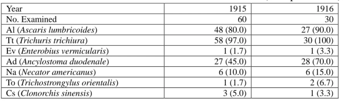
As shown in Table 8, in 1915-1916, the infection rates of Al among students of Taichung Middle School were 80-90%, those of Tt 97-100%, and those of hookworm (combing the two kinds) 55-80%. Moreover, there were 2 persons (2%) infected with pinworm, 3 persons (3%) with To, and 4 persons (4%) with Cs.
Table 8: The Parasitic Infection of Students at Taichung Middle School, 1915-1916 (% in parenthesis) Year 1915 1916 No. Examined 60 30 Al (Ascaris lumbricoides) 48 (80.0) 27 (90.0) Tt (Trichuris trichiura) 58 (97.0) 30 (100) Ev (Enterobius vermicularis) 1 (1.7) 1 (3.3) Ad (Ancylostoma duodenale) 27 (45.0) 28 (70.0) Na (Necator americanus) 6 (10.0) 6 (15.0) To (Trichostrongylus orientalis) 1 (1.7) 2 (6.7) Cs (Clonorchis sinensis) 3 (5.0) 1 (3.3)
Source: Ooi Tsukasa, 1919, pp. 819-821, Tables 1-3; Ooi Tsukasa, 1916, pp.356-357.
4.3 Vocational School Students
There are three reports concerning parasitic infection of vocational school students. The first report was presented in 1920 by Kojima Daiji and Ni Chiang-hai of the Taiwan Government-general’s Medical School. They conducted examinations with 298 students in October 1919. Of these students, 268 were Taiwanese (included Chinese) and 30 were Japanese. The age of the Taiwanese student ranged 14-27 and that of the Japanese 18-30. The native places of the Taiwanese students were as follows: 25 from Taipei, 8 from Taoyuan, 13 from Hsinchu, 72 from Taichung, 11 from Nantou, 36 from Chiayi, 65 from Tainan, 17 from Akou, and 2 from Penghu (澎
湖). And there were 17 from China.53
The second report was present in 1925 Yamazaki Shigeru, who conducted examinations with 161 Taiwanese and Japanese students at the coeducated Private Commercial and Industrial School from February 6
to March 4, 1925.54 The third report was presented by Morioka, Imaizumi, and Kao
51
Ooi Tsukasa, 1915, p. 819-821.
52
Ooi Tsukasa, 1916, “On the distribution of Necator americanus and Trichostrongylus orientalis in Taiwan,” JFMA, No. 163-164, pp. 355-363.
53
Kojima Daiji and Ni Chiang-hai, 1920, “The results of examinations on intestinal parasites with students at Medical School of Taiwan Government-general,” JFMA, No. 206-207, pp. 117-120. Table 1 on page 118 listed the number of Taiwanese students examined as 268, but the total number listed in Table 3 on page 120 was 266.
54
19
in 1935 with examinations of 260 students at Taipei Medical School, of which 121 were Japanese and 139 were Taiwanese.55 Since the third report was a lecture note with no details of statistics, here only the results of the first two are listed in Table 9.
Table 9: The Parasitic Infections of Vocational School Students, 1919-1925 (% in parenthesis)
School Medical School Commercial and Industrial School
Year 1919 1925
Student Taiwanese Japanese Taiwanese Japanese
No. Examined 268 30 126 35 No. Infected 207 (77.2) 16 (53.3) -- -- Al (Ascaris lumbricoides) 123 (45.8) 15 (50.0) 67 (53.0) 19 (54.0) Tt (Trichuris trichiura) 148 (55.2) 14 (46.6) 103 (82.0) 27 (77.0) Ev (Enterobius vermicularis) 1 ( 0.37) 0 1 ( 0.8) 0 Ad (Ancylostoma duodenale) 46 (17.2) 0 64 (51.0) 8 (23.0)
Tso (Taenia solium) 1 ( 0.37) 0 0 0
To (Trichostrongylus orientalis) 0 0 1 ( 0.8) 1 ( 3.0) Cs (Clonorchis sinensis) 0 0 1 ( 0.8) 4 (11.0)
Ad and Al 12 ( 4.5) 0 0 0
Al and Tt 69 (25.7) 4 (13.3) 0 0
Ad, Al and Tt 21 ( 7.8) 0 0 0
Source: Kojima Daiji and Ni Chiang-hai, 1920, p. 118, Table 1; Yamazaki Shigeru, 1925, p. 1130, Table 2.
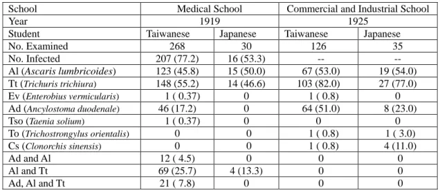
Table 9 shows that the infection rates of Al among Taiwanese and Japanese students were rather close, Medical School students had 46% and 50% and Commercial and Industrial School students had 53% and 54% respectively. However, the infection rates of Tt and Ad were higher among Taiwanese students. The infection rates of Tt among Taiwanese students in the two schools were 55% and 82% and among Japanese students were 47% and 77% respectively. The infection rates of Ad among Taiwanese students in the two schools were 17% and 51% and among Japanese students were 0% and 23% respectively. Moreover, the infection of Cs was found only among students of the Commercial and Industrial School, Japanese had 11% and Taiwanese only 0.8%.
In their 1935 report, Morioka et al. pointed out that at the Taipei Medical School, the infection rates of Al among Taiwanese and Japanese were quite similar; but the rates of hookworm (Ad and Na together) among Japanese were 3 times higher than among Taiwanese; on the contrary, the infection rate of Tt was higher among Taiwanese than among Japanese. They also noted that there were 3 Japanese students infected with Cs before they came to Taiwan. Among the 3 students infected with Metagonimus yokogawai there was 1 Taiwanese and this reflected that the habit of eating Japanese food had gradually become popular. In addition, Morioka et al. also
55
Morioka Kouichi, Imaizumi Kyouhei, and Kao Chi-tien, 1935, “The results of stool examinations among students of Taipei Medical School,” JFMA, No. 369, pp. 2184-2185.
20
reported the result of examinations with protozoa. There were 95 students (36.5%) infected, of which Taiwanese accounted for 37.4% and Japanese 35.5%. They also noted that the infection rate of Eh among Taiwanese was 3 times higher than that of Japanese students.56
5. Inhabitants in General
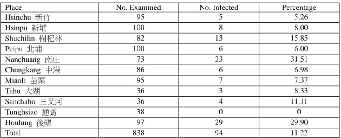
The earliest report concerning parasitic infection among inhabitants in Taiwan was presented by Matsuo Minetarou and Yokogawa Sadamu in 1912. They conducted examinations from February 23 to March 6 in Hsinchu sub-prefecture with the assistance of staff at district offices and public and private medical doctors. The object of examinations included sputum of 838 Taiwanese and stool of 500 Taiwanese. The samples of sputum were collected from 50 males and 50 females aged 15 and above at each district and those of stool from 25 males and 25 females. The results were reported separately with lung fluke (Pw) and intestinal parasites.57 Here, the infection rates of lung fluke are listed in Table 10 (see Map 2.1) and those of intestinal parasites in Table 11 (see Map 2.2-2.3).
Table 10: The Infections of Lung Fluke among Inhabitants of Hsinchu Sub-Prefecture, 1911
Place No. Examined No. Infected Percentage
Hsinchu 新竹 95 5 5.26 Hsinpu 新埔 100 8 8.00 Shuchilin 樹杞林 82 13 15.85 Peipu 北埔 100 6 6.00 Nanchuang 南庄 73 23 31.51 Chungkang 中港 86 6 6.98 Miaoli 苗栗 95 7 7.37 Tahu 大湖 36 3 8.33 Sanchaho 三叉河 36 4 11.11 Tunghsiao 通霄 38 0 0 Houlung 後壟 97 29 29.90 Total 838 94 11.22
Source: Matsuo Minetarou and Yokogawa Sadamu, 1912, p. 384, Table 1.
Table 10 shows that in 1911, the total infection rate of lung fluke among inhabitants in Hsinchu sub-prefecture was 11.2%, with Nanchuang 31.5%, Houlung 29.9%, and Shuchilin 15.9% ranking on the top. Matsuo and Yokogawa pointed out that these districts were located along rivers with easy access to irrigation. They also
56
Morioka Kouichi, Imaizumi Kyouhei, and Kao Chi-tien, 1935, p. 2185.
57
Matsuo Minetarou and Yokogawa Sadamu, 1912, “The statistical results of examinations on intestinal parasites among Taiwanese in the Hsinchu area,” JFMA, No. 114-115, pp. 382-387. Matuo Minetarou was the Health Advisor of Hsinchu Ting at that time; his report on this investigation to the Prefect of Hsinchu was included in the Official Documents of Taiwan Government-general, Vol. 5450, No. 3, pp. 25-38.
21
reported that with age differentiation, the highest infection rate was 21.7% among those aged 10-20 and the next was 19.0% among those aged 21-30. In terms of
occupational differentiation, students had 47.4% and merchants had 19%.58
It should be noted that Maxwell said that Paragonimus westermanii was first described by Patrick Manson (1844-1922) with a specimen from northern Taiwan. In some villages in central Taiwan, the large majority of the inhabitants, Chinese and especially aborigines, harbored this parasite.59
Table 11: The Infection Rates of Intestinal Parasites among Inhabitants in Hsinchu Sub-Prefecture, 1911 (% in parenthesis) Place Hsin-chu Hsin-pu Shuchi lin Peipu Nan- Chuang Chung-kang
Miaoli Tahu Sancha- ho Tung- hsiao Hou- lung Total No. Examined 54 40 51 50 36 50 36 61 34 53 35 500 No. Infected 54 (100) 35 (87) 51 (100) 49 (98) 26 (72) 48 (96) 34 (94) 54 (88) 28 (82) 50 (94) 35 (100) 464 (93) Al (Ascaris lumbricoides) 54 (100) 34 (85) 49 (96) 48 (96) 23 (64) 45 (90) 33 (92) 49 (80) 26 (76) 43 (81) 34 (97) 438 (88) Tt (Trichuris trichiura) 21 (39) 20 (50) 37 (73) 20 (40) 9 (25) 32 (64) 7 (19) 19 (31) 7 (21) 27 (51) 28 (80) 227 (45) Ad (Ancylostoma duodenale) 10 (19) 12 (30) 11 (22) 20 (40) 3 (8) 6 (12) 5 (14) 14 (23) 4 (12) 11 (21) 8 (23) 104 (21) Ev (Enterobius vermicularis) 2 (4) 0 0 0 0 0 0 0 0 2 (4) 2 (6) 6 (1) Intestinal ell 1 (2) 1 (3) 1 (2) 0 0 0 4 (11) 0 0 0 0 7 (1) Al only 26 11 12 17 15 14 19 24 18 12 4 172 (35) Tt only 0 0 2 1 3 2 0 2 2 1 0 13 (3) Ad only 0 1 0 0 0 0 1 3 0 5 1 11 (2) Al and Tt 15 11 25 11 5 26 6 14 4 24 21 162 (32) Al and Ad 5 3 2 12 2 2 3 8 3 4 2 46 (9) Al and Ev 2 0 0 0 0 0 0 0 0 2 0 4 (0.8) Al and Intestinal ell 0 0 0 0 0 0 4 0 0 0 0 4 (0.8) Al, Tt and Ad 5 8 9 8 1 4 1 3 1 2 5 47 (9) Al, Tt and Ev 0 0 0 0 0 0 0 0 0 0 2 2 (0.4) Al, Ev, and
Intestinal ell
1 1 1 0 0 0 0 0 0 0 0 3
(0.6)
Source: Matsuo Minetarou and Yokogawa Sadamu,1912, p. 383[387], Table 4.
Table 11 lists the statistics concerning infection of intestinal parasites among inhabitants of Hsinchu sub-prefecture in 1911. The total infection rate of intestinal parasites was 93%; specifically, Al 88%, Tt 45%, and Ad 21%. The highest infection rate was 100% found at Hsinchu, Shuchilin and Houlung and the lowest rate was 72% at Nanchuang. There was no significant dirrerence between males and females as both had 92%. In terms of occupation, those engaged in restaurant business had the highest
58
Matsuo Minetarou and Yokogawa Sadamu, 1912, p. 383, p. 385, p. 386.
59
22
rate at 100%, followed by agriculture 97%, laborers 96%, commerce 95%, miscellaneous 93%, and handicraft 92%.60
Map 2.1: The Infection Rate of Lung Fluke in Hsinchu Sub-prefecture, 1911
Map 2.2: The Infection Rate of Intestinal Parasites in Hsinchu Sub-prefecture, 1911 Map 2.3: The Infection Rate of Various Intestinal Parasites in Hsinchu Sub-prefecture,
1911
60
23
In addition, many cases of multiple infections were discovered. In terms of single infection, there were 172 (34%) infected with Al, 13 (3%) with Tt, and 12 (2%) with Ad. In the cases of double infection, there were 162 (32%) infected with Al and Tt and 46 (9%) infected with Al and Ad. As for triple infection, there were 47 (9%) infected with Al, Tt and Ad. Moreover, there were a few cases infected with pinworm and intestinal ell-like worm, but they were not found in the cases of single infection. Both adult and larva of the ell-like worms were discovered, but it was not certain whether they were Strongyloides stereoralis (Bavay, 1876) or Strongylus subtilis (Loos. 1895).61
In 1927, Ooi, serving at the Hualienkang Hospital, presented a report on the parasitic infection among Japanese resided at colonist villages. He undertook examinations with Japanese inhabitants at Yoshino 吉野 village from February to May and at Toyota 豐田 and Hayata 林田 villages from September to October, 1926.62 Among these three Japanese villages, Yoshino was established first around 1 li (1 Japanese league = 2.44 miles) southwest to Hualienkang. From February 1910 onward, several immigrations were recruited from Japan and the village was turned to the jurisdiction of Hualienkang by Taiwan Government-general in 1916. The inhabitants of Yoshino Village came from 21 counties outside of Hokkaido, with the largest number from Tokushima followed by Hiroshima, Fukuoka, Kagawa, Kumamoto, Saga, and Yamaguchi. At the end of 1925 there were 331 households with 1,768 persons at Yoshino Village. The Yoshino villagers mostly engaged in agriculture; the main crops were rice and sweet potato and the side lines were tobacco and other garden crops.
Toyota Village was established in April 1913. Its opening was more difficult than Yoshino because of geographical conditions. However, with appropriate arrangements by the authority, immigrations gradually arrived. At the end of 1925 there were 179 households with 911 persons at Toyota Village. The production of Toyota Village was similar to that of Yoshino.
The first immigrants of Hayata Village came in February 1914, just around the time when the railway connecting Hualienkang and Juishui 瑞穗 was completed. It was located 8 li south to Hualienkang. In the beginning, Hayata Village, similar to Toyota, was established at a wilderness grown with miscanthus reeds, at a juncture of plain woods and mountain woods, with wild animals swaggering around and very difficult to open up. Later, it was rather unstable because of epidemic diseases and natural calamities, but with tenacious encouragement of the authority and the efforts of villagers, the village developed gradually and the foundation was established.
61
Matsuo Minetarou and Yokogawa Sadamu, 1912, p. 383-384 [387-388].
62
24
Immigrants to Hayata Village came from 13 counties in one prefecture with the largest number came from Fukuoka followed by Kumamoto, Saga, and Yamaguchi. At the end of 1925, there were 167 households with 689 persons. The production of Hataya was similar to the above two villages.63
In addition to the three Japanese villages, Ooi also undertook examinations with
Okinawa fishermen resided at Milun Village 米崙庄 from April to May in 1925.64
Table 12 showed that in 1925, the parasitic infection rates of Japanese inhabitants at 3 colonist villages and Okinawa fishermen at Milun village were as high as 98%. Among various parasites, the infection rate of Tt was the highest; the average rate of the 3 village was 74.7% and that of Milun village 87.5%. The next highest was the infection rate of Al; the average rate of the 3 village was 58.5% and that of Milun village 45%. The third was the infection rate of Ad; the average rate of the 3 villages was 56% and that of Milun village 20%. In addition, the infection rates of To, Cs, My and Heterophyes were relatively small. Ooi pointed out that at the Japanese villages, the infection rate of malaria was also rather high, 50% at Yoshino,
21% at Toyota, 72% at Hayata, and the average was 48%.65 The situation of
coexisting ancylostomiasis and malaria at the same area was quite similar to that found with patients at the Red Cross Taipei Branch Hospital mentioned above.
Table 12: The Parasitic Infections among Japanese Inhabitants at Colonist Villages in Hualienkang Prefecture, 1926 (% in parenthesis)
Village Yoshino Toyota Hayata Total Milun
No. Examined 270 210 236 716 40 No. Infected 266(98.5) 205(97.6) 233(98.7) 704(98.3) 39(97.5) Tt (Trichuris trichiura) 212(78.5) 145(69.1) 178(75.4) 535(74.7) 35(87.5) Al (Ascaris lumbricoides) 164(60.7) 122(58.1) 135(57.2) 419(58.5) 18(45.0) Ev (Enterobius vermicularis) 1( 0.4) 1( 0.5) 2( 0.9) 4( 0.6) 0 Ad (Ancylostoma duodenale) 149(55.2) 113(53.8) 140(59.3) 402(56.2) 8(20.0) To (Trichostrongylus orientalis) 2( 0.7) 4( 1.9) 2( 0.9) 8( 1.1) 1( 2.5) Cs (Clonorchis sinensis) 2( 0.7) 1( 0.5) 1( 0.4) 4( 0.6) 0 My (Metagonimus yokogawai) 1( 0.4) 1( 0.5) 2( 0.9) 4( 0.6) 2( 5.0) Heterophyes 1( 0.4) 0 0 1( 0.1) 0
Tae (Cestoidea, Taenia sp.) 1( 0.4) 0 0 1( 0.1) 0 Source: Ooi Tsukasa, 1927, pp. 228-229, Table 1; p. 230, Table 2. The original tables listed the number of persons not infected; here it was re-calculated into the number infected.
The third report concerning parasitic infection among general inhabitants was presented in 1929 by Suzuki Sotoo, a technician at Taiwan Government-general and Central Research Institute. This report focused on rural area of Taichung prefecture, but also referred to similar topographical areas in other prefectures; it was a quite 63 Ooi Tsukasa, 1927, p. 227. 64 Ooi Tsukasa, 1927, p. 230. 65 Ooi Tsukasa, 1927, p. 293.
25
comprehensive study on the parasitic infection of rural Taiwan at that time.66
Suzuki commented on living conditions in rural Taiwan to provide a background for understanding the parasitic infection. He said, the staple food of Taiwan villagers was rice, or rice mixed with pieces or strips of sweet potato, usually cooked into dry or gruel meal and the proportions of rice and sweet potato varied according to the level of family wealth. Except for fruit, Taiwanese usually did not eat raw food. However, they used polluted water to wash utensils and did not care whether there were touches of flies or cockroaches. Drinking water was usually rather muddy and it was never used after filtrating but only simply precipitated. At occasions of ceremonies, villagers might have pork, mutton, chicken, duck and goose, but in daily life they were used to have salt fish to go with coarse food. Vegetables were cooked with animal or vegetable oil and seasoned with salt, seldom with sugar or soy sauce. Except for the day of ceremony, farmers were used to be barefoot all day long and they seldom took a bath. In the summer, they put a little warm water in a tray to wash the face, the body and the limb; in cold days, they almost did not wash. The toilet was usually shared with the neighbor; an earthen jar was laid down in a pit at open space around the house to serve for the purpose but there were no other devices around it and thus it was very easy to breed flies. People even could relieve the bowls at any time under the shadow or on the fields. Women would have a wooden pail for urine in the bedroom. Moreover, Taiwanese used to have their finger nails grown to very long and even boasted with the amount of filth harboring in them. They also liked to chew betel-nuts and threw up the juice at will; they used fingers to blow the nose and spread the snivel on the wall, on the post, or on the cloth.
Village houses were mostly Chinese style, roofed with tiles or thatches, lighting and ventilation were so poor that even in daytime it was dark and damp indoor. Around the house, there were cowshed, pigpen, and manure heap. And domestic
animals and fowls were raised everywhere. What a spectacular of living together! There was in general a deep superstition regarding diseases. Once getting ill,
they believed it was a penalty of god or a plague of devil and thus they engaged in saying prayers and charting incarnations to seek for a cure. When prayers and incarnations were ineffective, they took Chinese medicine made of roots of herbs and barks of woods. Their concept of health was rather naïve. They did not believe that parasites could be infected through mouth and skin; they used polluted water to wash food and utensils, spread night soil as fertilizer, tilled with barefoot, and lived together with domestic animals and fowls. The prevalence of parasites was just a natural
66
Suzuki Sotoo, 1929, “An investigation of the relationship between distribution of parasitic infection and age, sex, occupation, and topography at villages in rural Taiwan,” JFMA, No. 291, pp. 535-560; No. 292, pp. 717-770.