行政院國家科學委員會專題研究計畫 期中進度報告
內毒素前置處理對新生鼠腦缺氧缺血的神經保護之研究
(1/3)
計畫類別: 個別型計畫 計畫編號: NSC94-2314-B-006-053- 執行期間: 94 年 08 月 01 日至 95 年 07 月 31 日 執行單位: 國立成功大學醫學系小兒科 計畫主持人: 黃朝慶 報告類型: 精簡報告 報告附件: 出席國際會議研究心得報告及發表論文 處理方式: 本計畫可公開查詢中 華 民 國 95 年 6 月 1 日
行政院國家科學委員會補助專題研究計畫
□期中
進 度 報 告
內毒素前置處理對新生鼠腦缺氧缺血的神經保護之研究(1/3)
計畫類別:□ 個別型計畫 □ 整合型計畫
計畫編號:NSC 94-2314-B-006-053 -
執行期間:94 年 08 月 01 日至 95 年 07 月 31 日
計畫主持人:黃朝慶
共同主持人:
計畫參與人員:
成果報告類型(依經費核定清單規定繳交):□精簡報告 □完整報告
本成果報告包括以下應繳交之附件:
□赴國外出差或研習心得報告一份
□赴大陸地區出差或研習心得報告一份
□出席國際學術會議心得報告及發表之論文各一份
□國際合作研究計畫國外研究報告書一份
處理方式:除產學合作研究計畫、提升產業技術及人才培育研究計畫、
列管計畫及下列情形者外,得立即公開查詢
□涉及專利或其他智慧財產權,□一年□二年後可公開查詢
執行單位:國立成功大學
中 華 民 國 95 年 05 月 31 日
Keywords: endotoxin, eNOS, hypoxic-ischemia
Abstract
There are still no effective therapies against neonatal hypoxic-ischemic brain injury. A sublethal stress before a lethal injury can reduce the neuronal death, a phenomenon called “preconditioning”. Elucidating the underlying mechanisms of preconditioning might provide potential therapy for neonatal hypoxic-ischemic brain injury. Studies in adult brain have shown that systemic LPS pretreatment at a dose range of 0.3-1 mg/kg induced eNOS expression, which was suggested to play an important role in the brain tolerance induced by LPS preconditioning. Here we examined whether the neuroprotective preconditioning effect could be established in neonatal rats at a lower dose (0.05 mg/kg) of systemic LPS. We also tested whether eNOS expression is required in this low-dose LPS preconditioning model, and more importantly, and whether pharmacological activation of eNOS is neuroprotective against hypoxic-ischemic brain injury in newborns..
Rat pups pretreated with lipopolysaccharide (LPS) at a low dose of 0.05 mg/kg 24 hours before hypoxic-ischemia had a significantly lower mortality rate during hypoxic-ischemia compared with those pretreated with a higher dose of 0.3 mg/kg LPS or normal saline. The low-dose LPS preconditioning was able to provide significantly long-term neuroprotective effects at pathological and behavioral levels compared with the saline-pretreated group on a neonatal rat model of brain hypoxic-ischemia. The LPS-preconditioned rats had significantly reduced
expression of proinflammatory cytokines, microglia activation, nuclear factor κB (NF-κB) activation, and reactive oxygen species (ROS) production in the brain after hypoxic-ischemia than the saline-pretreated rats. In addition, the expression levels of apoptosis markers, such as cleavaged form of caspase-8, caspase-9, and caspase-3, nuclear translocation of apoptosis inducing factor, and poly (ADP-ribose) polymerase (PARP) in the cortex after hypoxic-ischemia were significantly lower in the LPS-preconditioned rats compared with the saline-pretreated rats.
Low dose LPS preconditioning could selectively increase endothelial nitric oxide synthase (eNOS) expression predominately in vascular endothelial cells and neurons. Inhibition of eNOS expression by pharmacological constitutive NOS instead of nNOS or iNOS inhibitor or by antisense oligodeoxynucleotide abolished the neuroprotective effect of LPS preconditioning. Furthermore, eNOS overexpression by L-arginine, not by D-arginine, or by adenovirus- eNOS overexpression provided significant neuroprotection against hypoxic-ischemic injury. eNOS expression is found to play an important role not only in the low dose LPS
preconditioning-induced neuroprotection but also in providing neuroprotection against
hypoxic-ischemia in the neonatal rat brain. This study suggests that strategy targeting to increase eNOS expression is a potential approach for protection against neonatal hypoxic-ischemic brain injury.
關鍵詞:內毒素、缺血缺氧 中文摘要: 新生兒缺血、缺氧性腦病變是新生兒疾病中常見且嚴重的疾病,通常會造成癲癇、學 習障礙、心智障礙、腦性麻痺、甚至是新生兒死亡之臨床病徵。目前針對這樣的腦傷尚未 有良好的治療或預防措施。在我們的研究計畫中嘗試以一極低劑量的內毒素-脂多醣 ( lipopolysaccharide 0.05mg/Kg) 作為缺血、缺氧狀況之前置處理,發現當此前置處理於缺 血、缺氧前二十四小時進行,將明顯的減緩新生幼鼠之缺血、缺氧性腦傷的程度,同時也 減緩缺血、缺氧性腦傷所導致之學習及記憶障礙的情況。先前的研究報告指出缺血、缺氧 狀況引發細胞內氧化壓力增加,而氧化壓力敏感性的轉錄因子〈例如Nuclear Factor-kappa B or Activator Protein-1〉將會明顯的活化,並且啟動重要基因的表現其引含的機制包括:發 炎反應、細胞凋亡 …等等,最終導致腦傷的形成。為了確定內毒素前置處理所提供之腦部 保護機制的方向,在我們的研究計畫中嘗試以各種角度去觀察在內毒素的前置處理之後這 些形成腦傷之重要機制的變化。我們發現不論是發炎反應、細胞凋亡或是細胞在缺血、缺 氧後的氧化壓力都有明顯被抑制的情況。基於臨床應用之可行性,我們將著重於此種神經 保護機制的探討,嘗試從中找出最重要的樞紐因子,作為日後臨床應用之參考。
Introduction
The incidence of hypoxic-ischemic (HI) encephalopathy was reported to occur in 2–6/1,000 full-term newborns. Twenty percent of the affected infants die during newborn period, and additional 25% exhibit permanent neurodevelopmental handicaps (Johnston et al., 2001; Ferriero DM 2004; Chang et al 2006). Although HI brain injury can cause immediately injury, the final pattern and severity of the damage are determined by the cascades of biochemical and molecular events such as excitotoxicity, inflammation, oxidative stress, and apoptosis, that can extend for days to weeks after the insult (Johnston et al., 2001; Tasaki., 2002; Ferriero DM 2004; Tasaki., 2002; Xanthou et al., 2002; Johnston et al., 2001; Saliba et al., 2001). The pathogenesis of neonatal HI brain injury has considerably been delineated, however, there is still no effective therapy clinically. Although hypothermia was recently used clinically in the treatment of neonates with hypoxic-ischemic encephalopathy within 6 hours of birth in multicenter randomized trials, its overall beneficial effect was relatively small (Shankaran et al., 2005).
Nitric oxide (NO) generated by endothelial nitric oxide synthase (eNOS) plays a crucial role in vascular function and homeostasis. NO derived from eNOS is antithrombotic,
anti-inflammatory, and a potent vasodilator. Pharmacological and genetic approaches in adult animals of cerebral ischemia have clearly shown that eNOS and vascular NO plays a prominent role in maintaining cerebral blood flow and preventing neuronal damage. eNOS knockout mice show decreased blood flow in the ischemic border zone and develop larger cerebral infarction. In contrast, enhancing NO production by administration of the eNOS substrate L-arginine, physical activity or by prophylactic treatment of statin increased cerebral blood flow, improve functional brain activity and confers neuroprotection against stroke. Therefore, therapeutic modalities that target eNOS expression and endothelial NO bioavailability not only serve as preventive measures to reduce stroke incidence but also could represent novel treatment strategies for reducing brain injury during cerebral ischemia.
A sublethal stimulation can confer protection against the subsequent lethal stimulation, this phenomenon is called “preconditioning”. Elucidating the mechanisms of preconditioning can offer potential effective therapies against HI brain injury. Systemic LPS, a cell-wall component of gram-negative bacteria, administration in a dose range of 1-20 mg/kg has been used as in-vivo models of endotoxic shock study. Previous studies have showed that systemic LPS
preconditioning at a dosage range of 0.3-1 mg/kg could afford neuroprotection against focal cerebral ischemia in adult rats when administered 48 to 72 hours before ischemic insults (Ahmed et al., 2000; Bordet et al., 2000; Tasaki et al., 1997), and against hypoxic-ischemic brain injury in neonatal rats when administered 24 hours before insult (Yang et al., 2004; Coumans et al., 2003; Eklind et al., 2001; Eklind et al., 2005). Studies in adult brain have shown that systemic LPS pretreatment induced eNOS expression in major components of the blood-brain barrier (Iwase K et al 2000), and that upregulation of eNOS expression in cerebral blood vessels was suggested to play a role of trigger in the brain tolerance induced by LPS preconditioning (Puisieux et al 2000; Furuya K et al 2005). Reduction of local cerebral blood flow in the periinfarct area was also
reported to be significantly inhibited in the LPS preconditioned adult rats, which was correlated with eNOS expression (Furuya K et al 2005). Whether the neuroprotective preconditioning effect still could be established by an even lower dose (0.05 mg/kg) of systemic LPS in neonatal rats remains unknown. Whether eNOS expression is required in the low-dose LPS preconditioning model, and more importantly, whether pharmacological activation of eNOS is neuroprotective against hypoxic-ischemic brain injury in newborns remains to be examined.
By using an established rat pup model of HI brain injury, this study was: 1) to examine the long-term neuroprotective effect of the low-dose LPS (0.05 mg/kg) preconditioning at behavioral and pathological levels; 2) to determine the potential role of eNOS in the neuroprotective
mechanisms in this low-dose LPS preconditioning model, and 3) to test whether pharmacological upregulation of eNOS expression is neuroprotective against hypoxic-ischemic brain injury.
Materials and Methods
This study was approved by our university's Animal Care Committee. Ten to twelve
Sprague-Dawley male pups per dam were used and housed with a 12/12-hour light/dark schedule in a temperature- and humidity-controlled colony room. The pups were housed with their dams until weaning on postnatal (P) day 21, and then housed in groups of 4-5 per cage.
LPS preconditioning in neonate hypoxic-ischemic brain injury model
On P7 rat pups, unilateral common carotid artery ligation followed 1 hour later by 8% oxygen hypoxia for 2 hours produces selective damage in the cerebral hemisphere ipsilateral to the artery occlusion that resembles HI damage to the human neonatal brain (Vannucci et al., 1999). To establish the LPS preconditioning model, LPS (0.05 mg/kg, or 0.3 mg/kg, Escherichia coli 0111:B4 LPS, Sigma Chemical Co., St Louis) or normal saline (NS-HI group) was injected intraperitoneally in rat pups 3 hour or 24 hour before being exposed to unilateral carotid artery ligation followed by 2 hours of 8% oxygen hypoxia on P7. The animals were anesthetized with 2.5% halothane (balance, room air), and the right common carotid artery was surgically exposed and permanently ligated with 5-0 surgical silk. After surgery, the pups were returned to the incubator for a 1-hour recovery to keep their body temperature before hypoxia. Rats were placed in airtight 500-ml containers partially submerged in a 37°C water bath, and the humidified 8% oxygen (balance, nitrogen) was maintained at a flow rate of 3 L/min for 2 hours. After completion of hypoxia, the rat pups were returned to their cage. The rat pups that received normal saline injection only on P6 without subsequent exposure to HI was served as controls.
Outcome Measure
Behavioral assessment by Morris water maze (MWM) from P35 to P40. Rats undertook the MWM as described previously, but with some modifications. In brief, the pool was illuminated by room lights, four points on the perimeter of the pool were designated, and the pool was
divided into four quadrants. An 8 cm × 8 cm Plexiglas platform, onto which the rats could escape from the water, was positioned in the center of one of the quadrants, 1 cm below the water surface. Escape distance, escape latency, swimming speed, and swimming patterns of the rats were monitored by a camera mounted above the pool and connected to a computer program (EthoVision, Wageningen, The Netherlands). The MWM task was divided into five phases. Training and testing was conducted between 9:00 and 17:00.
Phase I: Acquisition phase (Days 1-2). Phase I consisted of 2 days of training with 2 sessions per day. Each session consisted of 4 trials with 4 different starting positions. After swimming from each of the starting positions and mounting the submerged platform, the animals were allowed to remain there for 60 seconds. If an animal did not find the platform within 120 seconds, it was placed directly on the platform and allowed a 60-second rest period.
Phase II: Probe test 1 (Day 3). On day 3, the platform was removed from the pool, and the rats were placed in the pool in the quadrant opposite the previous platform position. The rats were allowed 60 seconds of free swimming. Swimming time in each quadrant was recorded. The percentage of time spent in the target quadrant where the platform had been previously located was calculated.
Phase III: Reversal phase (Days 4-5). Phase III was performed by placing the platform at a new position in the quadrant opposite the one used in Phase I. This reversal training was performed using the same procedure as described in Phase I.
described in Phase II. The percentage of time spent in the new target quadrant was calculated for each animal.
Phase V: Cued version (Visible platform test) (Day 6). Two hours after the probe test on day 6, the visible platform test was performed to determine whether the experimental manipulation resulted in a crude change in visual motor performance. The animals were released from a specific location and required to locate a green-colored escape platform 2 cm above the water surface in a position not used during the former sessions. If the animal did not reach the platform within 120 seconds, it was recorded as 120 seconds. Swimming speed was measured across the training sections in the acquisition and reversal phase.
Degree of brain damage by hemispheric weight reduction at P42. After being anesthetized with pentobarbital (50 mg/kg, i.p.), rats were decapitated and the brain was removed. The brainstem and cerebellum were removed, the forebrain was sectioned at the midline, and left and right hemispheric weights were determined. The percentage of hemispheric weight reduction, measured as [(left hemisphere weight − right hemisphere weight)/left hemisphere weight], was used as the measure of cerebral injury, as described previously.
Histopathological Assessment
Nissl stain. Rats was anesthetized with pentobarbital and perfused with normal saline followed by 4% paraformaldehyde (PF). The brains were removed into 4%PF for post-fixation for 3 days for paraffin-embedded sections. The brains were dehydrated by 75%, 85%, 95%, 100% alcohol and embedded into paraffin. Coronal sections were cut at 10µm, and Nissl stain was performed from the brains 24 hours after HI insult. For the frozen section, the rat brain was dealt with the same perfusion procedure and put into 4%PF for post-fixation for 24 hours. The brain was dehydrated by 10%, 20%, 30% sucrose dissolved in 1× PBS and then embedded into OCT. The brains were cut for 14µm in coronal sections by cryostat microtome.
TUNEL Assay. The paraffin-embedded brain sections were used in the TUNEL stain by TdT-FragEL DNA fragmentation detection kit (Oncogene Research Products). After
deparaffinization and rehydration, proteinase K (20µg/ml) was applied to the slides at room temperature (RT) for 20 minutes. The endogenous peroxidase was inhibited by 3% H2O2 in methanol at RT for 5 minutes. The terminal deoxynucleotidyl transferase (TdT) labeling reaction mixture containing biotin-labeled deoxynucleotides (biotin-dNTP) and TdT enzyme was
incubated with sample slides at 37℃ for 90 minutes. During the labeling reaction, TdT enzyme could catalyze the addition of biotin-dNTP to 3’-OH end of fragmented DNA in apoptotic cells. Finally the signal was detected by a streptavidin-horseradish peroxidase (HRP) conjugate and diaminobenzidine (DAB) reaction was used to produce visualized-brown pigments at the site of DNA fragmentation. The slides were slightly counterstained with hematoxylin.
Immunoblotting
Tissue samples from the cortex were lysed by ice-cold lysis buffer (0.2M NaCl, 0.1M HEPES, 10% glycerol, 2mM NaF, 2mM Na4P2O7, 1mM EGTA, 2mM DTT, 0.5mM PMSF, 1mM
benzardine, 10 g/ml leupeptin, 400U/ml aprotinin, and 1 M microcystin) and homogenized by sonicator. The homogenates were centrifuged at 14,000 rpm for 30 min at 40C, and the pellet was removed. The supernatant protein concentration of the lysates was determined with protein assay dye reagent concentrate. Protein samples (50 g/lane) were separated by 8% or 10% SDS -PAGE and then electrotransfered onto PVDF membrane (pore size: 0.45 m). The membrane was blocked with 5% nonfat dry milk and subsequently incubated with a following primary antibody including rabbit polyclonal anti-rat caspase 9 antibody (1:1000 dilution, Cell Signaling
Technology, Inc); a rabbit polyclonal anti-rat caspase 3 antibody (1:1000 dilution, Cell Signaling Technology, Inc); a mouse monoclonal anti-human caspase 8 antibody (1:800 dilution,
Calbiochem Corp); a rabbit polyclonal anti-rat PARP antibody (1:1000 dilution, Cell Signaling Technology, Inc); a rabbit polyclonal anti-rat TNF-α antibody (1:250 dilution, 100 g
100 g protein/lane, R&D Systems, Inc), a rabbit polyclonal anti-rat eNOS antibody (1:800 dilution, Santa Cruze Biotechnology, Inc.), or a rabbit polyclonal anti-rat iNOS antibody (1:800 dilution, Santa Cruze Biotechnology, Inc.). The immunoreactivity was detected by
horseradish-conjugated secondary antibody and visualized with the enhanced ECL (Pierce
Biotechnology, Inc). Chemiluminescent signals were detected on x-ray films and quantified using the MacBas bioimage analyzer. Every membrane was stripped by western stripping buffer and incubated with anti mouse β-actin antibody (1:1000) to check the protein loading.
Immunohistalchemistry staining
The paraffin-embedded coronal brain sections were de-paraffin by xylene followed by re-hydration with 100%; 95%; 85%; 75%, 50% alcohol progressively and finally infused in phosphate-buffered saline (PBS). After inhibition of endogenous peroxidase activity by
0.3%H2O2 in methanol for 30 minutes, the sections were subjected to antigen unmasking by bring
slides to boiling in 10mM sodium citrate buffer (pH 6.0). The sections were then incubated with the primary antibody diluted with PBS containing 0.5% normal goat serum overnight at 40C. A primary antibody used was a rabbit polyclonal anti-rat TNF-α antibody (1:50 dilution, Bender MedSystems.) or a mouse monoclonal anti-rat monocyte and marcrophage antibody (1:200 dilution, BioSource International, Inc.) or a rabbit polyclonal anti-rat eNOS antibody (1:50 dilution, Santa Cruze Biotechnology, Inc.). After a 2-hour incubation with the biotinalated secondary antibody obtained from the ImmunoPure ABC Peroxidase Staining Kit (Pierce
Biotechnology, Inc.), the sections were further incubated with a mixture of avidin and horseradish peroxidase-conjugated biotin for 1 hour. Then peroxidase activity was visualized using
3-3’-diaminobenzidine as a substrate. Sections were slightly counterstained with hematoxylin, and were mounted.
Immunofluorescence staining
The procedure of the paraffin-embedded slides was similar to that in immunohistochemistry and just different in the used secondary antibody as described following. The frozen sections were fixed with the mixture of acetone and methanol (1:1) for 5 minutes and air-dry followed by PBS-washing. Then the slides were infused with 0.3% H2O2 in methanol for 30 minutes to inhibit
the endogenous peroxidase activity. As a primary antibody, a polyclonal rabbit
anti-apoptosis-inducing factor (AIF) antibody (1:50 dilution, Cell Signaling Technology, Inc.) or a goat monoclonal anti- rat IL-6 antibody (1:50 dilution, R﹠D Systems, Inc.) or a monoclonal R-phycoerythrin (R-PE) conjugated hamster anti-rat/mouse TNF-α antibody (1:300 dilution, BD Biosciences, Inc.) or a rabbit polyclonal anti-rat eNOS antibody (1:100 dilution, Santa Cruze Biotechnology, Inc.) or a mouse monoclonal anti-rat eNOS (1:50 dilution, BD transduction lab) was applied to the slides at 40C overnight followed by incubation with fluorescence conjugated secondary antibody, such as a FITC-conjugated anti-mouse IgG antibody (1:500 dilution, Jackson ImmunoResaearch Laboratories, Inc.) or a TRITC-conjugated goat anti-rabbit IgG antibody (1:500dilution, Jackson ImmunoResaearch Laboratories, Inc.), at room temperature for one hour. The double staining analysis was carried out to identify the LPS induced eNOS-expressing cells in the sections previously immunoreacted for eNOS as described above. For neuronal
identification, sections were further incubated with a monoclonal mouse anti-neuronal nuclei (NeuN) antibody (1:100 dilution, Chemicon international a Serologicals Corp.) and a
FITC-conjugated goat anti-mouse IgG antibody (1:500 dilution, Jackson ImmunoResaearch Laboratories, Inc.). For astroglial identification, a polyclonal anti-GFAP antibody (1:1000
dilution, Dakopatts, Denmark, Inc.) was used and followed by incubation of a TRITC-conjugated goat anti-rabbit IgG antibody (1:500dilution, Jackson ImmunoResaearch Laboratories, Inc.). A monoclonal mouse anti- rat CD31 antibody (1:25 dilution, BD Biosciences Pharmigen, Inc.) used for vascular endothelial cell labeling and a mouse monoclonal anti-rat monocytes /marcrophages antibody (1:200 dilution, BioSource International, Inc.) used for microglial cells idetification were followed by a FITC-conjugated goat anti-mouse IgG antibody (1:500 dilution, Jackson
ImmunoResaearch Laboratories, Inc.) incubation. Double-stained sections were analyzed using a Nikon Eclipse E600 epifluorescence microscope.
Reactivate oxygen species (ROS) production
The cortex from lesioned hemisphere was weighted and then homogenized with 0.2 ml NS. ROS production was detected by chemiluminescence (CL) detector. The CLwas measured in a completely dark chamber of the ChemiluminescenceAnalyzing System. After 100-second background level determination,1.0 ml of 0.1 mM luminol in NS was injected into the sample. The CL was monitored continuouslyfor an additional 500 seconds. The total amount of CL was calculatedby integrating of the area under the curve and subtracting itfrom the background level. The assay was expressed as CL counts/10 seconds/mg of the brain tissue. The mean ± SEM of the CL level of each sample wascalculated.
RNA isolation and semi-quantitative reverse transcription (RT)-PCR
The total RNA was extracted from each hemispheric cortex by TRIZOL reagent (Invitrogen, San Diego, CA). A 5µg of total RNA and 1.5µg oligo-dT primer were mixed and pre-incubated at 700Cfor 10minutes. This mixture was cool down to room temperature gradually and mixed with RT reagent containing 25units MMLV-reverse transcriptase (Promega, Madison, WI), (Promega, Madison, WI), 10 µl 5× reaction buffer and 0.5mM dNTP. Finally, each RT mixture filled with nuclease-free distilled water to a final volume of 50µl and was incubated at 370C for 2 hours followed by denatured at 950C for 10 minutes. Each PCR mixture contained 5µl of RT product, 1 unite of Taq DNA polymerase (Viogene, Taiwan), 2µl 10 × PCR buffer plus MgCl2, 0.2mM dNTP, 0.5µM gene specific primers. The amplified reaction was performed by a thermocycler for a single 3-minute initial denaturation at 940C followed by gene-specific number of cycle under the conditions: denaturation at 940C for 20 seconds, annealing at each primer-specific annealing condition, and extension at 720C for 20 seconds and single final extension at 720C for 4 minutes. The PCR products were separated in 1.5% agarose gels containing ethidium bromide and
quantified by densitometry. All of PCR products were normalized to that the GAPDH PCR product in each sample. Gene specific primer sequences and PCR conditions: TNF-α: forward 5’-CAT GAT CCG AGA TGT GGA ACT-3’, reverse 5’-CTC CTG GTA TGA AAT GGC AAA-3’, product size: 486 bp, PCR condition: 940C 30”, 500C 30”, 720C 60”; IL-6: forward 5’-TGT GCA ATG GCA ATT CTG AT-3’, reverse 5’-GGT TTG CCG AGT AGA CCT CA-3’, product size: 472 bp, PCR condition: 940C 30”, 570C 30”,720C 30”; IL-10: forward 5’-CTG GAG TGA AGA CCA GCA AAG-3’, reverse 5’-TTC ATG GGC TTG TAG ACA CCT-3’, product size: 435 bp, PCR condition: 940C 30”, 600C 30”, 720C 45”; MIP-1α: forward 5’-ACT GAG CTG GAA CTA AAT GC-3’, reverse 5’-AAT GTG CCC TGA GGT CTT TC-3’, PCR condition: 940C 30”, 600C 59”, 720C 30”; GAPDH: forward 5’-ACA TTG TTG CCA TCA ACG AC-3’, reverse 5’-ACG CCA GTA GAC TCC ACG AC-3’, product size: 216 bp, PCR condition: 940C 30”, 580C 30”, 720C 30”.
Electrophoretic mobility shift assay
The hemispheric cortex was dissected out using a Dounce grinder with a loose pestle in ice-chilled buffer (15mMHEPES, 60mM KCl, 1mM NaCl, 0.25M sucrose, 5mM EDTA, 1 mM EGTA, 1 mM PMSF, 10µg/ml aprotinin, 15µg/ml leupeptin, 2mM NaF, and 1 mMsodium orthovanadate). The homogenate was centrifuged for 10 minutes at 2000 rpm, and pellet was resuspended in buffer (10mM HEPES, pH 7.2, 15 mM MgCl2, 10 mM KCl, 1 mM PMSF, 2 mM NaF, 15µg/ml leupeptin, and 1mM sodium orthovanadate). After brief vortexing, they were incubated on ice for 10 minutes and lysed with the tight pestle. The homogenate was centrifuged at 4000 rpm for 10 minutes. The pelleted nuclei were resuspended in 40µl of final extraction buffer (100mM HEPES, pH 7.2, 1.5 mM MgCl2, 1 mM EDTA, 0.8 M NaCl, 15% glycerol, 2 mM NaF, 1mM PMSF, 15µg/ ml leupeptin, 1 mM sodium orthpvanadate) and were incubated on ice for 2 hours. The nuclear suspension was centrifuged at 14000 rpm for 30 min at 4℃ and the supernatant was conserved. Protein concentration was assessed with the protein assay reagent
(Bio-Rad). Thirty pmol of the forward and reverse NF-κB oligonucleotides (forward:
5’-AGTTGAGGGGACTTTCCCAGG-3’; reverse: 5’-GCCTGGGAAAGTCCCCTCAA-3’) placed in a volume of 23 µl Klenow (DNA polymerase) working buffer were heated at 94℃ for 2 minutes and annealed at RT for 30 min. The annealed double-stranded oligonucleotides were end-labeled by fill-in reaction using 1 unit of Klenow enzyme (DNA polymerase, Promega) and 40µCi of [α32P] dTTP (PerkinElmer Life Sciences) was added. This mixture was incubated at
30℃ for 15 minutes. The labeled double-stranded oligoneucleotides were purified by Sephadex G-50 column (Amersham Biosciences). A 20µg nuclear extract and 0.5mg/ml poly(dI-dC) (Amersham Biosciences.) were incubated at 4℃ in binding buffer (10mM HEPES, pH7.2,
50mKCl, 0.1mM EDTA, 2.5mM DTT, 10%glycerol and 0.05% NP-40) for 10minutes. The 2×10⁴ cpm of 32P-labeled double-stranded oligonucleotides were added to the binding reaction mixture and stay on ice for 30min . Sampleswere analyzed on a 4% polyacrylamide gel
(acrylamide/bisacrylamide29:1 in 0.5 x Tris borate-EDTA buffer) at 10 V/cm for 2.5 hrs.The gel was dried and analyzed by autoradiography.
Statistics
Statistical significance (p < 0.05) was determined using one-way analysis of variance (ANOVA) to compare hemispheric weight reduction, and probe test and visual motor test results of the water maze between experimental groups. Multivariate analysis of variance (MANOVA) was used to compare escape time over the learning phase of the water maze. Post-hoc comparisons using Tukey's method were employed in one-way ANOVA, and Bonferroni's method in MANOVA. Continuous data were represented as mean ± SEM, unless indicated otherwise.
Results
Long-term behavioral and neuropathology outcome of a low-dose LPS preconditioning model in neonatal rats
Systemic LPS, in the dose range of 1-20 mg/kg, administration has been used in the in vivo model study of endotoxic shock. Previous studies have showed that LPS preconditioning against focal cerebral ischemia in adult rats could only be induced 48 to 72 hours after systemic LPS administration at a dosage range of 0.2-0.9 mg/kg (Ahmed et al., 2000; Bordet et al., 2000; Tasaki et al., 1997). In neonatal rats, a similar dose of LPS pretreatment 6 hours or 72 hours before hypoxic-ischemia worsened the degree of brain injury, in contrast, significant neuroprotection was observed when LPS was administered 24 hours before hypoxic-ischemia (Eklind et al., 2005). In this study, we examined whether an even a lower-dose of LPS (0.05 mg/kg) administration could induce neuroprotection against hypoxic-ischemia in neonatal rat brain.
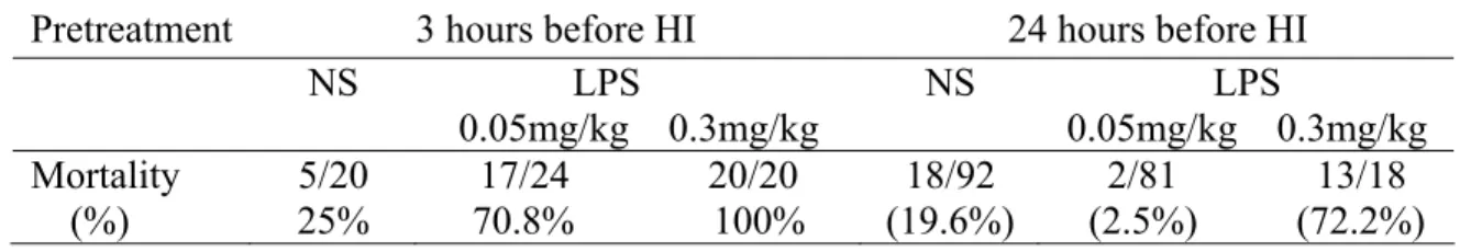
Mortality rate during hypoxic-ischemia after LPS pretreatment in neonatal rats. First, we pretreated rat pups with two different doses of LPS (0.05 mg/kg or 0.3 mg/kg, i.p.) or normal saline 3 hours before HI at P7. We found that both the LPS-pretreated groups had significantly higher mortality rates during HI than the NS-pretreated groups (high-dose LPS group, p < 0.0001; low-dose LPS group, p < 0.05). The mortality during HI 3 hours after LPS pretreatment was comparable between the high-dose (100%) and low-dose (70.8%) LPS-pretreated group.
Next, we examined the effect of different dose of LPS (0.05 mg/kg or 0.3 mg/kg, i.p.) administration 24 hours before HI. We found that the low-dose LPS-pretreated group had a significantly lower mortality (2.5%) during HI compared with the NS-pretreated rats (19.6%) (p < 0.05). In contrast, the high-dose LPS pretreated rats had a significantly higher HI mortality (72.2%) than the low-dose LPS or normal saline pretreated rats (both p < 0.05) (Table 1). The findings that high-dose LPS is associated with a high mortality rate during HI when administered 3 or 24 hours before HI and low-dose LPS induced a high mortality rate when administered 3 hours but a very low mortality when administered 24 hours before HI suggests that the potential neuroprotective effect could be examined 24 hours after low-dose LPS treatment.
Low-dose LPS preconditioning 24 hours before hypoxic-ischemia induced long-term neuroprotection at behavioral and morphological levels. We first examined whether preconditioning with a low-dose LPS (0.05 mg/kg) 24 hours before HI had long-term neuroprotective effects at behavioral and pathological levels.
During the learning and reversal-learning phases of Morris water maze, the LPS-HI, NS-HI, and control rats all learned to find the submerged platform (all p < 0.001), however, there were significant differences among the three groups (all p < 0.001). Post-hoc multiple analyses showed that the LPS-HI rats spent significantly less time finding the submerged platform than the NS-HI rats (both p < 0.001) in both the learning and the reversal-learning phase. There was no
In the memory phase of Morris water maze, there was significant difference among the three groups at probe test 1 and test 2 when the platform was removed from the pool. Post-hoc multiple analysis showed that the LPS-HI group spent significantly more time in the target quadrant than the NS-HI groups (p < 0.05 in probe test 1, and p < 0.001 in probe test 2), while no significant difference was found between the LPS-HI and control groups. There are no notably differences among the three groups in their swimming speed measured across the training sections. In the visual motor test, the NS-HI rats spent significantly more time finding the visible platform than the LPS-HI (p < 0.05) or vehicle groups (p < 0.001). No significant difference was found between the LPS-HI and control groups.
The degree of brain injury, measured by brain weight reduction on P42, showed significant difference among the three groups (p < 0.001). The LPS-HI rats had significantly less degree of brain damage than the NS-HI rats (7.0±2.1% vs 46.3±2.9%, p < 0.001). No significant difference was found between the control and LPS-HI groups.
Twenty-four hours after HI injury, histopathological examination by Nissl staining showed extensive neuronal injury and loss in the hippocampus and cortex of the lesioned hemisphere in the NS-HI group, while the degree of neuronal injury or loss was obviously less in the LPS-HI rats. TUNEL staining examination also showed extensive TUNEL-(+) cells in the cortex and hippocampus in the NS-HI group, and scattered distribution of TUNEL-(+) cells in the cortex and hippocampus in the LPS-HI group.
Low-dose LPS preconditioning significantly reduced reactive oxygen species production and activation of apoptosis pathways after hypoxic-ischemic injury in the neonatal rat brain ROS production. Overproduction of ROS may cause cell damage (Hudome et al., 1997). ROS production is up-regulated, and plays a critical factor for inducing brain damage after
hypoxic-ischemic brain injury in neonatal brain (Ferriero DM, 2004). By using a
chemiluminescence detector, we examined the temporal course of cortical ROS production after HI. Increased cortical ROS production was seen 3 hours to 72 hours after HI in both NS-HI and LPS-HI groups. However, the LPS-HI group had significantly less ROS production (29.34 ± 4.11/ mg/10 sec) 24 hours after HI injury than the NS-HI group (125.3 ± 33.02/ mg/10 sec) (p < 0.001) .
Caspase-dependent and -independent pathways of apoptosis after hypoxic-ischemia. Apoptosis plays a prominent role in the evolution of hypoxic-ischemic injury in the neonatal brain and may be more important than necrosis after injury. Caspase activation was reported to play important roles in neonatal HI brain damage (Cheng et al., 1998; Wang et al., 2001; Johnston et al., 2001; Saliba et al., 2001; Tasaki., 2002; Xanthou et al., 2002; Ferriero DM 2004;). The caspases are a group of cysteine proteases that exist within the cells as inactive pro-forms and can be cleavaged to form the active caspase in the early stage of apoptosis (Cheng et al., 1998; Wang et al., 2001). We first examined the activation of caspase-8 and caspase-9, the “initiator” caspases after HI brain injury between rat pups with and without LPS preconditioning. The LPS-HI rats had
significantly less accumulation of the cleavaged forms of caspase 9 (40 kDa and 17 kDa subunits) and caspase 8 (44 kDa and 28 KDa subunits) 24 hours and 72 hours after HI than the NS-HI rats.
There were no significant differences in the levels of pro-caspase 8 and 9 after HI between the two groups. We next examined the expression of the downstream caspase, the “terminator” caspase- caspase-3 implicated in the execution step of apoptosis (Tanaka et al., 2004). We found that there were no significant difference in the levels of procaspase-3 72 hours after HI between the LPS-HI and NS-HI rats. The LPS-HI rats showed significantly less cleavaged form of caspase-3 (29 kDa and 17 kDa subunit) 24 hours and 72 hours after HI than the NS-HI rats.
Poly (ADP-ribose) polymerase (PARP), the DNA repair and surveillance enzyme (Tanaka et al., 2004), is one of the target proteins of activated caspase 3, and the cleavage of PARP is a hallmark of apoptosis and harmful to the neuron. We then examined the expression levels of PARP between the LPS-HI and NS-HI groups. The cleavage of PARP (86 kDa) peaked at 24 hours after HI in both LPS-HI and NS-HI rats. The expression of cleavaged form of PARP was significantly less in the LPS-HI rats compared with NS-HI rats 24 hours and 72 hours after HI. The expression of caspase-independent apoptosis factor, apoptosis inducing factor (AIF) was examined by immunofluoresence microscopy 24 hours after HI. Evidence of nuclear
translocation of AIF in the cortex was significantly increased in the NS-HI group compared with the LPS-HI rats. Taken together, these findings suggest that activation of caspase-dependent and –independent pathways of apoptosis after HI in the neonatal rat brain was both significantly decreased by low-dose LPS preconditioning.
Low-dose LPS preconditioning significantly reduced pro-inflammatory cytokine responses, microglial and NF-κB activation after HI in the neonatal rat brain
Hypoxic-ischemia induces an inflammatory response in the neonatal brain that is important for development of brain injury (Ferriero DM 2004). Here, we first measured the
pro-inflammatory cytokine responses after HI in the LPS-HI and NS-HI groups. Significantly increased IL-6 and TNF-α mRNA expressions in the ipsilateral cerebral cortex were seen 3 hours and 24 hours after HI in the NS-HI group, and LPS preconditioning could significantly reduce the increased IL-6 and TNF-α mRNA expression at these time points. Increased macrophage
inhibitory protein-1α (MIP-1α) mRNA expression was seen in both the NS-HI and LPS-HI groups after HI, and there was no significant difference between the two groups. Significantly higher levels of IL-10 mRNA expression was observed in the NS-pretreated group. Western blotting showed that LPS preconditioning significantly reduced the increased IL-6 expression levels 3 hours and 24 hours after HI. LPS preconditioning also significantly reduced the increased TNF-α levels 24 hours and 72 hours after HI compared with the NS-HI group. Further
immunohistochemistry showed that the NS-HI group had more TNF-α immunoreactive cells in the ipsilateral cortex 24 hours after HI than the LPS-HI group.
Activated inflammatory cells such as microglia are implicated in the degree of ischemic brain injury by producing various bioactive mediators. Compared with the NS-HI group, the LPS-HI group had significantly reduced cell numbers of activated microglia, as demonstrated by ED-1 staining, in the cortex and hippocampus 24 hours after HI. NF-κB activation is a key mediator involved in the inflammatory responses. We therefore tested whether the decreased
inflammatory responses after HI by LPS preconditioning involved alteration of NF-κB activation. Evaluation of NF-κB activation by measuring DNA-binding activity of cortical nuclear extracts with electrophoretic mobility shift assay (EMSA) showed increased DNA binding of NF-κB 3 hours and 24 hours after HI, and LPS-preconditioning greatly attenuated the NF-κB activation during the periods.
Discussion
In this study, we found that systemic administration of a low-dose LPS (0.05 mg/kg) 24 hours before HI could not only dramatically decrease the mortality rate during HI but also induce long-term neuroprotective effects at behavioral and pathological levels in a neonatal rat model of HI brain injury. In this low-dose LPS preconditioning model, we found that 1) the
neuroprotective mechanisms after HI involved reduced ROS production, decreased activation of microglia and proinflammatory cytokines, decreased NF-κB activation, decreased activation of caspase-dependent and -independent pathways of apoptosis, and decreased PARP activation. In this study, we found that low-dose LPS preconditioning protected the neonatal rat brain against HI, and, concurrently preserved brain function. Further work will examine whether 1) eNOS instead of iNOS expression was upregulated in neurons and vessels by low-dose LPS, and inhibition of eNOS expression by pharmacological method or by antisense ODN abolished the neuroprotective effect of LPS preconditioning, and 2) increased eNOS expression by L-arginine instead of D-arginine, or by adenovirus overexpression provided significant neuroprotection against HI brain injury in neonatal rats.
Previous studies in neonatal rats have shown that systemic LPS (at a dose of 1 mg/kg) pretreatment increased brain damage when given 4 hours before hypoxic-ischemia, in contrast, the same dose showed neuroprotection when administered 24 hours before hypoxic-ischemia despite early inflammatory molecules and apoptotic responses was observed (Eklind et al., 2005;Ikeda et al 2006; Hagberg et al 2005). Our study showed significantly different responses and outcomes between 0.3 mg/kg LPS and 0.05 mg/kg LPS pretreatment in neonatal rats. Both dosages sensitized the animals and resulted in a very high mortality during hypoxic-ischemia when given 3 hours before hypoxic-ischemia. The higher dose of LPS given even 24 hours before hypoxic-ischemia had a long-lasting effect and still sensitized the neonatal rats with a high
mortality during hypoxic-ischemia by enhancing microglia activation, and increasing
proinflammatory cytokine production and iNOS expression in the brain. In contrast, the lower dose of LPS induced significantly less inflammatory responses and not only lowered the
mortality during hypoxic-ischemia but also provided neuroprotection. Although different animal species and LPS products from different bacteria would cause either sensitizing or
preconditioning effect, our findings showed that neonatal rat brain is very sensitive to and could be preconditioned with a very low-dose of systemic LPS to induce long-term neuroprotective effects against hypoxic-ischemic injury.
Acute inflammatory responses and ROS production triggered by hypoxic-ischemia could exacerbate primary brain hypoxic-ischemic damage (Johnston et al., 2001; Saliba et al., 2001; Tasaki., 2002; Xanthou., 2002; Kariko et al 2004; Ferriero DM, 2004; Hagberg et al 2005). The neonatal brain is particularly susceptible to oxidative stress-induced injury because of its
underdeveloped scavenging systems and high availability of formation of free radicals (Blomgren et al 2006). Proinflammatory cytokines, such as TNF-α, IL-1β, IL-6, and microglia activation could exacerbate brain damage in acute hypoxic-ischemic or excitotoxic injury model. Biological
effects of these cytokines that influence the progression of brain injury include stimulating the synthesis of other cytokines and neuronal injury mediators such as NOS, inducing leukocyte infiltration and the expression of adhesion molecules. We found that the ROS production, the pro-inflammatory cytokine responses, such as TNF-α and IL-6, and microglial activation in the neonatal rat brain after HI could be greatly reduced by low-dose LPS preconditioning.
Diminished production of proinflammatory cytokines and activation of microglia contributed to neuroprotective effect induced by the low-dose LPS preconditioning. MIP-1α is a
well-characterized monocyte chemoattractant and its expression coincided closely with regions where HI neuronal injury was most pronounced and microglia/monocytes accumulated. The upregulation of MIP-1α mRNA expression after HI showed no significant difference between rat pups with and without LPS preconditioning.
NF-κB is an inducibletranscription factor that regulates inflammation, immune and stress-related responses, and controls cellular survival and apoptosis in all tissues, including nervous system [30•,31•,32••]. NF-κB is a major inducer of proinflammatory responses in microglial cells. NF-κB decoy treatment downregulated proinflammatory cytokines in rat pup hippocampus after HI injury [Qiu 2004]. Although the role of NF-κB in perinatal brain injury needs to be further evaluated, the timing of reduced activation of NF-κB DNA binding activity after HI injury in this LPS preconditioning, might be correlated with the timing of diminished proinflammatory cytokine production and microglia activation after HI injury.
The immature brain retains its apoptotic machinery to a larger extent than the adult brain, and therefore may be more prone to apoptotic death after injury (Cheng et al., 1998; Wang et al., 2001). Two main pathways lead to apoptosis of neurons- the extrinsic or death receptor-pathway, and the intrinsic or mitochondrial pathway. Unlike cytochrome c, AIF acts in a caspase-independent manner. AIF translocates to the nucleus and induces chromatin condensation and large-scale DNA fragmentation. The number of AIF-(+) nuclei showed a positive correlation with the infarct volume 72 hrs after HI. We found that low-dose LPS preconditioning greatly reduced the activation of caspase-8 and -9, and the nuclear translocation of AIF after HI injury, suggesting low-dose LPS preconditioning can extensively suppress death-signaling activation not only in the extrinsic and intrinsic apoptotic pathways but also in the caspase-dependent and
caspase-independent pathways. The very-low dose of LPS preconditioning also reduces the cleavage form of caspase-3, a crucial apoptosis execution caspase, and PARP, the target protein of caspase-3. Further studies are needed to delineate the signaling pathways by which the low-dose of LPS preconditioning induces extensively endogenous apoptosis brakes against HI-induced neuronal death in the developing brain.
References
1. Ahmed SH, He YY, Nassief A, Xu J, Xu XM, Hsu CY. (2000) Effects of lipopolysaccharide priming on acute ischemic brain injury. Stroke 31:193-9.
2. Angele M. K, Schwacha M G, Smail N, Catania R A, Ayala A, Cioffo W G, Chaudry I H (1999) Hypoxemia in the absence of blood loss upregulates iNOS expression and activity in
macrophages. Am J Physiol. 276(2 Pt 1):C285-90.
3. An H, Yu Y, Zhang M, Xu H, Qi R, Yan X, Liu S, Wang W, Guo Z, Guo J, Qin Z, Cao X. (2002) Involvement of ERK, p38 and NF-kappaB signal transduction in regulation of TLR2, TLR4 and TLR9 gene expression induced by lipopolysaccharide in mouse dendritic cells.
Immunology. 106(1):38-45.
4. Asehnoune K, Strassheim D, Mitra S, Kim JY, Abraham E. (2004) Involvement of reactive oxygen species in toll-like receptor 4-dependent activation of NF- kappaB. J Immunol. 15;172(4):2522-9.
5. Bordet R, Deplanque D, Maboudou P, Puisieux F, Pu Q, Robin E, Martin A, Bastide M, Leys D, Lhermitte M, Dupuis B. (2000) Increase in endogenous brain superoxide dismutase as a potential mechanism of lipopolysaccharide- induced brain ischemic tolerance. J Cereb Blood Flow Metab. 20(8):1190-6.
6. Brucklacher RM, Vannucci RC, Vannucci SJ (2002) Hypoxic preconditioning increases brain glycogen and delays energy depletion from hypoxia- ischemia in the immature Rat. Dev Neurosci 24:411-417
7. Cheng Y, Deshmukh M, D’Costa A, Demaro JA, Gidday JM, Shah A, Sun Y, Jacquin MF, Johnson EM, Holtzman DM. (1998) Caspase inhibitor affords neuroprotection with delayed administration in the rat model of neonatal hypoxic-ischemic brain injury. J Clin Invest 101:1992-9.
8. Coumans AB, Middelanis JS, Garnier Y, Vaihinger HM, Leib SL, Von Duering MU, Hasaart TH, Jensen A, Berger R. (2003) Intracisternal application of endotoxin enhances the susceptibility to subsequent hypoxic-ischemic brain damage in neonatal rats. Pediatr Res. 53(5):770-5.
9. Couturier JY, Ding-Zhou L, Croci N, Plotkine M, Margaill I. (2003) 3-Aminobenzamide reduces brain infarction and neutrophil infiltration after transient focal cerebral ischemia in mice. Exp Neurol. 24(2):973-80.
10. Cuzzocrea S, Mazzon E, Costantino G, Serraino I, Dugo L, Calabro G, Cucinotta G, De Sarro A, Caputi AP. (2000) Beneficial effects of n-acetylcysteine in ischaemic brain injury. Br J Pharmacol. 130(6):1219-26.
11. Eklind S, Mallard C, Leverin AL, Gilland E, Blomgren K, Baltzer IM, Hagberg H. (2001) Bacterial endotoxin sensitizes the immature brain to hypoxic-ischaemic injury. Eur J Neurosci 13: 1101–1106.
12. Gidday JM, Fitzgibbons JC, Shah AR (1995) Reduction in cerebral ischemic injury in newborn rat by potentiation of endogenous adenosine. Pediatr Res 38:306-311.
13. Gong KZ, Zhang ZG, Li AH, Huang YF, Bu P, Dong F, Liu J.(2004) ROS-mediated ERK activation in delayed protection from anoxic preconditioning in neonatal rat cardiomyocytes. Chin Med J (Engl). 117(3):395-400.
14. Gu Z, Jiang Q, Zhang G.. (2001) Extracellular signal-related kinase and c-Jun N-terminal protein kinase in ischemic tolerance. Neurochem 12(16):3487-91.
14. Han BH, Holtzman DM. (2000) BDNF protects the neonatal brain from hypoxic-ischemic injury in vivo via the ERK pathway. J Neurosci 20:5775-5781.
15. Horiguchi T, Kis B, Rajapakse N, Shimizu K, Busija DW. (2003) Opening of mitochondrial ATP-sensitive potassium channels is a trigger of 3-nitropropionic acid-induced tolerance to transient focal cerebral ischemia in rats. Stroke. 34(4):1015-20.
17. Hudome S, Palmer C, Roberts RL, Mauger D, Housman C, Towfighi J. (1997) The role of neutrophils in the production of hypoxic-ischemic brain injury in the neonatal rat. Pediatr Res. 41(5):607-16.
18. Johnston MV, Trescher WH, Ishida A, Nakajima W. (2001) Neurobiology of hypoxic- ischemic injury in the developing brain. Pediatr Res.49(6):735-41.
19. Krams M, Lees KR, Hacke W, Grieve AP, Orgogozo JM, Ford GA; ASTIN Study Investigators.(2003) Acute Stroke Therapy by Inhibition of Neutrophils (ASTIN): an adaptive dose-response study of UK-279,276 in acute ischemic stroke. Stroke. 34(11):2543-8.
20. Kyosseva SV. (2004) Mitogen-activated protein kinase signaling. Int Rev Neurobiol. 59:201-20. Review.
21. Lee SJ, Lee S. (2002) Toll-like receptors and inflammation in the CNS. Curr Drug Targets Inflamm Allergy. 1(2):181-91.
22. Liu D, Lu C, Wan R, Auyeung WW, Mattson MP. (2002) Activation of mitochondrial ATP-dependent potassium channels protects neurons against ischemia-induced death by a mechanism involving suppression of Bax translocation and cytochrome c release. Cereb Blood Flow Metab. 22(4):431-43.
23. Liu S, Gallo DJ, Green AM, Williams DL, Gong X, Shapiro RA, Gambotto AA, Humphris EL, Vodovotz Y, Billiar TR. (2002) Role of toll-like receptors in changes in gene expression and NF-kappa B activation in mouse hepatocytes stimulated with lipopolysaccharide. Infect Immun. 70(7):3433-42.
24. Nakai Y, Horimoto H, Mieno S, Sasaki S. (2001) Mitochondrial ATP-sensitive potassium channel plays a dominant role in ischemic preconditioning of rabbit heart. Eur Surg Res. 33(2):57-63.
25. Nelson CW, Wei EP, Povlishock JT, Kontos HA, Moskowitz MA. (1992) Oxygen radicals in cerebral ischemia. Am J Physiol. 263(5 Pt 2): H1356-62.
preconditioning. Antioxid Redox Signal. 6(2):449-69.
27. Palmer C, Roberts RL, Young PI. (2004) Timing of neutrophil depletion influences long-term neuroprotection in neonatal rat hypoxic-ischemic brain injury. Pediatr Res. 55(4):549-56. 28. Peters O, Back T, Lindauer U, Busch C, Megow D, Dreier J, Dirnagl U. (1998) Increased
formation of reactive oxygen species after permanent and reversible middle cerebral artery occlusion in the rat. J Cereb Blood Flow Metab. 18(2):196-205.
29. Poltorak A, He X, Smirnova I, Liu MY, Van Huffel C, Du X, Birdwell D, Alejos E, Silva M, Galanos C, Freudenberg M, Ricciardi-Castagnoli P, Layton B, Beutler B. (1998) Defective LPS signaling in C3H/HeJ and C57BL/10ScCr mice: mutations in Tlr4 gene. Science. 11;282(5396):2085-8.
30. Rajapakse N, Shimizu K, Kis B, Snipes J, Lacza Z, Busija D. (2002) Activation of
mitochondrial ATP-sensitive potassium channels prevents neuronal cell death after ischemia in neonatal rats. Neurosci Lett. 26;327(3):208-12.
Table 1. The dosage and timing effect of lipopolysaccharide pretreatment on the mortality during hypoxic-ischemia in neonatal rats
Pretreatment 3 hours before HI 24 hours before HI
NS LPS 0.05mg/kg 0.3mg/kg NS LPS 0.05mg/kg 0.3mg/kg Mortality (%) 5/20 25% 17/24 20/20 70.8% 100% 18/92 (19.6%) 2/81 13/18 (2.5%) (72.2%) HI: hypoxic-ischemia 3 hours: NS vs 0.05 mg/kg LPS: RD 45.8% 95% CI=16.2%~65.7% NS vs 0.3 mg/kg LPS: RD 75% 95% CI=47.8%~88.8% 0.3 mg/kg vs 0.05 mg/kg LPS: RD -29.2% 95% CI=-49.2%~-7.7% 24 hours: NS vs 0.05 mg/kg LPS: RD -17.1% 95% CI=-26.5%~ -8% NS vs 0.3 mg/kg LPS: RD 52.7% 95% CI=27.8%~69.4% 0.3 mg/kg vs 0.05 mg/kg LPS: RD -69.8% 95% CI=-85.1%~-45.9%