RpA, an extracellular protease similar to the metalloprotease of serralysin family, is required for pathogenicity of Ralstonia pickettii
Chih-Ming Chen1, Jau-Jin Liu2, Chih-Wei Chou3, Chih-Ho Lai 2,* and Lii-Tzu Wu2,*
1Division of Infectious Disease, Department of Internal Medicine, Tungs’ Taichung MetroHarbor Hospital, Taichung, Taiwan; 2The Institute of Medical Science and Department of Microbiology, China Medical University; Taiwan, Republic of China;
and 3Department of Cosmeceutics, College of Pharmacy, China Medical University, Taiwan
*To whom correspondence should be addressed: Lii-Tzu Wu and Chih-Ho Lai, The Institute of Medical Science and Department of Microbiology, China Medical
University, No 91, Hsueh-Shih Road, 404 Taichung, Taiwan, R.O.C.
TEL: 886-4-22053366-2169 Fax: 886-4-22053764 Lii-Tzu Wu, E- mail: [email protected] Chih-Ho Lai, E-mail: [email protected] Running Title: Cytotoxin production of R. pickettii 1
2
3 4 5 6 7 8 9 10 11
12 13
14 15 16 17 18
Abstract
Aims: To investigate the biochemical and functional properties of an extracellular
protease RpA in Ralstonia pickettii WP1 isolated from water supply systems.
Methods and Results: An extracellular protease was identified and characterized
from R. pickettii WP1. A mutant strain WP1M2 was created from strain WP1 by mini-Tn5 transposition. The culture filtrates from WP1M2 had a lower cytotoxic effect than the parental WP1 on several mammalian cell lines. Cloning and sequence analysis revealed the Tn5 transposon inserted at a protease gene (rpA) which is 81%
homologous to prtA and aprX genes of Pseudomonas fluorescens. The rpA gene encodes a 482-residue protein showing sequence similarity to metalloproteases of the serralysin family. The RpA protein was expressed in Escherichia coli by using a pET expression vector and purified as a 55 kDa molecular weight protein. Furthermore, the
protease activity of RpA was inhibited by protease inhibitor and heat treatment.
Conclusions: The in vitro cytotoxic activity of R. pickettii culture filtrates was
attributed to RpA protease.
Significance and Impact of the study: An extracellular protease, RpA was identified
from R. pickettii WP1 isolated from water supply system. The RpA metalloproteases is required for pathogenicity of R. pickettii to mammalian cell lines.
Keywords: Ralstonia pickettii WP1, culture filtrates, cytotoxic effect, metalloprotease 19
20
21 22 23 24 25 26 27 28 29 30
31 32
33 34 35 36 37 38
Introduction
Extracellular proteases play a key role as hydrolytic enzymes to help meet regulate nutritional needs, and as virulence factors in microorganisms, and are widely used in food and pharmaceutical industries (Adekoya and Sylte 2009). The extracellular proteases of bacterial pathogens are required for normal growth and to facilitate multiple modes of bacterial infection (Hase and Finkelstein 1993). Among the secreted proteases, elastase and alkaline protease of Pseudomonas aeruginosa (Hase and Finkelstein 1993; Matsumoto 2004) are two well-characterized metalloproteases.
Structurally, the metalloproteases contain one or two metal ions in the active centers, most of which are zinc-containing proteins. In fact, many zinc-containing bacterial proteases have a worldwide distribution among microorganisms, such as the AprX extracellular metalloprotease of many fluorescent pseudomonads (Ahn et al. 1999;
Dufour et al. 2008; Zhang et al. 2009), members of the Bacillus neutral protease family (Hase and Finkelstein 1993; Wu and Chen 2011), the SmP metalloprotease (serralysin) of Serratia marcescens (Baumann 1994; Ishii et al. 2014), and alkaline protease of fungi (Monod et al. 1993; Sirakova et al. 1994). Due to the significant potential for application in the medical, physiologic, and commercial use of these
enzymes, screening and isolating various aspects of the strains is necessary.
R. pickettii is a gram-negative bacterium that has been identified in ultrapure 39
40 41 42 43 44 45 46 47 48 49 50 51 52 53 54 55 56 57
water in industrial systems (Kulakov et al. 2002) and is a ubiquitous microorganism that grows well in moist environments, such as soils, rivers, and lakes (Phillips et al.
1972; Pickett and Greenwood 1980). R. pickettii has been shown to have biodegradative abilities, demonstrating its large metabolic diversity (Ryan et al.
2007). R. pickettii is not considered to be a major pathogen and its virulence is thought to be low; however, it has been isolated from various clinical sources and causes many unusual infections (Ryan et al. 2006; Ryan and Adley 2014). R. pickettii is often associated with pseudobacteremia or asymptomatic colonization of patients, particularly amongst immunocompromised patients, patients with cystic fibrosis (McCarthy 2000; Mikulska et al. 2009), and patients with severe diseases, such as meningitis, septic arthritis, and osteomyelitis (Ryan et al. 2006). Most of the infections with R. pickettii are acquired via contamination of water supplies, skin disinfectants, saline solutions, or medical equipment (Ryan et al. 2006; Szymanska
2007).
There have been numerous reports suggesting that R. pickettii is potentially more pathogenic than previously thought (Coenye et al. 2003; Ryan et al. 2006; Pellegrino et al. 2008; Ryan and Adley 2014); however, there is currently no information
available on the cytotoxicity or principal pathogenic component of R. pickettii. In the current study we have used genetic and biochemical approaches to investigate an 58
59 60 61 62 63 64 65 66 67 68 69 70
71 72 73 74 75 76
extracellular protease (RpA) in the culture supernatant of R. pickettii to determine the cytotoxicity of culture filtrates against human hepatoma (HepG2), HeLa, and animal
Vero cells.
Material and Methods
Bacterial strains and culture conditions
The R. pickettii WP1 used in thisstudy was isolated from a water supply pool (the source of Taiwan’s drinking water). R. pickettii strain WP1M2 is a mutant of R.
pickettii WP1 derived by Escherichia coli S17-1(pUT-miniTn5Tc) (de Lorenzo et al.
1990) transposition. Luria-Bertani (LB) agar and nutrient Agar (Difco, Detroit, MI, USA) were the media used for bacterial growth at appropriate temperatures with aeration. The antibiotics used included ampicillin (50 µg ml-1), chloramphenicol (15 µg ml-1), kanamycin (50 µg ml-1), and tetracycline (15 µg ml-1) (Sigma-Aldrich, St.
Louis, MO, USA). The proteolytic activities of all the screening isolates at various
temperatures for 48 h were compared with the transparent zones surrounding the colony in skimmed milk agar. The skimmed milk mediumwas composedof 10 g of skimmed milk powder (Oxoid, Basingstoke, UK), 2·5 g of yeast extract, 5 g of tryptone, 1 g of glucose,and 15 g of agar (Becton, Dickinson Diagnostics) per liter. E.
coli strain DH5 (Hanahan 1983) was used as a cytotoxicity negative control and as a cloning strain. Purified cultures of selected isolates were streaked on LB agar slants 77
78
79 80 81 82 83 84 85 86 87 88 89 90 91 92 93 94 95
and stored at -20°C in nutrient broth (Difco, Detroit, MI, USA) with 30% glycerol.
The use of optical density (600 nm) readings was to determine the growth rates of the
bacteria in liquid cultures.
Identification of selected isolate
The phenotypic analysis of select isolates was identified on the basis of microbiological methods and verified using a VITEK system (BioMerieux Vitek Inc., Hazelwood, MO, USA). Genotypic analysis was performed using i) species-specific- PCR and ii) PCR and sequencing of the 16S-23S rRNA interspatial region (ISR), as
described previously (Coenye et al. 2002, 2003; 2005; Ryan et al. 2011).
Isolation of mutants by mini-Tn5-Tc transposition
Conjugal transfer of the mini-Tn5-Tc transposon was performed by triparental mating. The help strain of E. coli HB101 (pRK2013) (Figurski and Helinski 1979) was grown overnight in LB medium containing 50 μg of kanamycin/ml. Overnight cultures of the donor cells (E. coli S17-1)[pUT-miniTn5-Tc] (de Lorenzo et al. 1990) were grown with shaking at 37°C in LB medium containing 50 μg of ampicillin/ml and 15 μg of tetracycline/ml. The recipient cells (R. pickettii WP1) were grown for 48 h at 25°C in fresh LB broth (30 ml in 250 ml flasks) with an initial OD600 of 0·2, and grown until the OD600 was 1·0. The cells were mixed (donor : helper : recipient [1 : 10 : 10]) and spun down (5000 × g for 5 min). The pellet was resuspended in a minimal 96
97
98 99 100 101 102 103
104 105 106 107 108 109 110 111 112 113 114
volume of LB broth that was sufficient to suspend the cells. The cell suspension was dropped onto a piece of nylon membrane (4 cm2) on an LB agar plate. After incubating at 25°C for 16 h, the conjugation mixture was washed once with 0·01 mol l-1 MgSO4, then the transconjugants were selected on an LB agar plate containing 15 µg ml-1 of chloramphenicol and 15 µg ml-1 of tetracycline (Hung et al. 2002).
Tetracycline-resistant transconjugants were screened for the loss of the ability of transparent zones surrounding the colony on skimmed milk medium by replica
plating.
Preparation of culture filtrates
R. pickettii WP1 and the WP1M2 mutant strain were cultured to stationary phase at 25°C for 48 h. E. coli in liquid LB medium(Difco, Detroit, MI, USA) was cultured at 37°C overnight with shaking on a rotary platformshaker. The cultures were harvested by centrifugationat 10000 g for 10 min at 4°C. The supernatants were immediately filter-sterilized by passage through Millipore 0·22-µm-pore-size filters (Millipore®, MA, USA). After repeating the centrifugation and filtration steps three times, the culture filtrates were tested for the absence of bacteria by incubating in liquid LB medium at 25°C for 48 h or 37°C for 24 h to confirm separation of bacteria. The bacteria-free culture filtrates were divided into aliquots and stored at 4°C, during which the conditions for cytotoxic activity did not detectably diminish for at least 2 115
116 117 118 119 120 121
122 123 124 125 126 127 128 129 130 131 132 133
weeks. The supernatants were assayed for total protein content and proteolytic activity. The protein content used the Bio-Rad protein assay reagent, with bovine serum albumin as a standard. The filtrates were refrigerated prior to immediate use or
stored at −70°C.
Molecular analysis
The insertion site of mini-Tn5 was identified by cloning the transposon resistance marker, Tc, and by sequencing the flanking WP1 genomic DNA. Standard methods for DNA manipulation, as described by Sambrook et al and Hung et al. (Sambrook et al. 1989; Hung et al. 2002), were used for preparing genomic and plasmid DNAs.
DNA templates for inverse PCR were prepared from approximately 1 µg of genomic DNA from WP1M2 digested with a series of restriction enzymes (these restriction enzymes do not digest mini-Tn5Tc) and examined by Southern blot hybridization analysis in which probes internal to the mini-Tn5Tc transposon were used. A 4-kb KpnI cross-hybridizing fragment encoding mini-Tn5Tc and flanking DNA was
identified and cloned by generating a sub-genomic library of KpnI fragments ranging from 3-6 kb long in pOK12 (Vieira and Messing 1991) and plating the library on LB agar containing 15 μg of tetracycline per ml to select for mini-Tn5Tc-encoded
tetracycline resistance.
Multiple alignment of protein sequences was carried out using ClustalW2.
134 135 136
137 138 139 140 141 142 143 144 145 146 147 148 149 150 151 152
Phylogenetic tree and bootstrap confidence values for each branch were constructed based on the complete genomes using the MEGA program (version 6·06) with a neighbor-joining method, maximum composite likelihood model, and bootstrap confidence value of 1000 replicates. Conserved metalloprotease domains were identified employing the Conserved Domain Database (CDD) program from NCBI
(http://www.ncbi.nlm.nih.gov/Structure/cdd/cdd.shtml).
Expression, purification and renaturation of recombinant RpA
The 1449-bp DNA fragment containing the complete RpA sequence was amplified by PCR using WP1 chromosomal DNA as the template and primers RpA NotI-F (5’- AAGGAAGCGGCCGCTTGCAAACAAGGAAGTAC-3’) and RpA XhoI-R (5’- GTCGCCTCGAGGCTTTACATCACGCCACG-3’), with the underlined nucleotides representing novel NotI and XhoI recognition sites, respectively. The amplicon was cloned into pGEM-T easy vector (Promega Corporation, WI, USA). Positive clones were selected by double digestion and to identify the RpA-containing DNA fragment by sequencing; the correct insert was excised using NotI and XhoI and ligated into pET32. The resulting plasmid pET-rpA was introduced into E. coli strain BL21(DE3)pLysS for expression of recombinant RpA, which had six His residues
attached to the C terminus.
Cells of E. coli BL21(DE3)(pET-rpA) were grown at 30℃ with shaking to an 153
154 155 156 157
158 159 160 161 162 163 164 165 166 167 168 169 170 171
OD600 of 0·5. Isopropyl b-D-thiogalactopyranoside (IPTG) was added to a final concentration of 0·1 mol l-1 and growth was continued for 4 h at 30℃. Cells were harvested by centrifugation at 10000 g for 15 min, resuspended in buffer A (0·05 mol l-1 Tris–HCl [pH 8·0], 0·001 mol l-1 EDTA, 0·1 mol l-1 NaCl, 0·001 mol l-1 dithiothreitol [DTT], and 0·0001 mol l-1 phenylmethylsulfonyl fluoride [PMSF]) and sonicated on ice. After centrifugation at 12000 g for 30 min, the resulting supernatant was filtered through a 0·45-mm membrane filter and applied to a TALONTM Metal Affinity Resin column (Clontech Laboratories, Mountain View, CA, USA). The column was washed with buffer B (0·05 mol l-1 Tris–HCl [pH 8·0], 0·002 mol l-1 EDTA, 0·5 mol l-1 NaCl, 0·00001 mol l-1 ZnCl2, 0·0001 mol l-1 PMSF, and 4 mol l-1 urea). Recombinant RpA bound to the resin was eluted from the column using 0·3 mol l-1 imidazole. The protein was pooled and renatured by sequential dialysed overnight against renaturation buffer A [0·05 mol l-1 Tris/HCl (pH 8·0)/0·002 mol l-1 EDTA/0·5 mol l-1 NaCl/0·0001 mol l-1 PMSF/4 mol l-1 urea] at 4°C and sequentially dialysed with renaturation buffer B [0·05 mol l-1 Tris/HCl (pH 8·0)/0·0001 mol l-1 PMSF] containing 0%, 15% and 25% (v/v) glycerol (Hsiang et al. 1998). The purified protein was freeze at −70°C or in liquid nitrogen. The purified protein was used to electrophorese in a 0.1% sodium dodecyl sulfate (SDS) –12% polyacrylamide gel and stained with Coomassie brilliant blue. Protein concentrations were quantified with a 172
173 174 175 176 177 178 179 180 181 182 183 184 185 186 187 188 189 190
Bradford assay as described previously.
Protease assay
The protease activities of the WP1/WP1M2 supernatants or purified protease were quantified using azocasein (Sigma Chemical Company, St. Louis, MO, USA) as a substrate. Briefly, 120 μl of suitable dilution of enzyme solution was incubated with 480 μl of 1% azocasein in reaction buffer (0·1 mol l-1 Tris-HCl buffer [pH 8·0] and 0·0005 mol l-1 MgCl2) at 30 °C for 30 min. Adding 600 μl of 10% trichloracetic acid stopped the reaction and the mixture was left for 30 min on ice. After centrifugation of the reaction mixture at 15000 g at 4°C for 10 min, absorbance of the supernatant was measured at 340 nm. Samples were assayed in duplicate and the activity was expressed in relative enzymatic units (EUs). One unit of enzyme activity was defined as the amount which yielded an increase in 0·001 at A340 under the assay conditions.
Six hundred microliters of the non-incubated sample plus 600 μL of 10% TCA was
used as a blank.
Cell culture
The hepatoma (HepG2 [human hepatoma cells]), HeLa, and Vero cells were obtained from the Food Industry Research and Development Institute (Hsinchu, Taiwan, ROC). The cells were cultured at 37°C under 5% CO2 and 90% humidityin 25-cm2 tissue culture flasks (Corning Costar Corp., Cambridge,MA, USA) using Dulbecco's 191
192 193 194 195 196 197 198 199 200 201 202 203
204 205 206 207 208 209
Modified Eagle Medium (DMEM; Sigma, St. Louis, MO, USA) supplemented with 10% (vol/vol) fetal bovine serum, 1% (vol/vol) L-glutamine (Gibco BRL, Grand Island, NY, USA), and 5000U of penicillin/ml and 0·85% streptomycin (Invitrogen, Carlsbad, CA, USA). Twenty-four hours prior to each experiment, 96-well tissue culture plates were seeded with 4·0 × 104 cells/well.
Cytotoxicity assay
The MTT assay was used to assess the viability of the cells after the treatment with the various dilutions of culture filtrates. All Ralstonia strains were cultivated to stationaryphase in LB broth at 25°C for 48 h, and E. coli was grown at 37°C for 24 h.
The cell cultures were harvested by centrifugation and the supernatants were immediately passedthrough 0·2-µm pore size filters. Serial dilutions ofeach culture filtrate were applied to monolayers of different animal cells as indicated for each experiment and were incubated for24 h at 37°C under 5% CO2. The culture medium was removed from the wells. Subsequently, the cells were washed with 0·05 mol l-1 phosphate buffer solution (PBS), then incubated with 50 μg ml-1 of MTT (3-[4,5- dimethylthiazol-2-yl]-2,5-diphenyltetrazolium bromide) for 4 h at 37°C. The MTT solution was dissolved in isopropanol. After 20 min, the absorbance of the samples was measured at 570 nm, and the viability was calculated as a percentage of the control as described previously (Wang et al. 2006).
210 211 212 213 214 215 216 217 218 219 220 221 222 223 224 225 226 227 228
Metalloprotease inhibitors and heat treatment of purified protease
For inhibitors studies, proteins of purified protease were treated with 0·01 mol l-1 ethylene-diaminetetraacetic acid (EDTA), ethylene glycol tetraacetic acid (EGTA), 1,10-phenanthroline monohydrate, or PBS (pH 7·2 [control]) for 24 h at 4°C, then 1 h at 37°C. For thermostability testing, the purified proteins were incubated at 37°C, 50°C, or 100°C for 15 min and were immediately placed on ice until they were further analyzed. Afterwards, the residual purified protease activity was assayed with 1%
azocasein as a substrate as described above.
Statistical analysis
Data analyses were conducted using a Student’s paired t-test.A P value < 0·05 was
considered statistically significant.
Nucleotide sequence accession number.
The nucleotide sequences of 1449 bp and the 16S-23S spacer region have been deposited in GenBank under accession numbers KP225110 and KM488204.
229 230 231 232 233 234 235 236
237 238
239 240 241 242
Results
Isolation of R. pickettii strain with extracellular protease activity
A total of 128 water samples were collected from water supply systems in south Taiwan, including surface water, groundwater, and well water. Samples were screened separately for the presence of a clear zone on skim milk agar plates. The plates were incubated separately at 20, 25, 30, and 37°C for 48 h. Eleven colonies were isolated after three consecutive assays forming transparent zones and designated as WP1-11. WP1, which was isolated from a surface water sample and formed the clearest zone surrounding the colony in a skimmed milk plate, was selected for further study (Fig. 1A). The WP1 isolate had a maximum growth at temperatures of 30°C, close to the minimum at 20°C and 25°C, and relatively lower growth was observed at 37°C for 48 h (Fig. 2). The WP1 isolate was a Gram-negative non-fermentative rod, and oxidase- and catalase-positive based on biochemical characteristics. PCR and sequencing of the of WP1 16S-23S spacer region (GenBank accession no.
KM488204) showed that WP1 strain belonged to the genus Ralstonia, and had a 99%
sequence similarity to R. pickettii (GenBank accession no. AM501945) (Ryan et al.
2011).
Creating of WP1 Mutants
To isolate extracellular protease-defective mutants of R. pickettii, WP1 was 243
244 245 246 247 248 249 250 251 252 253 254 255 256 257 258
259 260 261
mutagenized with mini-Tn5Tc; approximately 3000 tetracycline-resistant transconjugants (Tcr) were obtained. Several transconjugants were unable to form lysis zones on plates of skimmed milk (proteaseactivity), indicating the absence of extracellular protease activity (Ashdown and Koehler 1990). One of the transconjugants, designated WP1M2, was chosen for further study. WP1M2 did not show any lysis zone as compared with WP1, even after incubation at 25°C for 3 days (Fig. 1B). This indicated that the synthesis of extracellular protease were hampered by the mutation. However, WP1M2 exhibited a similar growth curve to WP1 under
different culture temperatures (data not shown).
Identification of the gene interrupted by the transposon in the WP1M2 mutant
A Tcr cartridge pOK12-Tc was electroporated into the WP1M2 chromosome. The chromosomal DNA from the resultant strain (WP1M2:pOK12-Tc) was digested with KpnI, followed by self-ligation. Transformed Escherichia coli cells were spread on
LB plates containing tetracycline and kanamycin for screening of cloned DNA inserts containing Tn5-tagged regions. The plasmid obtained, containing a 2.5-kb region from the WP1 chromosome, was designated as pOWK2.5. Plasmid DNA from Tcr and Kmr clones was sequenced with forward Tc-R primer and the M13-reverse primer. The M13-reverse primer is a universal primer cloned in pOK12 (Hung et al.
2002).
262 263 264 265 266 267 268 269
270 271 272 273 274 275 276 277 278 279 280
The insertion site of the transposon was defined by sub-cloning and sequence analysis. A 1221-bp transposon that included a tetracycline-resistant gene was inserted at the 95th nucleotide of the hypothetical extracellular protease gene in the reverse orientation. This gene harbors a 1449-bp ORF and encodes a hypothetical extracellular protease that is 482 amino acids in length (RpA; Genbank accession no.
KP225110). This protein showed 76% and 73% sequence identity with the proteases, PrtA and AprX, of the P. fluorescens strains, SIK W1 and CY091, respectively (Liao and McCallus 1998; Ahn et al. 1999), 52% with the metalloprotease, SmP of S.
marcescens (Baumann 1994), and 58% with the alkaline protease AprA of P.
aeruginosa (Guzzo et al. 1990). A conserved domain search identified a zinc-
dependent metalloprotease domain (residues 187–195) in RpA with a well-defined zinc-binding consensus sequence (xxxQTLTHEIGHxxGLxHPx) and a peptidase domain (residues 349–390). Within the peptidase domain is a calcium-binding site that contains four tandem repeats of a motif with the pattern, GGxGxD, which is characteristic of the serralysin family protease. The multiple alignment of the deduced amino acid of RpA with PrtA, SmP, and AprA proteins is shown in Fig. 3. A phylogenetic tree constructed on the basis of multiple sequence alignment reflects the
evolutionary relationships among these homologous proteins (Fig. 4).
Expression and purification of the recombinant RpA 281
282 283 284 285 286 287 288 289 290 291 292 293 294 295 296 297 298 299
For expression of recombinant protein, E. coli BL21(DE3)pLysS harboring pET-rpA plasmid was cultured at 30°C in the presence of IPTG. After induction with IPTG, one major band of 55-kDa fusion protein was observed by SDS–PAGE analysis (Fig.
5). Therefore, recombinant protein was carefully purified with a TALON affinity
column resin under denaturing conditions.
Analysis of the extracellular protease activity of WP1 and WP1M2
To compare the extracellular protease activity between WP1 and WP1M2, the two strains were grown at 25°C in standard LB medium. The extracellular protease activity of WP1 increased with growth and reached a maximum at 40 h by protease assay (Fig. 6). WP1M2 exhibited a similar trend, but was much lower than WP1 at each of the selected time points after 40 h (Fig. 6). The extracellular protease activity
of WP1 exhibited no further loss of activity for up to 2 weeks at 4°C.
Cytotoxic effects of culture filtrates
The cytotoxicity of R. pickettii WP1 culture filtrates on mammalian cells was identified using the well-established MTT assay. In the presence of 0.1, 0.5, 1, 1.5, and 2 mg of total protein/ml of filtrate, viability was inhibited by 21, 25, 88, 95, and 96% for HeLa cells and by 29, 48, 58, 88, and 95% for HepG2 cells and by 25, 53, 75, 88, and 96% for Vero cell, respectively. HeLa cells were the most sensitive cells to vacuolation following treatment with dilutions of the culture supernatants, with 1.5 300
301 302 303
304 305 306 307 308 309 310
311 312 313 314 315 316 317 318
mg/ml resulting in a 95% of loss of cell viability (Fig. 7A). It was shown that the culture filtrate of this recently obtained environmental isolate, WP1, exhibited a dose- dependent cytotoxicityagainst mammalian cells, with maximum effects observed at concentrationsof approximately 1-2 mg of protein/ml of culture filtrate.In contrast, identical concentrations of culture filtrates prepared from WP1M2 and a non- pathogenic E. coli (DH5α) did not induce morphologic changes within monolayers
and were significantly less cytotoxic towards HeLa, HepG2 and Vero cells.
Inhibition of the caseinolytic activity of the purified protease
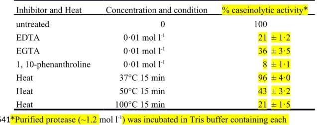
To furtherexplore the possibility that the caseinolytic activity of RpA, purified recombinant proteins were extensively incubated with EDTA, EGTA, or 1,10- phenanthroline, or were heat-treated. In the case of EDTA, a final concentration(0.01 mol l-1) was used because this concentration had previously been demonstrated to be sufficient to completely inhibit extracellular protease activity. Pre-treatment with EDTA, EGTA, or 1,10-phenanthroline, or heat treatment significantly reduced the caseinolytic activity of purified recombinant RpA to the non-treatment control level (Table 1). EDTA, EGTA and 1,10-phenanthroline were effective in blocking the protease activity of thehighest concentration of purified protease tested (1.2 g ml-1).
These results suggest that the majority of the divalent cation-dependent cytotoxic activityobserved in purified recombinant RpA is due to the activityof a heat-liable 319
320 321 322 323 324
325 326 327 328 329 330 331 332 333 334 335 336 337
exo-enzyme.
338
Discussion
Our primary purpose focused on identifyingbacterial-secreted factors within culture filtrates thatcontribute to cytotoxic activity. Some pathogens have been shown to be capable of secreting many known cytotoxic factors in culture filtrates (Finlay and Falkow 1997). A lethal exotoxin to cells in culture (cytolethal toxin [CLT]) was identified in culture filtrates of Burkholderia pseudomallei, the causative organism of melioidosis (Haase et al. 1997). S. marcescens culture filtrates contain a 56-kDa metalloprotease that contributes significantly to in vitro cytotoxic activity (Marty et al. 2002). Culture filtrates from select Moraxella (Branhamella) ovis field isolates
have been shown to have hemolytic activity on bovine erythrocytes and cytotoxic activity on bovine peripheral blood mononuclear and corneal epithelial cells (Cerny et al. 2006). How factors in all bacterial strains interact with host cells will becritical to understanding bacterial pathogenesis. Herein we identified one strain, R. pickettii WP1, that was isolated from aquatic environments and grew well at temperatures between 25°C and 30°C, but not at 37°C. The culture filtrates contributed to cellular
cytotoxicity by exerting damaging effects upon host cells.
As a part of studies toward better understanding cytotoxic factors in culture filtrates of R. pickettii WP1, WP1M2, a mini-Tn5 derivative of WP1, was constructed and the sequence was determined for a proteolytic defective clone. Nucleotide 339
340 341 342 343 344 345 346 347 348 349 350 351 352 353
354 355 356 357
sequence analysis revealed that the mutation site was encoding an open reading frame (rpA) highly homologous (81%) to the sequenced P. fluorescens prtA genome (Genbank accession no. AAD09851) (Ahn et al. 1999). The rpA gene has an ORF of 1449 nucleotides that is capable of coding a 482-amino acid protein of approximately 55 kDa. Bioinformatic analysis revealed that protein RpA shares high identity with protease PrtA (76%) and metalloprotease AprX (73%) of P. fluorescens (Liao and McCallus 1998). The homology of a putative zinc-binding sequence (HExxHxxGxxH) and a calcium-binding domain (GGxGxD) between RpA and bacterial metalloproteases are also conserved, which support the RpA of metalloprotease (Guzzo et al. 1990; Baumann 1994; Liao and McCallus 1998; Ahn et al. 1999). This protein is expressed in E. coli using a pET expression vector and
identification of this protein was confirmed by SDS-PAGE, in which a RpA band of
55 kDa is similar to the size predicted from the nucleotide sequence.
R. pickettii has been shown an ability to survive in environments with a very low
concentration of nutrients. It is easy contamination in ultrapure water in industrial systems (Kulakov et al. 2002). It is also a cause of infection from the environment,
the majority are due to contaminated solutions such as water, saline and sterile drugs (Ryan and Adley 2014). We investigated R. pickettii isolate from the water supply systems and determined the proteolytic activity, focusing in particular on enzyme- 358
359 360 361 362 363 364 365 366 367 368 369
370 371 372 373 374 375 376
secretion activity of cytotoxic to mammalian cells. We also demonstrated decreased cytotoxicity to mammalian cells in a Tn5-induced mutant strain deficient in RpA production. The WP1M2 with reduced RpA protease production has a similar growth rate as WP1 when cultured at 25°C. Although the role of this protease is unclear, the bacteria-secreted proteases could digest tissues and support the in vivo proliferation during infection, as it seems to occur in aquaculture pathogens, such as Vibrio vulnificus (Valiente et al. 2008) and Aeromonus salmonicida (Gunnlaugsdottir and
Gudmundsdottir 1997). The above findings suggest that the in vitro cytotoxic activities of R. pickettii exo-enzyme RpA are predominant protein and present in
cultural filtrates.
Metalloproteases secreted by pathogenic organisms play important roles in virulence (Hase and Finkelstein 1993). It has been demonstrated that the cytotoxic activity in the case of anthrax, botulinum, tetanus (Menard et al. 1996; Haase et al.
1997), and S. marcescensculture filtrates (Marty et al. 2002) is eliminated by using metalloprotease inhibitors. In purified protease of R. pickettii strain WP1, the caseinolytic activity is attenuated by metal chelators (EDTA, EGTA, or 1,10- phenanthroline), which is strongly supported by the hypothesis that RpA protease is a serine protease (Hase and Finkelstein 1993). In this study, an extracellular protease, RpA, which is involved in caseinolytic activity and the biochemical mechanism is the 377
378 379 380 381 382 383 384 385
386 387 388 389 390 391 392 393 394 395
same as divalent-cation-dependent metalloprotease identified in the purified protease of WP1. Furthermore, the RpA activity observed in heat pre-treated WP1 purified protease significantly reduced the caseinolytic activity. This is the first evidence that when expressed, the 55-kDa RpA is sufficient to confer a cytotoxic phenotype on
culture filtrates of WP1.
In summary, we have confirmed that culture filtrates prepared from R. pickettii are cytotoxic to mammalian cells and demonstrate that RpA is a virulence factor that contributes to bacterial infection. This information will be useful in assessing the risk of infection with R. pickettii from contaminated solutions, including water for drinking and medicinal products. This work focused on R. pickettii cytotoxin production as the main factor contributing to in vitro cytotoxicity. Proteolytic activity
by metalloprotease in R. pickettii culture filtrates was first detected in this study.
Acknowledgments
We thank Drs. C-Y Hsiang and C-H Lee of China Medical University, who provided the techniques for expression and purification of recombinant protein, and the
laboratory instruments.
Conflict of Interest
The authors declare that there are no conflicts of interest.
396 397 398 399
400 401 402 403 404 405 406
407 408 409 410
411 412 413
References
Adekoya, O.A. and Sylte, I. (2009) The thermolysin family (M4) of enzymes:
therapeutic and biotechnological potential. Chem Biol Drug Des 73, 7-16.
Ahn, J.H., Pan, J.G. and Rhee, J.S. (1999) Identification of the tliDEF ABC
transporter specific for lipase in Pseudomonas fluorescens SIK W1. J Bacteriol 181, 1847-1852.
Ashdown, L.R. and Koehler, J.M. (1990) Production of hemolysin and other
extracellular enzymes by clinical isolates of Pseudomonas pseudomallei. J Clin Microbiol 28, 2331-2334.
Baumann, U. (1994) Crystal structure of the 50 kDa metallo protease from Serratia marcescens. J Mol Biol 242, 244-251.
Cerny, H.E., Rogers, D.G., Gray, J.T., Smith, D.R. and Hinkley, S. (2006) Effects of Moraxella (Branhamella) ovis culture filtrates on bovine erythrocytes, peripheral
mononuclear cells, and corneal epithelial cells. J Clin Microbiol 44, 772-776.
Coenye, T., Goris, J., De Vos, P., Vandamme, P. and LiPuma, J.J. (2003) Classification of Ralstonia pickettii-like isolates from the environment and clinical samples as Ralstonia insidiosa sp. nov. Int J Syst Evol Microbiol 53,
1075-1080.
Coenye, T., Spilker, T., Reik, R., Vandamme, P. and Lipuma, J.J. (2005) Use of PCR analyses to define the distribution of Ralstonia species recovered from patients 414
415
416 417 418
419 420 421
422 423
424 425 426
427 428 429 430
431 432 433
with cystic fibrosis. J Clin Microbiol 43, 3463-3466.
Coenye, T., Vandamme, P. and LiPuma, J.J. (2002) Infection by Ralstonia species in cystic fibrosis patients: identification of R. pickettii and R. mannitolilytica by
polymerase chain reaction. Emerg Infect Dis 8, 692-696.
de Lorenzo, V., Herrero, M., Jakubzik, U. and Timmis, K.N. (1990) Mini-Tn5 transposon derivatives for insertion mutagenesis, promoter probing, and
chromosomal insertion of cloned DNA in gram-negative eubacteria. J Bacteriol 172, 6568-6572.
Dufour, D., Nicodeme, M., Perrin, C., Driou, A., Brusseaux, E., Humbert, G., Gaillard, J.L. and Dary, A. (2008) Molecular typing of industrial strains of Pseudomonas spp. isolated from milk and genetical and biochemical
characterization of an extracellular protease produced by one of them. Int J Food Microbiol 125, 188-196.
Figurski, D.H. and Helinski, D.R. (1979) Replication of an origin-containing derivative of plasmid RK2 dependent on a plasmid function provided in trans.
Proc Natl Acad Sci U S A 76, 1648-1652.
Finlay, B.B. and Falkow, S. (1997) Common themes in microbial pathogenicity
revisited. Microbiol Mol Biol Rev 61, 136-169.
Gunnlaugsdottir, B. and Gudmundsdottir, B.K. (1997) Pathogenicity of atypical 434
435 436
437 438 439 440
441 442 443 444 445
446 447 448
449 450 451 452
Appl Microbiol 83, 542-551.
Guzzo, J., Murgier, M., Filloux, A. and Lazdunski, A. (1990) Cloning of the Pseudomonas aeruginosa alkaline protease gene and secretion of the protease
into the medium by Escherichia coli. J Bacteriol 172, 942-948.
Haase, A., Janzen, J., Barrett, S. and Currie, B. (1997) Toxin production by
Burkholderia pseudomallei strains and correlation with severity of melioidosis. J
Med Microbiol 46, 557-563.
Hanahan, D. (1983) Studies on transformation of Escherichia coli with plasmids. J Mol Biol 166, 557-580.
Hase, C.C. and Finkelstein, R.A. (1993) Bacterial extracellular zinc-containing
metalloproteases. Microbiol Rev 57, 823-837.
Hsiang, C.Y., Ho, T.Y., Hsiang, C.H. and Chang, T.J. (1998) Recombinant
pseudorabies virus DNase exhibits a RecBCD-like catalytic function. Biochem J 330 ( Pt 1), 55-59.
Hung, C.H., Wu, H.C. and Tseng, Y.H. (2002) Mutation in the Xanthomonas campestris xanA gene required for synthesis of xanthan and lipopolysaccharide
drastically reduces the efficiency of bacteriophage (phi)L7 adsorption. Biochem Biophys Res Commun 291, 338-343.
Ishii, K., Adachi, T., Hamamoto, H. and Sekimizu, K. (2014) Serratia marcescens suppresses host cellular immunity via the production of an adhesion-inhibitory 454
455 456
457 458 459
460 461
462 463
464 465 466
467 468 469 470
471 472 473
factor against immunosurveillance cells. J Biol Chem 289, 5876-5888.
Kulakov, L.A., McAlister, M.B., Ogden, K.L., Larkin, M.J. and O'Hanlon, J.F. (2002) Analysis of bacteria contaminating ultrapure water in industrial systems. Appl Environ Microbiol 68, 1548-1555.
Liao, C.H. and McCallus, D.E. (1998) Biochemical and genetic characterization of an extracellular protease from Pseudomonas fluorescens CY091. Appl Environ Microbiol 64, 914-921.
Marty, K.B., Williams, C.L., Guynn, L.J., Benedik, M.J. and Blanke, S.R. (2002) Characterization of a cytotoxic factor in culture filtrates of Serratia marcescens.
Infect Immun 70, 1121-1128.
Matsumoto, K. (2004) Role of bacterial proteases in pseudomonal and serratial
keratitis. Biol Chem 385, 1007-1016.
McCarthy, M. (2000) Pseudomonas genome reveals a formidable foe. Lancet 356,
918.
Menard, A., Papini, E., Mock, M. and Montecucco, C. (1996) The cytotoxic activity of Bacillus anthracis lethal factor is inhibited by leukotriene A4 hydrolase and
metallopeptidase inhibitors. Biochem J 320 ( Pt 2), 687-691.
Mikulska, M., Durando, P., Pia Molinari, M., Alberti, M., Del Bono, V., Dominietto, A., Raiola, A.M., Van Lint, M.T., Bregante, S., Orengo, G., Bacigalupo, A. and Viscoli, C. (2009) Outbreak of Ralstonia pickettii bacteraemia in patients with 474
475 476
477 478 479
480 481 482
483 484
485 486
487 488 489
490 491 492
haematological malignancies and haematopoietic stem cell transplant recipients.
J Hosp Infect 72, 187-188.
Monod, M., Paris, S., Sanglard, D., Jaton-Ogay, K., Bille, J. and Latge, J.P. (1993) Isolation and characterization of a secreted metalloprotease of Aspergillus fumigatus. Infect Immun 61, 4099-4104.
Pellegrino, F.L., Schirmer, M., Velasco, E., de Faria, L.M., Santos, K.R. and Moreira, B.M. (2008) Ralstonia pickettii bloodstream infections at a Brazilian cancer
institution. Curr Microbiol 56, 219-223.
Phillips, I., Eykyn, S. and Laker, M. (1972) Outbreak of hospital infection caused by
contaminated autoclaved fluids. Lancet 1, 1258-1260.
Pickett, M.J. and Greenwood, J.R. (1980) A study of the Va-1 group of
pseudomonads and its relationship to Pseudomonas pickettii. J Gen Microbiol 120, 439-446.
Ryan, M.P. and Adley, C.C. (2014) Ralstonia spp.: emerging global opportunistic
pathogens. Eur J Clin Microbiol Infect Dis 33, 291-304.
Ryan, M.P., Pembroke, J.T. and Adley, C.C. (2006) Ralstonia pickettii: a persistent
gram-negative nosocomial infectious organism. J Hosp Infect 62, 278-284.
Ryan, M.P., Pembroke, J.T. and Adley, C.C. (2007) Ralstonia pickettii in
environmental biotechnology: potential and applications. J Appl Microbiol 103, 754-764.
494
495 496 497
498 499 500
501 502
503 504 505
506 507
508 509
510 511 512 513
Ryan, M.P., Pembroke, J.T. and Adley, C.C. (2011) Genotypic and phenotypic diversity of Ralstonia pickettii and Ralstonia insidiosa isolates from clinical and environmental sources including High-purity Water. Diversity in Ralstonia pickettii. BMC Microbiol 11, 194.
Sambrook, J., Fritsch, E.F. and Maniatis, T. (1989) Molecular cloning: Cold spring
harbor laboratory press New York.
Sirakova, T.D., Markaryan, A. and Kolattukudy, P.E. (1994) Molecular cloning and sequencing of the cDNA and gene for a novel elastinolytic metalloproteinase from Aspergillus fumigatus and its expression in Escherichia coli. Infect Immun 62, 4208-4218.
Szymanska, J. (2007) Bacterial contamination of water in dental unit reservoirs. Ann Agric Environ Med 14, 137-140.
Valiente, E., Lee, C.T., Lamas, J., Hor, L. and Amaro, C. (2008) Role of the virulence plasmid pR99 and the metalloprotease Vvp in resistance of Vibrio vulnificus
serovar E to eel innate immunity. Fish Shellfish Immunol 24, 134-141.
Vieira, J. and Messing, J. (1991) New pUC-derived cloning vectors with different
selectable markers and DNA replication origins. Gene 100, 189-194.
Wang, S.C., Chung, J.G., Chen, C.H. and Chen, S.C. (2006) 2- and 4-Aminobiphenyls induce oxidative DNA damage in human hepatoma (Hep G2) cells via different 514
515 516
517 518
519 520 521 522
523 524
525 526 527
528 529
530 531 532
Wu, J.W. and Chen, X.L. (2011) Extracellular metalloproteases from bacteria. Appl Microbiol Biotechnol 92, 253-262.
Zhang, W.W., Hu, Y.H., Wang, H.L. and Sun, L. (2009) Identification and characterization of a virulence-associated protease from a pathogenic Pseudomonas fluorescens strain. Vet Microbiol 139, 183-188.
534
535 536 537 538
Table 1. Effect of protease inhibitors and heat on activity of the purified recombinant RpA
Inhibitor and Heat Concentration and condition % caseinolytic activity*
untreated 0 100
EDTA 0·01 mol l-1 21 ± 1·2
EGTA 0·01 mol l-1 36 ± 3·5
1, 10-phenanthroline 0·01 mol l-1 8 ± 1·1
Heat 37°C 15 min 96 ± 4·0
Heat 50°C 15 min 43 ± 3·2
Heat 100°C 15 min 21 ± 1·5
*Purified protease (~1.2 mol l-1) was incubated in Tris buffer containing each
treatment. Each value represents the mean ± standard deviations of three experiments.
The activity in the reaction mixture containing no inhibitor was considered 100%
activity.
539 540
541 542 543 544 545
Figure legends
Figure 1 Spot tests with two strains of Ralstonia pickettii that were incubated in the
top skim milk plate. The results showed a clear lawn of WP1 and no clear lawn of WP1M2 at 25℃ for 24 h (A) and 72 h (B).
Figure 2 Bacterial growth of WP1 strain of Ralstonia pickettii in LB medium at the
temperature of 20 (), 25 (), 30 (▲)and 37 ()℃. The WP1 cells were diluted into 30 ml LB medium to OD600 about 0.15 and grown at different temperatures.
Figure 3 Alignment of the deduced amino acid of RpA with zinc proteases of PrtA
(Pseudomonas fluorescens SIK W1), SM (Serratia marcescens), AprA (Pseudomonas aeruginosa PAO1). Conserved motif HEXXH and four calcium- binding domain are indicated below the alignment. The symbol * - single represent fully conserved residue, : - conservation of strong groups, . - conservation of weak groups and none of - no consensus.
Figure 4 Phylogenetic relationships between zinc proteases from WP1 and related genera. The numbers in parentheses were their Genbank accession number.
Figure 5 Analysis of recombinant protein pET- RpA by 12% SDS-PAGE, M:
546 547 548
549 550 551 552 553 554 555 556 557 558 559 560 561 562 563 564 565
Molecular weight protein marker, Lane 1; pET-RpA induced by IPTG for 4 h, Lane 2, purified recombinant of RpA after eluted from the resin column, Lane 3.
Figure 6 Extracellular protease activity of WP1 () and WP1M2 (). Supernatants
were collected from WP1 and WP1M2 cultured in LB medium at different time and assayed for protease activity using azocasein as the substrate.
Figure 7 Cytotoxic effect of the culture filtrates prepared from R. pickettii WP1 ( ),
WP1M2 ( ) and DH5 ( ) with 0·1, 0·5, 1 and 2 mg/ml protein, respectively.
Cytotoxicity effect was tested on Hela cell (A), HepG2 cell (B), and Vera cell (C), P values < 0·005. Error bars indicate standard deviations.
566 567 568 569 570
571 572 573 574 575 576 577