• 沒有找到結果。

Correlations of Anatomical and Chemical Leaf Characteristics of Eucalyptus Clones with Spontaneous Leaf Spot Disease Severity Associated with Phaeophleospora Fungi

N/A
N/A
Protected

Academic year: 2021

Share "Correlations of Anatomical and Chemical Leaf Characteristics of Eucalyptus Clones with Spontaneous Leaf Spot Disease Severity Associated with Phaeophleospora Fungi"

Copied!
11
0
0

加載中.... (立即查看全文)

全文

(1)

Correlations of Anatomical and Chemical Leaf Characteristics of Eucalyptus Clones with Spontaneous Leaf Spot Disease

Severity Associated with Phaeophleospora Fungi

Sri Rahayu,

1,2)

Ifert Ehrlick Tudon,

1)

Eko Bhakti Hardiyanto

1)

【 Summary】

Recently, leaf spot disease caused by Phaeophleospora spp. fungi has become a severe problem in eucalyptus clonal plantations. These pathogens can kill Eucalyptus trees, and cases can be found in different stages of tree development, ranging from seedlings in nurseries to trees in the field. Before deploying a large-scale Eucalyptus clonal plantation, selecting clones resistance to disease is impor- tant in addition to selecting for optimal growth and wood properties. In this research, we explored an- atomical and chemical leaf characteristics associated with potential leaf spot disease resistance. The study was conducted on a 6-mo-old eucalypt clonal plantation at a forestry company in South Suma- tra, Indonesia. Three selected clones, i.e., clones 79 and 80 (E. pellita × E. brassiana), and clone 47 (pure E. pellita), were assessed for their growth, severity of spontaneously occurring disease, and leaf characteristics (the stomatal density, stomatal size, thickness of adaxial and abaxial palisade meso- phyll, and phenol contents). Clone 79 was susceptible, while clones 47 and 80 were more resistant to the disease. The stomatal size and density and leaf phenol contents assessed from healthy clones were not good indicators for determining resistant clones. The thickness of the abaxial palisade parenchy- ma, however, was negatively correlated with disease severity. Comparing palisade mesophyll thick- ness is suggested to be a quick, simple, and cheap approach for a preliminary assessment of potential resistance against leaf spot disease among different Eucalyptus clones.

Key words: Clonal forestry, leaf disease, palisade mesophyll, Eucalyptus pellita, Eucalyptus brassiana.

Rahayu S, Tudon IE, Hardiyanto EB. 2020. Correlations of anatomical and chemical leaf charac- teristics of Eucalyptus clones with spontaneous leaf spot disease severity associated with Phaeophleospora fungi. Taiwan J For Sci 35(3):239-49.

1)

Faculty of Forestry, Universitas Gadjah Mada, Jalan Agro No. 1, Bulaksumur, Yogyakarta 55281, Indonesia.

2)

Corresponding author, e-mail:sri.rahayu2013@ugm.ac.id 通訊作者。

Received July 2020, Accepted December 2020. 2020年7月送審 2020年12月通過。

(2)

研究報告

桉樹營養系的解剖及化學特徵與

Phaeophleospora spp.真菌

所引起的自發性葉斑病嚴重程度的關係

Sri Rahayu

1,2)

Ifert Ehrlick Tudon

1)

Eko Bhakti Hardiyanto

1)

摘 要

近年來,Phaeophleospora spp.真菌所引起的葉斑病已對桉樹(Eucalyptus)營養系人工林地造成嚴重 影響。從苗圃內的苗木到野外的樹木,均可見到病原菌造成不同生長時期桉樹死亡的案例。在進行大規 模的桉樹造林前,除了考量生長和木材性質良好的性狀外,篩選具抗病性的營養系也顯得相當重要。

本研究中,我們探討了與潛在葉斑病抗性相關的葉片解剖和化學特徵。該研究是在印度尼西亞南蘇門 答臘的一家林業公司之 6個月大桉樹營養系人工林中進行。針對挑選的3個營養系,即營養系79、80 (E.

pellita × E. brassiana)、和47 (E. pellita),分析它們的生長,發病嚴重程度和葉片特徵(氣孔密度、氣孔

大小、近軸和遠軸端柵狀葉肉厚度以及酚含量)。營養系79呈現感病,而營養系47和80則呈現較高抗病 性。健康營養系的氣孔大小,密度和葉酚含量,並不是評估營養系抗病性的良好指標。然而,遠軸端柵 狀葉肉的厚度與染病的嚴重程度呈負相關。比較柵狀葉肉厚度可做為初步評估不同桉樹營養系對葉斑病 潛在抗性一種快速、簡單且便宜的方法。

關鍵詞:營養系林業,葉病,葉柵葉肉,粗皮桉,吉桉。

短期標題:桉樹無性繁殖系的解剖與葉斑病的關係。

Sri Rahayu、Ifert Ehrlick Tudon、Eko Bhakti Hardiyanto。2020。 桉樹營養系的解剖及化學特徵與 Phaeophleospora spp.真菌所引起的自發性葉斑病嚴重程度的關係。台灣林業科學35(3):239-

49。

INTRODUCTION

Eucalyptus species are globally impor- tant planted trees due to their fast growth, valuable wood properties, and wide adapt- ability (Orwa et al. 2009). Several Eucalyptus species have long been grown in many tropi- cal regions, including Indonesia. However, in recent years, only E. pellita has been planted on a large scale across Indonesia due to its resistance or tolerance to pests and diseases (Nambiar et al. 2018). So far, E. pellita is the best alternative species to replace Acacia mangium which is very susceptible to Cera- tocystis disease and monkey and squirrel at- tacks after 3 or 4 successive rotations (Tarigan et al. 2011). Relying only on a single species of Eucalyptus is certainly a risky management

policy; thus other alternatives, i.e., different Eucalyptus species and hybrids, need to be tested for their productivity and resistance to pests and disease.

Eucalyptus hybrids are essentially a

cross-breed between 2 or more eucalypt

species which possess a combination of de-

sirable characteristics. Eucalyptus species

with potential to be developed as hybrids in

Indonesia are limited, as the majority of sites

for plantation development are located in

lowland areas with high rainfall and humidity,

where many eucalypts are unsuitable to be

grown, as they are susceptible to leaf diseases

or poor growth. However, there are a number

of species with high potential for hybrid de-

(3)

velopment, namely E. pellita, E. grandis, E.

brassiana, E. camaldulensis, and E. urophyl- la. Normally, E. pellita serves as the female parent in interspecific hybridizations, combin- ing its best traits, particularly its tolerance to disease, with desirable traits such as growth and wood properties of other species (Prasetyo et al. 2018).

In forestry companies that have adopted clonal forestry techniques, vegetative propaga- tion of the best selected Eucalyptus clones is carried out to a greater or lesser degree of so- phistication to capture the full genetic potential of the clones. The yield of Eucalyptus planta- tions will continue to increase as greater num- bers of improved clones are developed and best silvicultural practices are applied (Zobel 1993).

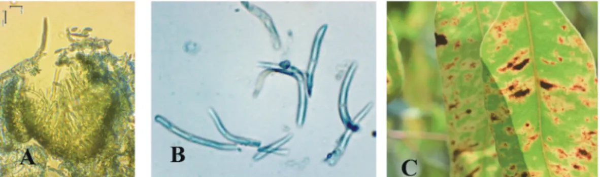
One of the virulent diseases in Euca- lyptus clonal plantations recently observed in South Sumatra is leaf spot disease caused by Phaeophleospora spp. Recently, leaf spot disease has emerged as a significant threat to eucalypt plantations in a number of South East Asian countries including Indonesia, Thailand, and Vietnam, and consequently the disease has been considered worthy of a proper management response.

Phaeophleospora spp. can cause the death of Eucalyptus trees, and they have been detected in different tree development stages, ranging from seedlings in nurseries to trees in the field with symptoms of reddish leaf patches and black spores on the leaf surface (Old et al.

2003, Videira et al. 2017). Before deploying a large-scale plantation of Eucalyptus and its hybrids, selection of clones resistant to disease is of paramount importance in addition to se- lecting for optimal growth and wood proper- ties. To date, all Phaeophleospora species are known to be associated with leaf spot diseases of plants (Taylor and Crous 1999). The asso- ciation between leaf anatomy and resistance to leaf pathogens was investigated in a number

of studies, including Mycosphaerella fijiensis M. Morelet on bananas (Craenen et al. 1997), frog-eye leaf spot (Cercospora sojina Hara) on soya beans (Yang, 2000), and Mycosphae- rella berkeleyi W.A. Jenkins on groundnuts (Jyosthna et al. 2004), as well as Terato- sphaeria leaf disease on Eucalyptus globulus (Smith et al. 2017). Stomata are located at the frontline between internal plant tissues and the surrounding environment, making them convenient gates for phytopathogens including Phaeophleospora spp. However, the infective process of Phaeophleospora spp. has not been studied in detail.

Stomata play an important role as a natu- ral entry point for pathogens; thus leaves with higher numbers of stomata per unit area will have greater chances that fungal mycelia can penetrate (Smith 2006). On the other hand, it was proposed that the first stage of the de- fense mechanism of plants involves a rapid accumulation of phenols at the infection site, which inhibits the growth of pathogens that penetrate into plant cells. Several phenols are regarded as a pre-infection inhibitors, pro- viding plants with a certain degree of basic resistance against pathogenic microorganisms (Satisha et al. 2008). A simple indicator of leaf properties can possibly be used to iden- tify susceptible clones to Phaeophleospora spp. before selected clones are released into the field as operational plantations. The aim of this study was to identify leaf properties (including leaf stomatal properties, the pro- portion of palisade mesophyll, and phenol leaf contents) potentially associated with leaf spot disease resistance of eucalypt clones of E.

pellita and its hybrids.

MATERIALS AND METHODS

Based on a previous preliminary study,

specific leaf spot symptoms caused by Pha-

(4)

eophleospora spp. were clearly observable on 6-mo-old Eucalyptus trees in the field.

Subsequently the study was conducted at a 6-mo-old eucalypt plantation belonging to PT Musi Hutan Persada, a forestry company growing eucalyptus in South Sumatra, Indo- nesia. Three clones, viz., clones 47, 79, and 80, were used in this research. A preliminary study on clones 79 and 80, both E. pellita × E. brassiana hybrids, indicated that they were respectively susceptible and resistant to Pha- eophleospora spp., while clone 47, a pure E.

pellita, was resistant.

The following characteristics were as- sessed: 1) leaf spot disease severity caused by Phaeophleospora spp.; 2) leaf stomatal attri- butes; 3) thickness of the palisade mesophyll;

and 4) constitutive total leaf phenols.

Disease severity of leaf spot caused by Phaeophleospora spp.

In order to assess the severity of spon- taneously occurring leaf spot disease caused by Phaeophleospora spp., an investigation was conducted on 6-mo-old trees in the field.

Trees were planted at a 3 × 2-m spacing. Ob- served traits were disease severity (DS) based on index scores as described in Table 1. Each clone was observed in 3 blocks, and in each block 40 trees were systematically selected, with 4 trees in every row. Leaf samples were systematically taken from 30% of the branch- es of every sample tree (from lower, middle,

and upper stems), with 10 leaves from every branch. Tree height was also measured in order to assess the effect of the disease on the growth of young eucalypt trees.

Disease severity was calculated as fol- lows (modified from Chester 1959):

where z1, z2, … z5 are index scores of leaf spot, n is the number leaves with scores of 1 to 5, N is the total number of leaves, and Z is the highest index score (5).

With regard to DS, the level of tree resis- tance was categorized as follows: resistant (R;

DS = 0~20%), somewhat resistant (SR; DS 21~40%), tolerant (T; DS = 41~60%), some- what susceptible (SS; DS = 61~80%), and susceptible (S; DS = 81~100%).

Leaf stomata

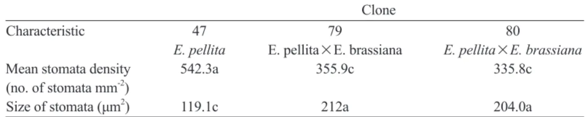
Leaf stomatal density, stomatal size, and stomatal proportion were assessed for clones 47, 80, and 79 at the age of 6 mo. Stomata were observed on 5 parts of each leaf lamina:

the tip, left, right, middle, and base. A thin layer of clear nail polish was spread on the bottom side of the leaf surface and then al- lowed to dry. Clear sticky tape was applied to the top of the nail polish and pressed down to make a good connection with the nail pol- ish and slowly released. The sticky tape was placed on a microscope slide. Slides were ob- Table 1. Index scores of leaf spot symptoms caused by Phaeophleospora spp. on 6-mo-old Eucalyptus clones in the field

No. Stage of symptomatic leaf Index score

1 Necrosis spots 1

2 Necrosis spots with chlorosis 2

3 Necrosis spots with chlorosis and spores emerging 3

4 Necrosis spots with chlorosis, blight, and spores emerging 4

5 Necrosis spots with chlorosis, blight, and spores emerging, followed 5

by leaf curling

(5)

served under an Olympus CX33 light micro- scope (Olympus, Tokyo, Japan) at 1000x and 400x magnifications. Images were taken with an OptiLab camera (Miconos Inc) to calculate and measure the number, size, and density of stomata in each clone.

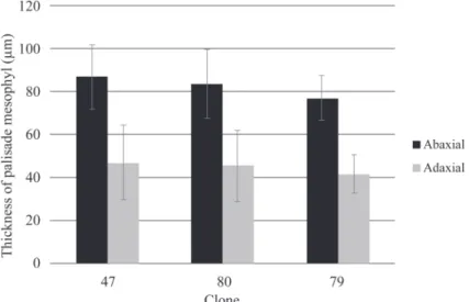
Measurement of palisade mesophyll Leaves were sampled from the 4th leaf pair from the shoot tip to assure that all leaf samples were of the same age. Five healthy leaves were sampled from each healthy 6-mo- old seedling. Leaf samples were prepared by standard free-hand sectioning. Sections were cut with smooth strokes and placed on a Petri plate with water to keep the sample fresh. The sample was then mounted on a microscope slide for observation. Slides were viewed under an Olympus CX33 light microscope.

Images were taken with an OptiLab camera to calculate the adaxial and abaxial palisade mesophyll lengths.

Constitutive total leaf phenols

One pair of healthy Eucalyptus leaves from the 1st 3 nodes was sampled per tree, with 3 trees from each clone. Leaf samples were prepared and extracted following the method outlined by Hagerman (1995).

Freeze-dried, ground leaf material (0.5 g) was placed in a glass test tube with 5 mL of 70% acetone. Samples were sonicated for 30 min at 4℃, and centrifuged at 2800 g min

-

1

for 10 min. The supernatant was decanted and stored at 4℃. The process was repeated a further 3 times with supernatants combined for each sample. Leaf phenolic compounds were determined using the modified Prus- sian blue assay for the total phenolic content (Graham 1992). The concentration of phe- nols (per unit of dry weight) was determined in relation to a gallic acid standard (G-7384 Sigma, St. Louis, MO, USA).

Statistical analysis

An analysis of variance was used to test the effect of different clones on leaf spot dis- ease severity, tree height, stomatal density, size of stomata, thicknesses of the abaxial and adaxial palisade mesophyll, and total leaf phenolic contents. A step-wise multiple linear regression analysis was used to determine which variables were significantly related to disease severity. All analyses were performed using the statistical package of R (vers. 3.6.1).

RESULTS

DS of leaf spot disease

The DS indicates the level of severity of leaf spot disease symptoms. Results from field observations showed that in the 6-mo- old plantation, clone 79 (DS = 76.4%) was somewhat susceptible to Phaeophleospora spp., while clones 80 (DS = 26.6%) and 47 (DS = 26.6%) showed some resistance. The growth performances of clone 79 and 80 at age 6 mo were almost the same, but that of clone 47 was significantly lower than the oth- er clones. Mean heights of clones 79, 80, and 47 were 2.6, 2.1, and 1.8 m, respectively. The correlation between tree height and leaf spot disease severity was moderate and positive (R

2

= 0.47). This indicates that the leaf spot DS tended to increase with increasing tree height. The disease did not seem to disturb the growth of the tree at younger than an age of 6 mo.

Leaf stomatal attributes

Based on the stomatal density and size

investigation (Fig. 1, Table 2), the stomata

density and size of clones 79 and 80 did not

significantly differ, but they significantly

differed from those of clone 47. The size of

stomata in clone 47 was smaller than and

significantly different from those of clone 79

(6)

and 80. These findings indicate that stomatal attributes are not good indicators for identify- ing susceptibility of E. pellita and its hybrid clones to leaf spot disease.

Thickness of the palisade mesophyll Results revealed that the thicknesses of the palisade mesophyll on both the adaxial and abaxial sides of clone 79 were thinner com- pared to those of clones 80 and 47 (Fig. 2). In addition, the DS and thickness of palisade me- sophyll of these 3 clones were strongly nega- tively correlated, with correlation coefficients (r) of 0.88 and 0.94 for the adaxial and abaxial palisade mesophyll thicknesses, respectively.

Leaves with a thicker palisade mesophyll tend- ed to have lower DS and thus may be more resistant to Phaeophleospora spp. infection.

The step-wise multiple regression analyses estimated that leaf spot DS on these 3 clones of Eucalyptus was only significantly related to the thickness of the abaxial palisade mesophyll through the following equation:

LS = 48 – 0.14 PA, with R

2

= 0.92;

where LS is leaf spot DS and PA is the ab- axial palisade mesophyll thickness.

Constitutive total leaf phenols

Results of the phenolic compound con- tent analysis expressed as gallic acid equiva- lents showed that clone 80 had a significantly higher phenolic content than did clone 79 (Fig. 4). In addition, the 2 clones (E. pellita x E. bassiana) had significantly higher phenolic contents than did clone 47 (pure E. pellita).

It is possible that clones 79 and 80 acquired a high content of phenol from E. brassiana;

however an analysis of phenolic compounds in leaves of E. brassiana was not carried out in this study. The correlation between phe- nolic contents and leaf spot DS on 6-mo-old trees was low and negative (r = -0.198). This finding suggests that the phenolic content is not a good indicator for selecting resistant clones to leaf blight spot disease, in spite of the fact that polyphenols are a part of the

Fig. 1. Leaf stomata of 6-mo-old clones: (A) 47 (pure Eucalyptus pellita), (B) Eucalyptus hybrid clone 79, and (C) clone 80 (all pictures were captured at 1000×).

Table 2. Stomatal density and size of stomata of 6-mo-old Eucalyptus clones Clone

Characteristic 47 79 80

E. pellita E. pellita×E. brassiana E. pellita×E. brassiana

Mean stomata density 542.3a 355.9c 335.8c

(no. of stomata mm

-2

)

Size of stomata (μm

2

) 119.1c 212a 204.0a

Notes: Numbers within the same row with the same letter do not significantly differ by the least sig-

nificant difference test at p = 0.05.

(7)

Fig. 2. Palisade mesophyll of 6-mo-old healthy leaves of Eucalyptus clones. (A) Clone 47, (B) clone 80, and (C) clone 79 (all pictures were captured at 400×).

Fig. 3. Average thickness of the palisade mesophyll layer (µm

2

) on 3 Eucalyptus leaf clones.

Bars indicate 1 standard error.

Fig. 4. Phenolic contents as gallic acid equivalents of healthy leaves of clones of Eucalyptus

pellita (47) and its hybrids (79 and 80).

(8)

complex immune system which can be acti- vated in tissues under stress.

DISCUSSION

As there might be a mixture of different Phaeophleospora spp. (Videira et al. 2017) associated with Eucalyptus leaf spot, isolation to obtain pure and specific Phaeophleospora spp. for artificial inoculation experiments will require sophisticated, expensive, and time- consuming assessments, including molecular analyses. Therefore, the assessment of leaf spot severity caused by Phaeophleospora spp. in the present study was conducted using spontaneous infection in the field. In addition, our preliminary study found that infection by Phaeophleospora spp. resulted in specific leaf spot symptoms only in trees aged 6~12 mo.

The most obvious symptom was observed in 6-mo-old trees (Fig. 5C). Subsequently, an evaluation of leaf spot DS based on spontane- ous infection in the field was conducted on 6-mo-old trees. It is suggested that evaluation of leaf spot DS using artificial inoculation with certain species Phaeophleospora can be carried out in the future.

Based on anatomical assessments, it is clear that most conidia of Phaeophleospora spp. entered leaf cells through the stomata,

while most pycnidia were formed on the lower side of the leaf epidermis (Fig. 5A). However, up to now, there is little information about the infection mode of Phaeophleospora spp. in leaves. Smith et al. (2017) reported that stoma- ta abundance of E. globulus had no significant role in determining resistance to Teratosphae- ria leaf disease, which was previously referred to as Mycosphaerella leaf disease.

Research on the response of Eucalyptus species to foliar pathogens is usually con- ducted at the cellular level of young leaves.

For example, in E. globulus, there is some evidence that leaf density (influenced by the internal leaf structure) may contribute to its resistance to Mycosphaerella species (Smith et al. 2006). A higher parenchyma cell density might enhance a plant’s resistance by prevent- ing infection of certain Mycosphaerella spe- cies due to their inability to penetrate closely packed palisade cells (Park et al. 2000). This theory is supported by studies associating the density of palisade parenchyma cells with resistance to several foliar diseases (Basra et al. 1985, Mayee and Suryawanshi 1995, Yang 2000). Although resistance to Mycosphaerella infection is likely due to a combination of several traits, there appears to be a relation- ship between resistance and the thickness of the palisade mesophyll (Smith et al. 2006).

Fig. 5. Conidia of Phaeophleospora spp. in vivo. (A) Pycnidium on the lower epidermis

surface (400x), (B) conidia (1000×) collected from (C) specific leaf spot symptom of 6-mo-old

young Eucalyptus hybrids in the field.

(9)

Higher palisade densities (or volume fractions) were associated with resistance to several leaf spot pathogens, including Cercospora species (Basra et al. 1985), and a tighter packing of palisade cells was also observed in resistant families of E. globulus compared to suscep- tible families. Evidence from past studies and the present study suggests that compact pali- sade layers may slow down or prevent hyphal development due to the inability of some My- cosphaerella species to penetrate and colonize tightly packed palisade cells (Park et al. 2000, Smith et al. 2006). Although deviations in the thickness of palisade mesophyll among resis- tant (clone 47), tolerant (clone 80), and suscep- tible (clone 79) clones appeared very small and might be significant only in direct comparison, most Phaeophleospora spp. pycnidia formed on the lower side of the leaves; thus the role of the abaxial palisade mesophyll appeared to be of paramount importance in preventing hyphal development in leaf cells. In this regard, it is suggested that the thickness of the palisade mesophyll of selected candidate clones be in- vestigated and quantified. Further testing of re- sistant or susceptible clones to leaf spot patho- gens will determine the suitability of using the palisade density and internal leaf structure as indicators for Eucalyptus clone selection.

Compounds such as phenols, salicylic acid, chloroisonicotinic acid, and benzolthi- adiazole-7-carbothioic acid S-methyl ester are able to induce systemic and acquired resistance against a wide range of microbial pathogens in a variety of plants (Sticher et al.

1997). Polyphenols are a part of the complex immune system, which can be activated in tis- sues under stress (Feucht 1994). The involve- ment of phenols in plant disease resistance is based to a large extent on their cytotoxic- ity, which is associated with their oxidation products (Aver’yonav and Lapikova 1994).

Results of our current study suggested that

the phenolic content is not a good indicator for selecting resistant clones to leaf spot dis- ease. In our study, leaf samples were taken from healthy trees. Since the accumulation of phenols normally occurs in infected leaves, the analysis of phenol contents in the future study should be conducted on infected leaves from the field or under artificial inoculation.

CONCLUSIONS

This pioneer study on correlations of anatomical and chemical leaf characteristics of Eucalyptus clones with spontaneous leaf spot disease severity associated with Phaeophleos- pora fungi reports important findings that can be summarized as follows. 1) Clones 79 and 80 (E. pellita × E. brassiana hybrids) and clone 47 (E. pellita) showed different responses to leaf spot disease caused by Phaeophleospora spp. Clone 79 was more susceptible then clone 80, while clone 47 was strongly resistant to leaf spot disease. 2) Leaf stomatal size and density of different clones were poorly cor- related with leaf spot disease severity. 3) The leaf phenolic content was not a good indicator for determining resistant clones. 4) The abaxial palisade mesophyll was the only good indica- tor of resistant clones. Assessing the thickness and number of palisade layers on greater num- bers of different E pellita clones and their hy- brids is recommended to confirm the findings of the current study.

ACKNOWLEDGMENTS

The authors are thankful to the Man-

agement of PT. Musi Hutan Persada for the

permission to conduct the study and its R&D

for providing helpful assistance both in the

field and in the laboratory. Universitas Gad-

jah Mada provided research grant support

through RTA (funding and support for gradu-

(10)

ate research student) in 2019.

LITERATURE CITED

Aver’yanov AA, Lapikova VP. 1994. Partici- pation of active forms of oxygen in the mecha- nism of ferulic acid toxicity. Biol Bull 4:352-7.

Basra RK, Kaur S, Dhillon M. 1985. Ana- tomical and biochemical studies of the resis- tance and susceptibility of the groundnut vari- eties to Cercospora leaf spot. Ann Biol 1:7-12.

Castro-Díez P, Puyravaud J, Cornelissen J.

2000. Leaf structure and anatomy as related to leaf mass per area variation in seedlings of a wide range of woody plant species and types. Oecologia 124:476-86. Doi: 10.1007/

PL00008873

Chester KS. 1959. How sick is the plant? In:

Horsfall JGH, Diamond A editors. Plant pa- thology, Vol. 1. New York: Academic Press.

ISBN: 9780123956774.

Craenen K, Coosemans J, Oritz R. 1997.

The role of stomatal traits and epicuticular wax in resistance to Mycosphaerella fijiensis in banana and plantain (Musa spp.). Tropicultura 15:136-40.

Crous PW, Wingfield MJ. 1997. New species of Mycosphaerella occurring on Eucalyptus leaves in Indonesia and Africa. Can J Bot 75:781-90.

Feucht W. 1994. The localization of phenols at the cellular and tissue level. Acta Horticult 381:803-15.

Graham HD. 1992. Stabilization of the Prus- sian blue colour in the determination of poly- phenols. J Agric Food Chem 40:801-5. Doi:

10.1021/jf00017a018.

Hagerman AE. 1995. Tannin analysis. 2nd ed.

Oxford, OH: Miami Univ.

Jyosthna MK, Reddy NPE, Chalam TV, Reddy GLK. 2004. Morphological and bio- chemical characterization of Phaeoisariopsis personata resistant and susceptible cultivars of

groundnut (Arachis hypogaea). Plant Path Bull 13:243-50.

Mayee CD, Suryawanshi AP. 1995. Struc- tural defence mechanisms in groundnut to late leaf spot pathogen. Ind Phytopathol 48:160-5.

Nambiar EKS, Harwood CE, Mendham DS. 2018. Paths to sustainable wood supply to the pulp and paper industry in Indonesia after diseases have forced a change of species from acacia to eucalypts. Aust For 81:148-61.

Old KM, Wingfield MJ, Yuan ZQ. 2003. A manual of diseases of eucalypts in South-East Asia. Jakarta, Indonesia: CIFOR, Available at http://www.patologiaforestal.com/descargas/

Enfermedades%20en%20Eucalipto.pdf. Ac- cessed 17 October 2019.

Orwa C, Mutua A, Kindt R, Jamnadass R, Anthony S. 2009. Agroforestree Database: a tree reference and selection guide version 4.0.

Available at http://www.worldagroforestry.org/

sites/treedbs/treedatabases.asp. Accessed 20 December 2019.

Park RF, Keane PJ, Wingfield MJ, Crous PW. 2000. Fungal diseases of eucalypt foliage.

In: Keane PJ, Kile GA, Podger FD, Brown BN, editors. Diseases and pathogens of euca- lypts. 153-239. Collingwood, Victoria, Austra- lia: CSIRO.

Prasetyo A, Aiso-Sanada H, Ishiguri F, Wa- hyudi I, Wijaya IPG, Ohshima J, Yokota S.

2018. Growth characteristics and wood proper- ties of two interspecific Eucalyptus hybrids de- veloped in Indonesia. For Prod J 68(4):436-44.

Satisha J, Doshi P, Adsule PG. 2008. Influ- ence of rootstocks on changing the pattern of phenolic compounds in Thompson seedless grapes and its relationship to the incidence of powdery mildew. Turk J Agric For 32:1-9.

Smith A. 2006. The development of strate-

gies for the management and research of foliar

pathogens on eucalypt plantations: using My-

cosphaerella as a case study. Hobart, Tasma-

nia: School of Agricultural Science, Univ of

(11)

Tasmania.

Smith AH, Gill WM, Pinkard EA, Moham- med CL. 2006. Anatomical and histochemical defence responses induced in juvenile leaves of Eucalyptus globulus and Eucalyptus nitens by Mycosphaerella infection. For Pathol. 37:

361–373. Doi: https://doi.org/10.1111/j.1439- 0329.2007.00502.x.

Smith AH, Potts BM, Ratkowsky DA, Pinkard EA, Mohammed CL. 2017. Associa- tion of Eucalyptus globulus leaf anatomy with susceptibility to Teratosphaeria leaf disease.

For Pathol 48(2). https://doi.org/10.1111/

efp.12395.

Stitcher L, Mauch Mani B, Metraux. 1997.

Systemic acquired resistance. Ann Rev Phyto- pathol 35:235-70.

Tarigan M, Roux J, van Week M, Tjahjono B, Wingfield MJ. 2011. A new wilt and die- back disease of A. mangium associated with

Ceratocystis manginecans and C. acaciivora sp. nov. in Indonesia. S Afr J Bot 77:292-304.

Taylor JE, Crous PW. 1999. Phaeophleos- pora faureae comb. nov associated with leaf spots on Faurea safigna (Proteaceae), with a key to the species of Phaeophleospora. Fungal Divers 3:153-8.

Videira SIR, Groenewald JZ, Nakashima C, U Braun, RW Barreto, PJGM De Wit, PW Crous. 2017. Mycosphaerellaceae – chaos or clarity? Stud Mycol 87. Doi: 10.1016/

j.simyco.2017.09.003.

Yang Q. 2000. Germplasm evaluation, inheri- tance, molecular marker and mechanism of resistance to frog-eye leaf spot. J Northeast Agric Univ 7:1-6.

Zobel BJ. 1993. Clonal forestry in the euca- lypts. In: Ahuja MR, Libby WJ, editors. Clonal forestry II. Berlin, Germany: Springer-Verlag.

ISBN: 9783642848155.

數據

Table 2. Stomatal density and size of stomata of 6-mo-old Eucalyptus clones  Clone
Fig. 3. Average thickness of the palisade mesophyll layer (µm 2 ) on 3 Eucalyptus leaf clones
Fig. 5. Conidia of Phaeophleospora spp. in vivo. (A) Pycnidium on the lower epidermis  surface (400x), (B) conidia (1000×) collected from (C) specific leaf spot symptom of 6-mo-old  young Eucalyptus hybrids in the field.

參考文獻

相關文件

In cases of odontogenic sinusitis, thorough examination is crucial as evidenced by the case reported in this study, where a sinusitis caused by a third ectopic molar associated with

194,195 In cases of medication- induced salivary gland hypofunction, the unstimulated whole saliva flow rate is usually significantly reduced, whereas the chewing- stimulated

Results We identified 19 new cases of oral syphilis (17 males, one female, and one case unknown sex) and described the clinical and histopathological features of this re-emerging

Coronavirus disease 2019 (COVID-19), caused by severe acute respiratory syndrome coronavirus 2 (SARS-CoV-2), is a viral infection with multiorgan manifestations that may a ffect the

(A) The scene where the heroine of Guardian: The Lonely and Great God places a maple leaf between the pages of a book was shot in Canada.. (B) The maple leaf on the Canadian

A discussion of the Dapagli flozin and Prevention of Adverse Outcomes in Chronic Kidney Disease (DAPA-CKD) trial, the Effect of Sotagli flozin on Car- diovascular Events in Patients

[Pat+17] Making deep neural networks robust to label noise: A loss correction approach,

provisional mission stations by the Sōtō School along with other schools in the Longshan Temple, had soon made the temple as “a hotly contested spot for monks.” These