行政院國家科學委員會專題研究計畫 成果報告
鈦金屬經水雷射處理後之生物相容性研究 研究成果報告(精簡版)
計 畫 類 別 : 個別型
計 畫 編 號 : NSC 95-2314-B-040-013-
執 行 期 間 : 95 年 08 月 01 日至 96 年 07 月 31 日 執 行 單 位 : 中山醫學大學牙醫學系
計 畫 主 持 人 : 李慈心 共 同 主 持 人 : 黃何雄
計畫參與人員: 學士級-專任助理:陳智華
碩士班研究生-兼任助理:莊羽喬
處 理 方 式 : 本計畫涉及專利或其他智慧財產權,2 年後可公開查詢
中 華 民 國 96 年 11 月 02 日
1. Introduction
The biocompatibility of implant material in the human body is related to the interaction between the living cells and implant material surface. Not all implant material surfaces have truly long-term biocompatibility, even though many biomaterials have been used for clinical implantation. Therefore, various modification techniques have been considered to increase the surface biocompatibility of titanium (Ti) 1-6 which is one of the most popular implant materials for clinical applications. To our knowledge, a simple but efficient method for implant surface modification is not well developed, and currently still under investigation.
Recently, a new generation of Erbium, Chromium: Yttrium, Scandium, Gallium, Garnet (Er,Cr:YSGG) laser uses a combination of laser energy, water, and air to ablate enamel, dentin, bone, and soft tissue 7-12. The wavelength (2780 nm) of the Er,Cr:YSGG laser has an affinity for water. The ablation is accomplished by hydrokinetic energy that prevents the temperature from rising. Although the Er,Cr:YSGG hydrokinetic laser system is widely used for dental purposes, its potential application to implant biocompatibility modification is still not available in the literature.
2. Purpose
This study proposed a fast and useful surface modification method, i.e.
Er,Cr:YSGG laser-powered hydrokinetic system, for improving the biocompatibility of Ti metal. The initial biocompatibility on the laser-treated Ti surface was evaluated.
3. Materials and methods
Commercially pure Ti disks were used as the test substrate (φ: 15 mm; thickness:
1 mm). The Ti substrate surface was ground with SiC paper to #1500 and had a surface roughness of around 0.15 μm. According to our previous study 13, the ground
#1500 Ti surface (Ra 0.15 μm) exhibits the optimal initial cell adhesion condition. An Er,Cr:YSGG-powered hydrokinetic system was utilized to modify the Ti surface using Waterlase® (Biolase Technology, Inc., San Clemente, CA). The Er,Cr:YSGG hydrokinetic laser system was used with a 500-µm laser tip, with the air pressure setting at 45% and the water spray at 20% according to the manufacturer’s advice.
During the laser treatment, the distance between the laser tip and specimen surface was controlled at a value of about 2 mm. In this study, two different laser energy densities, 125 and 190 J/cm2, were applied to the ground Ti substrate. The
corresponding specimens were designated as L-125 and L-190. The specimen without Er,Cr:YSGG laser treatment was used as control group and designated as L-0. Surface topography of the tested Ti specimens was observed using an S-3500 N scanning electron microscope (SEM) and a Nanoscope III atomic force microscope (AFM).
Human osteosarcoma U2-OS cells (American Type Culture Collection (ATCC) HTB-96) were used to imitate the behavior of osteoblasts because this cell line exhibits the osteoblast phenotype 14. Complete McCoy’s 5A medium was supplied to culture the U-2 OS cells with a density of 5 x 10⁴ cell no./cm2 on the test specimens in an incubator with a content of 5% CO2 + 95% air at 37℃. The trypan blue exclusion method and cell counter 15 were used to calculate the number of attached cells on the specimens after 1 and 3 days of cell culture. The difference in the attached cell number between 3- and 1-days cell culture was defined the initial cell proliferation index (CPI), statistically analyzed using one-way analysis of variance (ANOVA) for evaluating the applied laser energy density factor. Tukey’s test (α = 0.05) was chosen as the following multiple-comparison technique when necessary. The specimen number for each test group was five.
The U-2 OS cells were cultured on the Ti test specimens (density: 5 x 10⁴ cell no./cm2) for 1 day. The unattached cells and culture medium were then removed, followed by washing the specimens with phosphate-buffer saline. The cells attached onto the Ti test specimens were sequentially fixed and dehydrated. The surface spreading morphology of the attached cells was observed using a JSM-6700F field-emission scanning electron microscope (FE-SEM) after coating a thin platinum film onto the specimens.
4. Results and discussion
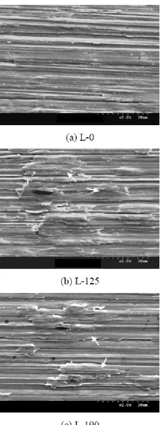
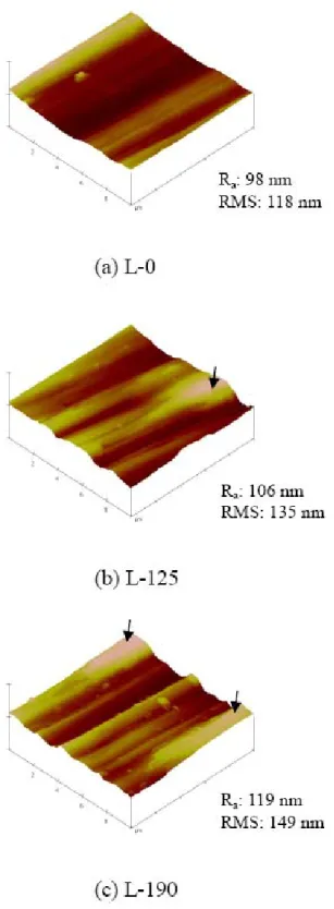
Figs. 1 and 2 show the SEM and AFM observations of the Ti specimens with and without Er,Cr:YSGG hydrokinetic laser treatment. The L-0 specimen (without laser treatment) revealed a surface morphology with parallel grooves ascribed to the mechanical grinding procedure during the specimen preparations. After laser treatment (specimens L-125 and L-190), locally melted morphology was observed on the specimen surface, as indicated by arrows in Figs. 1 and 2. By comparing specimens L-125 and L-190, it showed that increasing the applied energy density from 125 to 190 J/cm2 led to a slight increase in both the melted area (Fig. 1) and surface roughness (Fig. 2). When using the Er,Cr:YSGG hydrokinetic laser, the water molecules become energized and propelled by the laser light. A related article reported that the hydrokinetic laser with lower energy density of 5.6 J/cm2 can result in a macroscopic etched-like surface on enamel, whereas higher power energy results in
visible cavitations 16. In this study, the laser-energized water with the energy densities used could locally melt the Ti surface.
Table 1 shows the initial CPI, difference in the attached cell number between 3- and 1-days cell culture, for Ti specimens with and without Er,Cr:YSGG hydrokinetic laser treatment. One-way ANOVA results showed that the laser energy density had a statistically significant influence on the initial CPI for the Ti specimen (P < 0.001).
Furthermore, Tukey’s test results showed that there was no significant difference between the L-125 and L-190 specimens, although the L-190 specimen showed a slightly higher CPI than the L-125 specimen. It was obvious that the Er,Cr:YSGG hydrokinetic laser system was a potential candidate for improving the initial biocompatibility, in terms of CPI, of a titanium surface.
According to our recent article 13, the ground #1500 Ti with a Ra of 0.15 μm shows the optimal cell adhesion behavior with respect to either the rougher or smoother specimens. In this study, the laser-treated specimens apparently increased (1.2-1.3 times) the initial CPI (P < 0.001) with respect to the ground #1500 Ti specimen. Furthermore, during the Ti surface treatment process via Er,Cr:YSGG hydrokinetic laser system, the total operating period for each specimen was around 90
± 5 s. Compared with some recent Ti surface modification methods, chemical 17,18 or electrochemical 19-21, the laser-powered hydrokinetic system used in this study produced more rapid Ti surface biocompatibility modification.
Fig. 3 shows the FE-SEM observations of the Ti specimens, with and without Er,Cr:YSGG hydrokinetic laser treatment, after 1 day of cell culture: (a) L-0; (b) L-190; (c) and (d) higher magnifications of (a) and (b), respectively, as indicated by the arrow. As shown in Fig. 3, the attached cell with a flattened membrane was observed on the laser-treated L-190 specimen compared to the untreated L-0 specimen.
Similar phenomenon was also observed for the L-125 specimen. When the cell adhesion progresses on implant material surface, a thin membrane develops following the spread of filopodia on cell. The cell grows flattened onto the material surface, indicating high quality of cell growth and material biocompatibility 22. From the initial biocompatibility point of view, the attached cell morphology observation results (Fig. 3) are consistent with the CPI listed in Table 1.
In this study, the improved Ti surface biocompatibility mechanism after Er,Cr:YSGG hydrokinetic laser treatment is still not well understood. However, the physical and/or chemical variations, including the three dimensional change in the surface topography, on the outermost surface of the laser-treated Ti substrate might play an important role in the initial biocompatibility, and needs further investigation.
5. Conclusions
The initial biocompatibility of a Ti surface, in terms of the CPI and cell adhesion morphology, was improved using a fast and efficient surface modification technique, Er,Cr:YSGG laser-powered hydrokinetic treatment. The laser-treated Ti specimen had a higher (1.2-1.3 times) initial CPI (P < 0.001) and better cell spreading morphology.
The specimen with higher applied laser energy had somewhat better biocompatibility, but there was no statistical difference between the laser-treated specimens with dissimilar laser energy. For long-term clinical applications, further investigations on improving the cell differentiation behavior using Er,Cr:YSGG hydrokinetic laser treatment is suggested.
6. References
[1] Huang HH, Hsu CH, Pan SJ, He JL, Chen CC. Corrosion and cell adhesion behavior of TiN-coated and ion-nitrided titanium for dental application. Appl Surf Sci 2005;244:252-256.
[2] Faria AC, Beloti MM, Rosa AL. Nitric acid passivation does not affect in vitro biocompatibility of titanium. Int J Oral Maxillofac Implants 2003;18:820-825.
[3] Kim YH, Koak JY, Chang IT, Wennerberg A, Heo SJ. A histomorphometric analysis of the effects of various surface treatment methods on osseointegration. Int J Oral Maxillofac Implants 2003;18:349-356.
[4] De Maeztu MA, Alava JI, Gay-Escoda C. Ion implantation: surface treatment for improving the bone integration of titanium and Ti6Al4V dental implants.
Clin Oral Implants Res 2003;14:57-62.
[5] Sohmura T, Tamasaki H, Ohara T, Takahashi J. Calcium-phosphate surface coating by casting to improve bioactivity of titanium. J Biomed Mater Res 2001;58:478-485.
[6] Wieland M, Textor M, Spencer ND, Brunette DM. Wavelength-dependent roughness: a quantitative approach to characterizing the topography of rough titanium surfaces. Int J Oral Maxillofac Implants 2001;16:163-181.
[7] Miller RJ. Treatment of the contaminated implant surface using the Er,Cr:YSGG laser. Implant Dentistry 2004;13:165-170.
[8] Jacboson B, Berger J, Kravitz R, Patel P. Laser pediatric crowns performed without anesthesia: a contemporary technique. J Clin Pediatr Dent 2003;28:11-12.
[9] Rosenberg SP. The use of the erbium, chromium: YSGG laser in microdentistry. Dentistry Today 2003;22:70-73.
[10] Chen WH. YSGG laser root canal therapy. Dentistry Today 2002;21:74-77. H
[11] Lin S, Caputo AA, Eversole LR, Rizoiu I. Topographical characteristics and shear bond strength of tooth surfaces cut with a laser-powered hydrokinetic system. J Prosthet Dent 1999;82:451-455.
[12] Rizoiu I, Kohanghadosh F, Kimmel AI, Eversole LR. Pulpal thermal responses to an erbium,chromium: YSGG pulsed laser hydrokinetic system. Oral Surg Oral Med Oral Pathol Oral Radiol Endod 1998;86:220-223.
[13] Huang HH, Ho CT, Lee TH, Lee TL, Liao KK, Hsu CC. Effect of surface roughness of ground titanium on initial cell adhesion. Biomol Eng 2004;21,93-97.
[14] Nelissen JM, Torensma R, Pluyter M, Adema GJ, Raymakers RA, van Kooky Y, Figdor CG. Molecular analysis of its hematopoiesis supporting osteoblastic cell line U2-OS. Exp Hematol 2000;28:422-432.
[15] Freshney RI. Culture of animal cells- a manual of basic technique. New York:
Wiley-Liss; 2000. p. 310.
[16] Ü ümez S, Orhan M, Ü ümez A. Laser etching of enamel for direct bonding with an Er,Cr:YSGG hydrokinetic laser system. Am J Orthod Dentofacial Orthp 2002;122:649-656.
[17] Kim HM, Kokubo T, Fujibayashi S, Nishiguchi S, Nakamura T. Bioactive macroporous titanium surface layer on titanium substrate. J Biomed Mater Res 2000;52:553-557.
[18] Fujibayashi S, Neo M, Kim HM, Kokubo T, Nakamura T. Osteoinduction of porous bioactive titanium metal. Biomaterials 2004;25:443-450.
[19] Zhu X, Ong JL, Kim S, Kim K. Surface characteristics and structure of anodic oxide films containing Ca and P on a titanium implant material. J Biomed Mater Res 2002;60:333-338.
[20] Rosenberg R, Starosvetsky D, Gotman I. Surface modification of a low modulus Ti-Nb alloy for use in medical implants. J Mater Sci Lett 2003;1:29-32.
[21] Huang HH, Pan SJ, Lai YL, Lee TH, Chen CC, Lu FH. Osteoblast-like cell initial adhesion onto a network-structured titanium oxide layer. Scripta Mater 2004;51,1017-1021.
[22] Orsini G, Assenza B, Scarano A, Piatelli M, Piattelli A. Surface analysis of machined versus sandblasted and acid-etched titanium implants. Int J Oral Maxillofac Implants 2000;15:779-784.
7. 計劃成果自評
本研究計畫之成果已達預期目標,並有足夠之數據能發表於國際期刊,目前 正進行研究成果之彙整及撰寫論文。
Table 1 Initial cell proliferation index, difference in attached cell number between 3- and 1-days cell culture, of Ti specimens with and without Er,Cr:YSGG laser treatment: different letter (A and B) means significantly different; * standard deviations are given in parentheses.
Materials Cell Proliferation Index L-0 6.58 x 10⁴ (3.51 x 10³) A*
L-125 7.94 x 10⁴ (3.65 x 10³) B L-190 8.51 x 10⁴ (3.09 x 10³) B
Fig. 1 SEM observations of the Ti specimens with and without Er,Cr:YSGG hydrokinetic laser treatment.
Fig. 2 AFM observations of the Ti specimens with and without Er,Cr:YSGG hydrokinetic laser treatment.
Fig.3 FE-SEM observations of the Ti specimens, with and without Er,Cr:YSGG hydrokinetic laser treatment, after 1 day of cell culture: (a) L-0; (b) L-190;
(c) and (d) higher magnifications of (a) and (b), respectively, as indicated by the arrow.