Research Express@NCKU - Articles Digest
1 of 3
Research Express@NCKU Volume 23 Issue 9 - April 26, 2013
[ http://research.ncku.edu.tw/re/articles/e/20130426/4.html ]IL-20 is regulated by hypoxia-inducible factor and
up-regulated after experimental ischemic stroke
Ming-Shi Chang
Department of Biochemistry and Molecular Biology, College of Medicine, National Cheng Kung University
brianhsu47@gmail.com
The Journal of Immunology, 2009, 182: 5003–5012
I
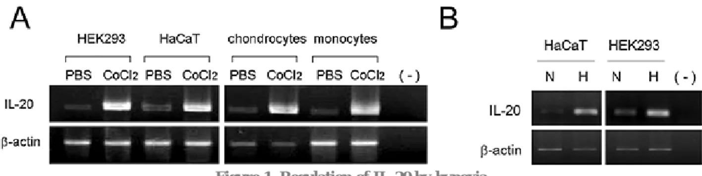
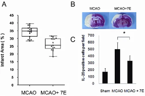
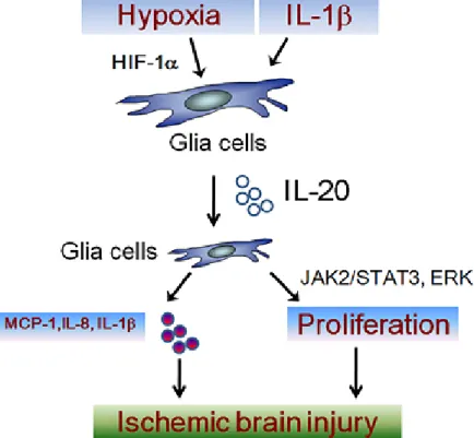
L-20, an IL-10 family member, is involved in various inflammatory diseases, such as psoriasis, rheumatoid arthritis, and atherosclerosis. Our previous study showed that IL-20 acts as a pro-atherogenic factor in atherosclerosis. In this study, we investigated whether hypoxia in vitro and an in vivo model of ischemic stroke would upregulate IL-20 expression. In vitro, IL-20 expression increased in hypoxic HaCaT, HEK293 cells, chondrocytes, and monocytes (Figure 1). We identified two putative hypoxia response elements in human il20-gene promoter. RT-PCR analysis showed that glioblastoma cellsGBM8901 cells expressed IL-20 and its receptors. IL-20 induced cell proliferation and also induced the production of IL-1β, IL-8, and MCP-1 in GBM8901 cells(Figure 2). In vivo, experimental ischemic stroke upregulated IL-20 in the sera and brain tissue of rats. IL-20 stained positively in glia-like cells in peri-infarcted lesions, but not in contralateral tissue. Administration of IL-20 monoclonal antibody ameliorated ischemia-induced brain infarction of rats after experimental ischemic stroke (Figure 3). We conclude that IL-20 is a pro-inflammatory cytokine and regulated by hypoxia. The upregulation of IL-20 may contribute to the pathogenesis of brain injury after ischemic stroke (Figure 4). Therefore, our findings provide evidence that IL-20 is a novel target, and that 7E may be a potential therapeutic for ischemic stroke.
Figure 1. Regulation of IL-20 by hypoxia
(A) HEK293, HaCaT, primary chondrocytes, and primary human monocytes were treated with PBS or CoCl2 (100 μM) for 6 hours and then RNA was extracted for RT-PCR analysis. (B) Normoxic (N, 21% O2) and
hypoxic (H, 1% O2) HaCaT and HEK293 cells were incubated for 24 hours, and then, using RT-PCR, IL-20 expression was analyzed. Representative results were obtained from 3 independent experiments.
Research Express@NCKU - Articles Digest
2 of 3
Figure 2. Induction of cytokines and chemokines in GBM8901 cells by IL-20
GBM8901 cells were treated with PBS, IL-20 (100 ng/ml, 200 ng/ml), anti-IL-20 mAb (7E, 1 μg/ml), 7E (1 μg/ml) plus IL-20 (200 ng/ml), IL-1β (30 ng/ml), and IL-1β (30 ng/ml) plus IL-20 (200 ng/ml) for 24 hours in
serum-free condition. The conditioned medium was collected and analyzed with ELISA kits for IL-1β, IL-8, MCP-1, and MIF. * < 0.05, compared to the PBS-treated group. # < 0.05, compared to the IL-20 (200
μg/ml)-treated group. Representative results were obtained from 3 independent experiments.
Figure 3. Amelioration of ischemic brain infarction in rat after MCAO by anti-IL-20 monoclonal antibody 7E
(A) Rats (n=15 each) underwent 90 minutes of MCAO and ischemic reperfusion were intravenously injected with PBS or anti-IL-20 mAb (10mg/kg). The brain slices were analyzed for the lesion areas 3 days after MCAO
using TTC staining. Lines inside the boxes represent the median. Differences between the 7E, and control groups are statistically significant (P<0.05). (B) Representative lesion areas analysis of the rat brain slices using TTC staining. (C) Quantification of the number of IL-20-positively stained cells in the ischemic-infarcted lesion
Research Express@NCKU - Articles Digest
3 of 3
Figure 4. Schematic working model of the role of IL-20 in ischemic stroke
IL-20 can be induced by hypoxia and IL-1β in glia cells. Glia cells are target cells for IL-20. IL-20 acts on glia cells induced cell proliferation through ERK and STAT3 dependent pathway, and induced production of
![HPSH [ 氧化數平衡反應式係數 ]](data:image/gif;base64,R0lGODlhAQABAIAAAP///wAAACH5BAEAAAAALAAAAAABAAEAAAICRAEAOw==)