Semi-Quantitative Expression of Tissue Inhibitor of
Matrix Metalloproteinase-2 in Cancer of Uterine Cervix
YAO-CHUNG WU, MD,1,2y PO-HUI WANG, MD, PhD,1,3,4* AMY TSAI, MD,3 SHUN-FA YANG, PhD,1,5,6y AND SHIUAN-CHIH CHEN, MD, PhD1,3,7y
1Institute of Medicine, Chung Shan Medical University, Taichung, Taiwan 2Department of General Surgery, Changhua Christian Hospital, Changhua, Taiwan 3School of Medicine, Chung Shan Medical University, Taichung, Taiwan 4Department of Obstetrics and Gynecology, Chung Shan Medical University Hospital, Taichung, Taiwan 5Department of Clinical Laboratory, Chung Shan Medical University Hospital, Taichung, Taiwan 6Department of Medical Research, Chung Shan Medical University Hospital, Taichung,
Taiwan
7Department of Family and Community Medicine, Chung Shan Medical University Hospital, Taichung, Taiwan
Background and Objective: Because matrix metalloproteinase-2 (MMP-2) is associated with tumor progression and tissue inhibitor of MMP-2 (TIMP-2) selectively inhibits MMP-2, we investigate the implication of TIMP-2 in carcinogenesis of uterine cervix.
Methods: Twenty-six cervical cancer tissues and their normal counterparts were collected to evaluate semi-quantitative mRNA expression of TIMP-2. Eighty-two cervical cancer, 26 high-grade and 26 low-grade dysplasia, and 26 normal tissues were collected to construct tissue microarrays for immunohistochemical study. We evaluated TIMP-2 immunoreactivity using H scores in cervical carcinogenesis. Semi-quanti- tative expression of MMP-2 was also detected for comparison.
Results: Cervical cancer tissues exhibited statistically lower semi-quantitative TIMP-2 (P ¼ 0.028) or higher MMP-2 (P ¼ 0.036) mRNA expression than their normal counterparts. None of cervical cancer tissues exerted elevated TIMP-2 and reduced MMP-2 mRNA expression simultaneously. Cancer tissues have significantly lower TIMP-2 or higher MMP-2 H scores than high-grade and low-grade dysplasia or normal tissues of uterine cervix.
K
EYW
ORDS: carcinogenesis of uterine cervix; semi-quantitative; real time polymerase
chain reaction; tissue microarrays; H score
INTRODUCTION
Cancer of uterine cervix is the most common cancer of lower genital tract and the fifth common type of malignancy in women in Taiwan. The 2007 age standardized incidence rate for cervical cancer in Taiwan was reported to be 12.2 per 1,00,000 women by Bureau of Health Promotion of the Department of Health in this country. Cer-vical intraepithelial neoplasia (CIN) is regarded as a precancerous lesion that may progress to invasive carcinoma. According to the potential of malignant transformation in ascending order from the Bethesda System, precancerous lesions are mainly categorized into low-grade squamous intraepithelial lesions (LSILs), including fea-tures of HPV infection and CIN 1 (confined to the basal 1/3 of the epithelium), which is pathologically compatible with mild dysplasia (low-grade dysplasia) and high-grade squamous intraepithelial lesions (HSILs), including CIN 2 (confined to the basal 2/3 of the epithelium) and CIN 3 (2/3 or full thickness of the entire epi-thelium), which are pathologically compatible with moderate dysplasia, severe dysplasia, and carcinoma in situ (high-grade dysplasia) [1].
The transformation from cervical preinvasive neoplasia to inva-sive carcinoma accompanies by focal disruption of the subepithelial basement membrane. Matrix metalloproteinases (MMPs), potent proteolytic enzymes, are known to play key roles in this process and able to degrade a variety of substrates those are major components of the basement membrane and extracellular membrane [2]. Overex-pression of several MMPs has been found in many kinds of cancers, such as lung cancer [3]. A 72-kDa type IV collagenase (MMP-2),
gelatinase A, cleaves collagen type IV, a major component of base-ment membrane. Its collagenolytic activity has been found highest among MMPs and been closely associated with tumor progression [4]. Tissue inhibitor of matrix metalloproteinases (TIMPs) function as key inhibitors of MMPs in tissues by binding to the active site and then forming stable enzyme-inhibitor complexes, which become inactive [5]. TIMPs bind MMPs in a 1:1 stoichi-ometry and four TIMPs (TIMP-1, TIMP-2, TIMP-3, and TIMP-4) have been identified in vertebrates [2]. Imbalances in the extracellu-lar activities of MMPs and TIMPs have been associated with pathological tissue destruction, which is found in cancer [6]. However, TIMPs differ in their specificity for MMPs inhibition, with TIMP-2 having higher affinity for MMP-2 [5].
Our previous study was focused on the clinical significance of MMP-2 for clinicopathological variables and prognosis of cervical
cancer patients [7]. Only lymph node metastasis and parametrium invasion are correlated with MMP-2 immunoreactivity. The study mentioned that MMP-2 is highly expressed in cervical cancer tissues at mRNA and protein levels as compared to their normal counter-parts. Because TIMP-2 selectively inhibits MMP-2 and MMP-2 expression is elevated in cervical cancer, we further delineate the expression of TIMP-2 in cervical carcinogenesis. We previously used reverse transcription-polymerase chain reaction (RT-PCR) to compare qualitative mRNA expression of MMP-2 between cancer tissues and their normal counterparts. To the best of our knowledge, few reports address the semi-quantitative expression of TIMP-2 using real time PCR and tissue microarrays in cancer of uterine cervix. We hypothesized that reduced TIMP-2 expression may be found in cervical tumorignesis. In this study, we emphasize on the implication of TIMP-2 in cervical carcinogenesis using semi-quantitative methods.
MATERIALS AND METHODS
Recruitment of Tissue Samples, Reverse
Transcription (RT), and Real Time
Quantitative Polymerase Chain Reaction
(PCR)
Twenty-six cancer tissues of uterine cervix were recruited from patients with cervical cancer, who were admitted to the Department of Obstetrics and Gynecology in Chung Shan Medical University Hospital and received the standard treatment protocols in the hospital between June 2001 and December 2008. Their normal counterparts, which were far away from cancer lesions and confirmed by pathology slides, were also collected from these patients. All fresh tissues were snap-frozen and stored in liquid nitrogen.
The Trizol (Life Technologies, Grand Island, NY) was used to extract total RNA from 100 mg of tissue according to manufacturer’s recommendations. Reverse transcription was applied to obtain cDNA by 100 units of moloney murine leukemia virus reverse transcriptase (MMLV, Promega) from 2 mg total cellular RNA with random primers (hexamers, Promega, Madison, WI). It was performed at 70 8C for 10 min and subsequently followed by 90 min at 42 8C to end the reaction. Real time quantitative PCR was carried out using 25 ml reaction mix. It comprised 5 ml of cDNA, 12.5 ml of 2× TaqMan Universal PCR Master Mix (Applied Biosystems, Foster City, CA), 0.5 ml of ROX buffer, 1.25 ml of 20× Assay-on-Demand gene expression assay mix (TIMP-2 probe: Hs00234278_m1, MMP-2 probe: Hs00MMP-2344MMP-2MMP-2_m1), and 5.75 ml of RNase-free H2O,
based
on manufacturer’s instructions. Then, the analysis was performed on ABI Perkin Elmer Prism 7700 Sequence Detection System (Applied Biosystems). Each data point was repeated thrice. The PCR reaction consisted of 50 8C 2 min and 95 8C 15 min, followed by 40 cycles, each contained 95 8C 15 sec and 50 8C 30 sec. Quantitative values were calculated from the threshold PCR cycle number (CT), where
the increase in signal associated with an exponential growth of PCR product became detectable. The relative mRNA levels of TIMP-2 and MMP-2 in each sample were normalized to their GAPDH content (GAPDH sense and anti-sense primer: 50 GCACCGTCAA
GGCTGAGAAC30 ; 30 GCCTTCTCCATGGTGGT-GAA50 ; GAPDH
probe: CAL Gold50 CCCATCACCATCTTCCAGGAGCGAG30 BHQ-1).
The expression levels of relative target gene were obtained using the formula: 2—DCT, DCT ¼ C
T (target gene) — CT (GAPDH).
Immunohistochemical Expression of
TIMP-2 in Cancer, High-Grade and
Low-Grade Dysplasia and Normal Tissues
Using Cervical Tissue Microarrays
We constructed the formalin-fixed, paraffin-embedded tissue microarrays (MaxArray tissue cores, Zymed, South San Francisco,
CA) composed of above 26 cancer and another 56 cancer tissue cores as well as 26 high-grade and 26 low-grade dysplasia, and 26 normal tissue cores of uterine cervix. These tissues were recruited from the histopathologic archives of the Pathology Departments in Chung Shan Medical University. Eighty-two patients with cervical cancer were staged IB or IIA based on the International Federation of Gynecology and Obstetrics classification system. All these patients had undergone radical abdominal hysterectomy and pelvic lymph node dissection or para-aortic lymph node sampling between June 2001 and December 2008. All patients received the routine treatment protocols established by the hospital. Twenty-six patients with HSIL had received large loop excisions of the transformation zone, total abdominal hysterectomy (TAH), or total vaginal hyster- ectomy (TVH). The pathological diagnosis of these samples revealed moderate, severe dysplasia, or squamous cell carcinoma (SCC) in situ (high-grade dysplasia of uterine cervix). Twenty-six patients had LSIL for which they received colposcopy-directed punch biopsies, all found by pathological studies to have mild dysplasia (low-grade dysplasia of uterine cervix). If these cases had discrepancy between cytologic and histologic diagnosis, the cervical tissue samples were excluded in this study. Twenty-six normal tissue specimens of uterine cervix were collected from patients receiving TAH or TVH for benign uterine disease without cervical lesions. Uterine cervix tissues were defined as normal if no precancerous lesions or invasive cancers were found. Chung Shan University Hospital Institutional Review Board has approved our study. Informed consent was obtained from each patient.
We used immunohistochemical method to evaluate the immunos-tainings of cervical specimens in our tissue microarrays. The tissue microarray slide, which was 5-mm thick, was de-waxed in xylene and re-hydrated in a series of ethanol solutions. Tissue sections
were incubated using anti-TIMP-2 [1:200 dilution, TIMP-2 Ab-5 (Clone 3A4) mouse monoclonal antibody; Thermo Fisher Scientific, Fremont, CA] or anti-MMP-2 (1:100 dilution, mouse monoclonal antibody; Zymed, CA) at 4 8C overnight. Following primary anti-body incubation, the microarray slides were stained using a LSAB kit by the streptavidin–biotin–peroxidase method (DAKO, Glostrup, Denmark). The chromagen (0.04% 30, 30-diaminobenzidine tetrahy- drochloride) was applied to produce brown color in stained cells. The inflamed colon tissues were used as positive controls for
TIMP-2 and MMP-TIMP-2 immunostainings. Negative control stainings were done by using control antibodies for the inflamed colon, which are same isotype immunoglobulins of TIMP-2 or MMP-2.
The immunostaining intensity of TIMP-2 or MMP-2 in cancer, precancerous and normal tissue cores was evaluated by the Pro Plus
6.1 image analysis program (Media Cybernetic, Inc., Silver Spring, MD). The immunoreactivity intensity was graded by assessing the mean of integrated optic density in a fixed field with a total pixel area of about 250 × 250 on a scale of 0–3; the higher the number the greater the immunoreactive intensity [7,8]. The mean signal intensity of the positive control (inflamed colon) for TIMP-2 or MMP-2 was 80.9 T 26.3 or 132.2 T 11.6 (mean T standard deviation). Mean value plus one standard deviation for TIMP-2 and MMP-2 were 107.2 and 143.8, respectively. The cervical tissues showing a mean digital density of more than 107.2 for TIMP-2 or
143.8 for MMP-2 were assigned a score of 3. Mean value minus one standard deviation for TIMP-2 and MMP-2 were 54.6 and 120.6, respectively. Cervical tissues showing a digital intensity between
54.6 and 107.2 for TIMP-2 or between 120.6 and 143.8 for MMP-2 were assigned a score of 2. The cervical tissues exhibiting a digital density of less than 54.6 for TIMP-2 or 120.6 for MMP-2 were
assigned a score of 1. When there was no staining, a score of 0 was assigned. Because less expression of MMP-2 and TIMP-2 may be found in fibroblasts (stroma cells) and vascular walls, only immunos-taining intensity, that was stronger than that of fibroblasts and vascular wall, was included into
analysis of Pro Plus 6.1 image analysis program. Positive cells were defined as cells those have stronger immunostaining than fibroblasts or vascular walls. The pro- portion of positive cells was calculated for each tissue core. Three
TABLE I. mRNA Levels
of Matrix Metalloproteinase-2 (MMP-2) and Tissue Inhibitor of Matrix Metalloproteinase-2 (TIMP-2) in 26 Pairs of Cancer Portions and
Their Normal
Counterparts of
Cancers of Uterine Cervix
images of representative areas were acquired and at least 300 cells per tissue core section were manually counted on a computer screen. The proportion of positive cells was assigned as none, 0; less than one tenth, 0.1; less than one half, 0.5; and greater than one half, 1 [7,8]. This proportion score was multiplied by the score of immunor- eactivity intensity to obtain a final semi-quantitative H score of TIMP-2 or MMP-2 immunostaining. The H score of TIMP-2 or MMP-2 for each tissue core was evaluated by two observers blindly.
Statist
ical
Analy
sis
We compared the mRNA expression ratio of TIMP-2/GAPDH or MMP-TIMP-2/GAPDH between 26 cervical cancer tissues and their normal counterparts using Wilcoxon signed-rank test by SPSS statistical package. Furthermore, a Kruskal–Wallis H test was used for non- parametric analysis of H score of TIMP-2 or MMP-2 among 26 nor- mal, 26 low-grade dysplasia, 26 high-grade dysplasia, and 82 cancer tissue cores. In response to Mann– Whitney U test for independent multiple comparisons, we adjusted the P values using Hommel’s method by WinPepi software. A P value of less than 0.05 was con- sidered significant.
MMP-2 mRNA levels (ratioa) in cancer portionsb MMP-2 mRNA levels (ratioa ) in norm al portio ns TIM P-2 mRN A levels (ratioa ) in cance r portio nsb TIMP-2 mRNA levels (ratio a) in norm al porti ons
R
E
S
U
L
T
S
2273.83 84.67 150.60 62.41 1419.64 197.52 90.14 255.16 187.44 29.24 75.63 14.35 13.83 362.33 33.17 267.66 22.74 96.20 3.61 154.36 44.94 58.32 13.29 151.25 462.16 48.77 126.82 109.87 524.83 1938.15 3228.81 4383.04 112.65 27.06 6.00 13.48 6792.87 45.91 619.36 21.04 1.37 63.36 1.21 132.22 120531.96 1240.20 357.04 1180.94 20225.06 32.88 16.59 10.74 772.37 101.48 92.35 97.00 110.91 48.80 45.79 89.47 1060.96 685.56 345.99 784.36 287.09 194.41 121.37 135.19 2617.32 61.36 1419.04 329.40 30451.06 100.00 17435.69 17452.62 71.51 231.72 31.91 399.56 1477.30 78.43 403.66 52.68 69.65 664.91 35.83 278.32 79.03 1988.09 74.97 2698.69 375.81 257.82 201.77 232.78 446.91 263.20 100.00 522.44The semi-quantitative mRNA expression ratio of MMP-2/GAPDH was found to be statistically higher in 26 cervical cancer tissues than in their normal counterparts using real time PCR (P ¼ 0.036;
25.87 389.40
13.11 1033.99
Table I). Whereas, the mRNA expression ratio of TIMP-2/GAPDH was found to be statistically
higher in normal tissues than in their cancer counterparts (P ¼ 0.028; Table I). In these 26 pairs of cervi- cal tissue specimens, 10 cancer portions had lower TIMP-2 and higher MMP-2; 7 higher TIMP-2 and MMP-2; and 9 lower TIMP-2 and MMP-2 mRNA expression than their normal counterparts. No cervical cancer sample simultaneously exhibited higher TIMP-2 and lower MMP-2 mRNA expression than their normal counterparts.
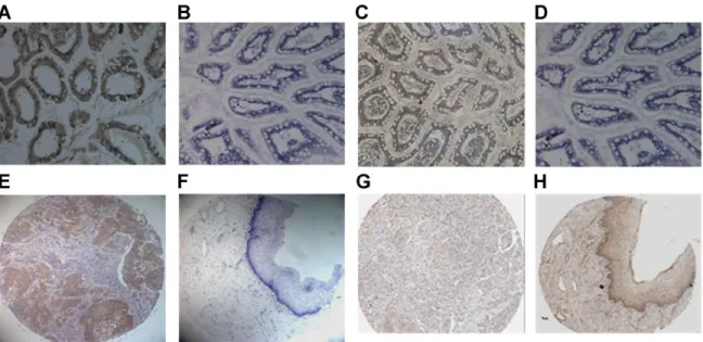
In the immunohistochemical study of TIMP-2 and MMP-2 expression, slides containing inflamed colon tissues were used as positive (incubated with anti-MMP-2 and anti-TIMP-2 mouse mono- clonal antibodies; Fig. 1A and C) and negative controls (incubated with goat serum instead of primary MMP-2 and TIMP-2 antibodies; Fig. 1B and D) of MMP-2 and TIMP-2 immunoreactivities based on manufacturers’ instructions. The immunostaining of MMP-2 was showed stronger in the cytoplasm of cancer cells (Fig. 1E) and may be found weaker in normal squamous epithelial cells of uterine cer- vix (Fig. 1F). However, the immunostaining of TIMP-2 was showed weaker in the cytoplasm of cancer cells (Fig. 1G) and may be found stronger in normal squamous epithelial cells (Fig. 1H).
The TIMP-2 and MMP-2 immunostainings of cancer and normal tissue cores in our cervical tissue arrays were cytoplasmic and had different H scores (Fig. 1E–H). There were different H scores for MMP-2 immunoreactivities among normal, low-grade and high-grade dysplasia, and cancer subgroups (P < 0.001; Fig. 2A). The significantly different H scores were found between 82 cancer and
26 normal (adjusted Hommel’s P ¼ 0.003), between 82 cancer and 26 low-grade dysplasia (adjusted Hommel’s P ¼ 0.007) and between
82 cancer and 26 high-grade dysplasia (adjusted Hommel’s
P ¼ 0.007) tissue cores. There
were also different H scores for TIMP-2 immunoreactivities among normal, low-grade and high- grade dysplasia, and cancer subgroups (P ¼ 0.001; Fig. 2B). The H scores of TIMP-2 immunostainings were significantly higher in 26 normal cervical tissue cores than 82 cancer tissue cores (adjusted
a
The relative mRNA level of MMP-2 or TIMP-MMP-2 in each sample is normalized to its GAPDH content. The values mean the ratio between TIMP-2 or MMP-2 and GAPDH mRNA and are presented after they are multiplied by 100 in the cancer portions and their normal counterparts.
bA significant difference (P < 0.05) of mRNA expression of MMP-2 or
TIMP-2 between cancer tissues and their normal counterparts. Statistic
analysis: Wilcoxon signed-rank test.
Hommel’s P ¼ 0.004). Twenty-six low-grade (adjusted Hommel’s P ¼ 0.003) and 26 high-grade dysplasia (adjusted Hommel’s P ¼ 0.021) tissue cores also had higher immunostainings as com- pared to 82 cancer tissue cores.
D
I
S
C
U
S
S
I
O
N
The semi-quantitative mRNA expression of TIMP-2 was found to be statistically lower in cancer tissues than their normal counterparts. In contrast, cervical cancer tissues displayed higher mRNA expres- sion of MMP-2. Our previous study found that MMP-2 is highly expressed in cervical cancer tissues at mRNA and protein levels as compared to their normal counterparts [7]. However, we previously used RT-PCR to compare qualitative mRNA expression of MMP-2 between cancer tissues and their normal counterparts. In this statisti- cal analysis, we used more specimens in real time PCR instead of RT-PCR to demonstrate decreased TIMP-2 mRNA amount semi-quantitatively in comparison with increased MMP-2 mRNA in cervi- cal cancer tissues. Wefound that cervical cancer tissues have lower TIMP-2 or higher MMP-2 mRNA expression than their normal counterparts. None of them has elevated TIMP-2 and reduced MMP- 2 simultaneously. Another study used only one parameter (integrated optical density) to demonstrate elevated MMP-2 immunoreactivity in cervical cancer tissue [9]. In order to delineate the role of TIMP-2 in cervical carcinogenesis, we therefore used H score, including two
Fig. 1. Immunohistochemical expression of MMP-2 and TIMP-2 protein in representative normal and squamous cell carcinoma (SCC) tissue cores in tissue microarrays of uterine cervix. (A) and (C): Positive controls of MMP-2 and TIMP-2. Tissue sections of colon were incubated using anti-MMP-2 and anti-TIMP-2 and then stained with streptavidin–biotin–peroxidase complex to produce brown color. (B) and (D): Negative controls of MMP-2 and TIMP-2. In tissue sections of colon, goat serum was substituted for anti-MMP-2 and anti-TIMP-2. A
blue color was produced after the sections were counterstained with hematoxylin. Stronger MMP-2 immunostaining was found in cervical SCC (E) than in normal cervix (F). Weaker TIMP-2 immunostaining was found in cervical SCC (G) than in normal cervix (H). Original magnification: A–D, ×200; E–H, ×100. MMP-2, matrix metalloproteinase-2; TIMP-2, tissue inhibitor of matrix metalloproteinase-2.
parameters immunostaining intensity and proportion of positive cells, to assess its immunoreactivity in normal, precancerous, and cancer tissues. Our results about semi-quantitative expression of mRNA (real time PCR) and protein (H score) of TIMP-2 or MMP-2 should be more accurate and was reported by few studies. Higher MMP expression has been demonstrated in colorectal cancer as compared
to normal mucosa [10]. In agreement with our findings, Figueira et al. showed that the relative mRNA expression levels of MMP-2 of tumor samples were significantly higher than their non-tumor counterparts in breast cancer [11]. Moreover, TIMP-2 gene expres-sion levels were significantly lower in tumor tissues than in their non-tumor counterparts. These findings may be explained by the fact
Fig. 2. The comparison of H scores of matrix metalloproteinase-2 (MMP-2) or tissue inhibitor of matrix metalloproteinase-2 (TIMP-2) immunoreactivities among 26 normal, 26 low-grade dysplasia (LSIL), 26 high-grade dysplasia (HSIL), and 82 cancer (Cx Ca) tissue cores in tissue microarrays of uterine cervix. (A) a,b,cSignificantly higher H scores of MMP-2 were found (P < 0.01) when cancer tissue cores were
compared with normal, low-grade, or high-grade dysplasia tissue cores, respectively. (B) a,b,cSignificantly lower H scores of TIMP-2 were found (P < 0.05) when cancer tissue cores were compared with normal, low-grade, or high-grade dysplasia tissue cores, respectively. An H score of MMP-2 or TIMP-2 immunoreactivity was obtained by the multiplication of proportion score of stain cells and their mean digital density. Statistical analysis: Kruskal–Wallis H test for non-parametric analysis. P values were adjusted after Mann–Whitney U test for independent multiple comparisons by Hommel’s method. LSIL, low-grade squamous intraepithelial lesion, low-grade dysplasia of uterine cervix; HSIL, high-grade squamous intraepithelial lesion, high-grade dysplasia of uterine cervix; Cx Ca, cervical cancer.
that tumor progression may select for cells expressing MMPs and do not express TIMP messages by promoting tumor cell growth [12]. Ryzhakova et al. found that cervical cell immortalization accompa-nies with increased MMP secreted activity but decreased level of free endogenous MMP inhibitors [13]. Contrary to previously documented anti-tumor effects of TIMPs [14], Jeffery et al. found that MMP-2 and TIMP-2 are co-expressed in a majority of prostate adenocarcinomas and TIMP-2 expression appears to enforce tumor formation [15]. However, we propose that simultaneous analysis of the expression status of MMP-2 and TIMP-2 will provide better parameters for the diagnosis of cervical cancer.
Because TIMP-1 and TIMP-2 selectively inhibit MMP-9 and MMP-2, respectively [2,5], we investigate the implication of MMP-2 and TIMP-2 in cervical carcinogenesis simultaneously. MMP-2 immunoreactivity was only elevated in cancer tissues as compared to normal, low-grade, or high-grade dysplasia tissues of uterine cervix. In line with our findings, MMP-2 was reported to be over-expressed in cervical cancer tissues using immunohistochemical studies [16]. Fernandes et al. also used immunohistochemical technique to corrob-orate that stromal cells play an important role in tumor progression by enhancing MMP-2 expression from CIN3 to advanced invasive tumor [17]. However, Nasr et al. found that MMP-2 immunoreactiv-ity was totally absent in control cervices and LSILs, while up-regula-tion in HSIL and invasive carcinoma [18]. Furthermore, Gaiotto et al. demonstrated that MMP-2 immunoreactivity gradually increases according to the degree of cervical intraepithelial neoplasia and cervical carcinoma [19]. We previously used enzyme-linked immunosorbent assay to find a significant elevation of plasma MMP-9 levels and MMP-MMP-9:MMP-2 ratio in high-grade CIN and SCC patients, which was not demonstrated for plasma MMP-2 levels [20]. High-grade dysplasia may progress to invasive cancer of uterine cer-vix after subepithelial basement membrane is destroyed. MMP-2 was demonstrated to degrade basement membrane [4]; therefore, MMP-2 may be highly expressed in invasive cancer tissues of uterine cervix.
The role of TIMP-2 in cervical carcinogenesis needs further elucidated. Our study found that the expression of TIMP-2 is significantly decreased in cervical cancer tissues as compared to nor-mal cervix or low-grade or high-grade cervical dysplasia tissues. As TIMP-2 has higher affinity for 2 and selectively inhibits MMP-2, it is reasonable to demonstrate that the TIMP-2 expression is lower in cancer tissues while compared with normal tissues and pre-invasive lesions of uterine cervix. Branca et al. supported our findings. They showed that TIMP-2 retained its normal expression until CIN3, with dramatic down-regulation in invasive disease of uterine cervix [16]. We suppose that MMP-2 is inhibited in normal or preinvasive cervical lesions because TIMP-2 may form a complex with MMP-2, designated large inhibitor of metalloproteinases [2,5]. In contrast, Sutinen et al. indicated that MMPs and TIMPs are clearly up-regulated during invasion in oral squamous cell carcinoma [21]. Zhai et al. used in situ hybridization techniques to analyze cervical tissue samples and found that expression of membrane type 1-matrix metalloproteinase (MT1-MMP) is very low or absent in normal cervix and LSILs. Furthermore, MT1-MMP expression is readily detectable in HSILs, and very strong in nearly all invasive carcinomas [22]. MT1-MMP may cleave the NH2-terminal prodo-main of pro-MMP-2, thus active MMP-2 enzyme [23–25]. In turn, MT1-MMP activity can be inhibited following interaction with TIMPs such as TIMP-2 [26,27]. This indirectly demonstrates that TIMP-2 may inhibit MMP-2 activity. Sheu et al. used immunohisto-chemical methods to demonstrate that the percentage of positive MMP-2 staining tumor cells in SCC was significantly greater than in HSIL, but absent or minimally expressed in LSIL and normal cervi-cal epithelium [28]. Although they found that TIMP-2 was expressed in 61% of SCC cases, it was rarely seen in HSILs and LSILs, which is contrast to our finding. Nair et al. used zymographic technique to
show a significant increase in the gelatinolytic activity of MMP-2 as tumors progressed from LSIL to SCC [29]. They also used immuno-histochemical analysis to reveal weak MMP-2 positivity in cervical normal epithelium and LSILs. However, HSILs and SCCs showed intense cellular and stromal reactivity for MMP-2. Normal cervical epithelium and LSILs exhibited intense TIMP-2 immunostaining, whereas >50% of HSILs showed positivity, and 95% of SCCs exerted intense stromal and cellular reactivity. However, some stud-ies reported TIMPs as multifunctional molecules [30]. Therefore, these molecules were both able to suppress and to promote tumor progression. It means that TIMP-2 may have different roles in different kinds of cancer, depending on the balance among MMPs, activators, and inhibitors [31].
The novelty of our manuscript is that we used tissue microarrays to detect TIMP-2 immunoreactivity semi-quantitatively (using Pro Plus 6.1 image analysis program) in cervical carcinogenesis. We demonstrated that uterine cervical cancer tissues had significantly lower TIMP-2 or higher MMP-2 H scores than high-grade and low-grade dysplasia or normal tissues. In addition, we used quantitative PCR to compare the semi-quantitative mRNA amount of TIMP-2 and MMP-2 between cancer and normal tissues of uterine cervix. We found cancer tissues exhibited statistically lower TIMP-2 or higher MMP-2 expression as compared to their normal counterparts, which was reported by few studies. These findings may be utilized as an adjuvant method to detect invasive cancer and differentiate it from high-grade dysplasia of uterine cervix. We used cervical tissue microarrays to analyze expressions of TIMP-2 and MMP-2. To the best of our knowledge, few studies had used tissue microarray to analyze TIMP-2 immunostaining in cervical carcinogenesis. Tissue microarray techniques allow simultaneously histological and immu-nohistochemical evaluation of tumors. When a large number of tissue cores evaluated on a microarray slide are stained by immunohisto-chemical methods at the same time under the same conditions, they are more reliable than individual cases, which are stained with a few samples at a time and may produce slightly different signal intensities under different environmental conditions at different times [32,33].
CONCLUSIONS
Low expression of TIMP-2 mRNA and protein is semi-quantitat-ively found in cancer of uterine cervix. TIMP-2 is implicated in cervical carcinogenesis.
ACKNOWLEDGMEN
TS
We acknowledge the grant support from Chung Shan Medical University Hospital and Changhua Christian Hospital (funding num-ber: 98-CCH-CSMU-08; no conflicts of interest).
REFERENCES
1. Petignat P, Roy M: Diagnosis and management of cervical cancer. Br Med J 2007;335:765–768.
2. Visse R, Nagase H: Matrix metalloproteinases and tissue inhibi-tors of metalloproteinases: structure, function, and biochemistry. Circ Res 2003;92:827–839.
3. Tokuraku M, Sato H, Murakami S, et al.: Activation of the precursor of gelatinase A/72kDa type IV collagenase/MMP-2 in lung carcinomas correlates with the expression of membrane-type matrix metalloproteinase (MT1-MMP) and with lymph node metastasis. Int J Cancer 1995;64:355–359.
4. Liotta LA, Tryggvason K, Garbisa S, et al.: Metastatic potential correlates with enzymatic degradation of basement membrane collagen. Nature 1980;284:67–71.
5. Goetzl EJ, Banda MJ, Leppert D: Matrix metalloproteinases in immunity. J Immunol 1996;156:1–4.
6. Liotta LA, Steeg PS, Stetler-Stevenson WG: Cancer metastasis and angiogenesis: an imbalance of positive and negative regula-tion. Cell 1991;64:327–336.
7. Wang PH, Ko JL, Tsai HT, et al.: Clinical significance of matrix metalloproteinase-2 in cancer of uterine cervix: a semiquantita-tive study of immunoreactivities using tissue array. Gynecol Oncol 2008;108:533–542.
8. Olaussen KA, Dunant A, Fouret P, et al.: DNA repair by ERCC1 in non-small-cell lung cancer and cisplatin-based adju-vant chemotherapy. N Engl J Med 2006;355:983–991.
9. Tee YT, Han CP, Ko JL, et al.: Evaluation of matrix metallopro-teinase-2 expression in cervical carcinogenesis using tissue array and integrated optical density for immunoreactivity. Reprod Sci 2007;14:719–726.
10. Pesta M, Holubec L Jr, Topolcan O, et al.: Quantitative esti-mation of matrix metalloproteinases 2 and 7 (MMP-2, MMP-7) and tissue inhibitors of matrix metalloproteinases 1 and 2 (TIMP-1, TIMP-2) in colorectal carcinoma tissue samples. Anti-cancer Res 2005;25:3387–3391.
11. Figueira RC, Gomes LR, Neto JS, et al.: Correlation between MMPs and their inhibitors in breast cancer tumor tissue speci-mens and in cell lines with different metastatic potential. BMC Cancer 2009;9:20–30.
12. Nuovo GJ, MacConnell PB, Simsir A, et al.: Correlation of the in situ detection of polymerase chain reaction-amplified metal-loproteinase complementary DNAs and their inhibitors with prognosis in cervical carcinoma. Cancer Res 1995;55:267–275. 13. Ryzhakova OS, Gureeva TA, Zhurbitskaia VA, et al.: Expression
of interstitial collagenase and its endogenous regulators in immortalized and transformed by E7 gene HPV16 fibroblasts. Biomed Khim 2007;53:322–331.
14. Gomez DE, Alonso DF, Yoshiji H, et al.: Tissue inhibitors of metalloproteinases: structure, regulation and biological func-tions. Eur J Cell Biol 1997;74:111–122.
15. Ross JS, Kaur P, Sheehan CE, et al.: Prognostic significance of matrix metalloproteinase 2 and tissue inhibitor of metalloproteinase 2 expression in prostate cancer. Mod Pathol 2003;16:198–205. 16. Branca M, Ciotti M, Giorgi C, et al.: Matrix
metalloproteinase-2 (MMP-metalloproteinase-2) and its tissue inhibitor (TIMP-metalloproteinase-2) are prognostic fac-tors in cervical cancer, related to iinvasive disease but not to high-risk human papillomavirus (HPV) or virus persistence after treatment of CIN. Anticancer Res 2006;26:1543–1556. 17. Fernandes T, de Angelo-Andrade LA, Morais SS, et al.: Stromal
cells play a role in cervical cancer progression mediated by MMP-2 protein. Eur J Gynaecol Oncol 2008;29:341–344. 18. Nasr M, Ayyad SB, El-Lamie IK, Mikhail MY: Expression
of matrix metalloproteinase-2 in preinvasive and invasive carcinoma of the uterine cervix. Eur J Gynaecol Oncol 2005;26: 199–202.
19. Gaiotto MA, Focchi J, Ribalta JL, et al.: Comparative study of MMP-2 (matrix metalloproteinase 2) immune expression in normal uterine cervix, intraepithelial neoplasias, and squamous
cells cervical carcinoma. Am J Obstet Gynecol 2004;190:1278– 1282.
20. Yang SF, Wang PH, Lin LY, et al.: A significant elevation of plasma level of matrix metalloproteinase-9 in patients with high-grade intraepithelial neoplasia and early squamous cell carcinoma of the uterine cervix. Reprod Sci 2007;14: 710– 718.
21. Sutinen M, Kainulainen T, Hurskainen T, et al.: Expression of matrix metalloproteinases (MMP-1 and -2) and their inhibitors (TIMP-1, -2 and -3) in oral lichen planus, dysplasia, squamous cell carcinoma and lymph node metastasis. Br J Cancer 1998; 77:2239–2245.
22. Zhai Y, Hotary KB, Nan B, et al.: Expression of membrane type 1 matrix metalloproteinase is associated with cervical carcinoma progression and invasion. Cancer Res 2005;65:6543–6550. 23. Murphy G, Knauper V, Cowell S, et al.: Evaluation of some
newer matrix metalloproteinases. Ann NY Acad Sci 1999;878: 25–39.
24. Seiki M: Membrane-type 1 matrix metalloproteinase: a key enzyme for tumor invasion. Cancer Lett 2003;194:1–11. 25. Lehti K, Lohi J, Juntunen MM, et al.: Oligomerization through
hemopexin and cytoplasmic domains regulates the activity and turnover of membrane-type 1 matrix metalloproteinase. J Biol Chem 2002;277:8440–8448.
26. Will H, Atkinson SJ, Butler GS, et al.: The soluble catalytic domain of membrane type 1 matrix metalloproteinase cleaves the propeptide of progelatinase A and initiates autoproteolytic activation. Regulation by TIMP-2 and TIMP-3. J Biol Chem 1996;271:17119–17123.
27. Zhao H, Bernardo MM, Osenkowski P, et al.: Differential inhi-bition of membrane type 3 (MT3)-matrix metalloproteinase (MMP) and MT1-MMP by tissue inhibitor of metalloproteinase (TIMP)-2 and TIMP-3 regulates pro-MMP-2 activation. J Biol Chem 2004;279:8592–8601.
28. Sheu BC, Lien HC, Ho HN, et al.: Increased expression and activation of gelatinolytic matrix metalloproteinases is associ-ated with the progression and recurrence of human cervical can-cer. Cancer Res 2003;63:6537–6542.
29. Nair SA, Karunagaran D, Nair MB, Sudhakaran PR: Changes in matrix metalloproteinases and their endogenous inhibitors during tumor progression in the uterine cervix. J Cancer Res Clin Oncol 2003;129:123–131.
30. Lambert E, Dasse E, Haye B, et al.: TIMPs as multifacial proteins. Crit Rev Oncolo Hematol 2004;49:187–198.
31. Hart IR, Saini A: Biology of tumour metastasis. Lancet 1992; 339:1453–1461.
32. Jin JS, Hsieh DS, Lin YF, et al.: Increasing expression of extra-cellular matrix metalloproteinase inducer in renal cell carci-noma: tissue microarray analysis of immunostaining score with clinicopathological parameters. Int J Urol 2006;13:573–580. 33. Lam JS, Belldegrun AS, Figlin RA: Tissue array-based
predictions of pathobiology, prognosis and response to treatment for renal cell carcinoma therapy. Clin Cancer Res 2004;10: 6304–6309.