Short Communication
Rapid positive confirmation of mosaicism for a small supernumerary marker chromosome
as r(8) by interphase FISH, QF-PCR and aCGH on uncultured amniocytes in a pregnancy
with fetal pyelectasis
Chih-Ping Chen a,b,c,d,e,f,g†*, Shuenn-Dyh Chang h†, Yi-Ning Su i, Ming Chen j,k,l, Schu-Rern Chern c, Jun-Wei Su b,m, Yu-Ting Chen c, Wen-Lin Chen b, Chen-Wen Pan b, Meng-Shan Lee b and Wayseen Wang c,n
a Department of Medicine, Mackay Medical College, New Taipei City, Taiwan
b Department of Obstetrics and Gynecology, Mackay Memorial Hospital, Taipei, Taiwan c Department of Medical Research, Mackay Memorial Hospital, Taipei, Taiwan
d Department of Biotechnology, Asia University, Taichung, Taiwan
e School of Chinese Medicine, College of Chinese Medicine, China Medical University, Taichung, Taiwan f Institute of Clinical and Community Health Nursing, National Yang-Ming University, Taipei, Taiwan g Department of Obstetrics and Gynecology, School of Medicine, National Yang-Ming University, Taipei, Taiwan
h Department of Obstetrics and Gynecology, Chang Gung Memorial Hospital, Lin-Kou Medical Center, Chang Gung University, Tao-Yuan, Taiwan
i Department of Medical Genetics, National Taiwan University Hospital, Taipei, Taiwan
j Department of Medical Research, Center for Medical Genetic, Changhua Christian Hospital, Changhua, Taiwan
k Departments of Genomic Medicine, Center for Medical Genetic, Changhua Christian Hospital, Changhua, Taiwan
l Department of Obstetrics and Gynecology, Changhua Christian Hospital, Changhua, Taiwan m Department of Obstetrics and Gynecology, China Medical University Hospital, Taichung, Taiwan n Department of Bioengineering, Tatung University, Taipei, Taiwan
* Correspondence to: Chih-Ping Chen, MD
Department of Obstetrics and Gynecology, Mackay Memorial Hospital 92, Section 2, Chung-Shan North Road, Taipei, Taiwan
Tel: +886-2-25433535; Fax: +886-2-25433642, +886-2-25232448 E-mail: [email protected]
† Chih-Ping Chen and Shuenn-Dyh Changcontributed equally to this work.
Abstract
Objective:
To present prenatal diagnosis and molecular cytogenetic characterization of a
small supernumerary marker chromosome (sSMC) derived from chromosome 8 by
fluorescence in situ hybridization (FISH), quantitative fluorescent polymerase chain
reaction (QF-PCR) and array comparative genomic hybridization (aCGH) on uncultured
amniocytes.
Materials, Methods and Results:
A 32-year-old woman underwent amniocentesis at 19 weeks
of gestation because of fetal pyelectasis. Amniocentesis revealed a de novo ring-shaped
sSMC in two of 21 colonies of cultured amniocytes. Repeated amniocentesis at 22 weeks
of gestation revealed a karyotype of 47,XY,+mar[8]/46,XY[32] in cultured amniocytes.
Spectral karyotyping and FISH confirmed that the sSMC was derived from chromosome 8.
She underwent a third amniocentesis at 26 weeks of gestation. Oligonucleotide-based
aCGH analysis on uncultured amniocytes demonstrated a 43-Mb genomic gain in
chromosome 8 encompassing 8p22q12.1. Polymorphic DNA marker analysis of the
uncultured amniocytes revealed a maternal origin of the sSMC and excluded uniparental
disomy 8. Interphase FISH analysis showed three D8Z2 signals in 8/40 (20%) of
uncultured amniocytes. The cultured amniocytes had a karyotype of 47,XY,+r(8)
(p22q12.1)[3]/ 46,XY[37]. The pregnancy was carried to term, and a 3300-g apparently
normal baby was delivered with mild hydronephrosis but no other phenotypic
abnormalities. The cord blood was found to have a karyotype of 47,XY,+r(8)(p22q12.1)
[2]/46,XY[38].
Conclusion:
Prenatal diagnosis of fetal pyelectasis should alert obstetricians of chromosome
aberration. Interphase FISH, QF-PCR and aCGH analyses on uncultured amniocytes are
helpful in rapid positive confirmation of an sSMC detected at amniocentesis.
Key words: aCGH, interphase FISH, prenatal diagnosis, QF-PCR, small supernumerary
marker chromosome, supernumerary ring chromosome 8
Introduction
Chromosome 8-derived small supernumerary marker chromosomes (sSMCs) have been reported in at
least 32 cases [1-3]. However, very few cases have been investigated by comprehensive molecular
characterization and identification of the genetic component of such supernumerary marker
chromosomes. Herein, we present our experience with mosaic chromosome 8-derived sSMC
associated with fetal pyelectasis where additional molecular work-up was performed on uncultured
amniocytes including interphase fluorescence in situ hybridization (FISH), quantitative fluorescent
polymerase chain reaction (QF-PCR) and array comparative genomic hybridization (aCGH).
Materials, Methods and Results
A 32-year-old, gravida 3, para 1, woman underwent amniocentesis at 19 weeks of gestation because
of fetal pyelectasis. Amniocentesis revealed a ring-shaped sSMC in two of 21 colonies of cultured
amniocytes. The karyotype was 47,XY,+mar[2]/46,XY[19]. The parental karyotypes were normal.
Repeated amniocentesis at 22 weeks of gestation found an increase of marker percentage with a
karyotype of 47,XY,+mar[8]/46,XY[32] in cultured amniocytes. The sSMC was characterized by
spectral karyotyping (SKY) using 24-color SKY probes (Applied Spectral Imaging, Carlsbad, CA,
USA) (Fig. 1) and the whole chromosome painting probe 8 (WCP8) (Cytocell, Adderbury,
Oxfordshire, UK) (Fig.2). The sSMC was derived from chromosome 8. At 26 weeks of gestation,
level II ultrasound revealed right hydronephrosis in the fetus. The fetal biometry was appropriate for
gestational age, and other internal organs were unremarkable. The patient and her family still felt
worried and fearful so they consulted another medical center and underwent a third amniocentesis for
further evaluation of the mosaic sSMC situation. Molecular cytogenetic techniques using aCGH,
interphase FISH and QF-PCR were applied to the uncultured amniocytes. The aCGH investigation
using CytoChip Oligo array (BlueGnome, Cambridge, UK) on uncultured amniocytes manifested a
43-Mb genomic gain in chromosome 8 encompassing 8p22q12.1 (18,100,180 – 61,104,884 bp)
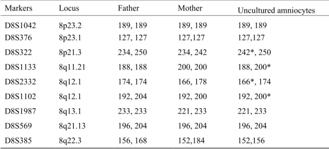
(UCSC hg18, NCBI Build 36, March 2006) (Fig. 3). Polymorphic DNA marker analysis of the
uncultured amniocytes using chromosome 8-specific microsatellite markers revealed a biparental
diallelic pattern for chromosome 8 (Fig. 4, Table 1). The microsatellite markers specific for the
region between 8p22 and q12.1 such as D8S322, D8S1133, D8S2332 and D8S1102 revealed gene
dosage increase in the maternal allele (Fig. 4, Table 1). Interphase FISH analysis on uncultured
amniocytes using an 8p11.1-q11.2-specific probe (Vysis CEP8, D8Z2) (Abbott Laboratories, Abbott
Park, IL, USA) showed three D8Z2 signals in 8/40 (20%) of uncultured amniocytes, and two signals
in 32/40 (80%) of uncultured amniocytes (Fig. 5). Cytogenetic analysis of the cultured amniocytes
revealed a karyotype of 47,XY,+r(8)(p221q12.1)[3]/46,XY[37] (Fig. 6). The parents decided to
continue the pregnancy. At 38 weeks of gestation, a male baby was delivered with a body weight of
3300 g. He was apparently normal except for mild bilateral hydronephrosis. Postnatal cytogenetic
analysis of the cord blood, umbilical cord and placenta revealed the karyotype of 47,XY,+r(8)
(p22q12.1)[2]/46,XY[38], 47,XY,+r(8)(p22q12.1)[2]/46,XY[18] and 47,XY,+r(8) (p22q12.1)
[4]/46,XY[36], respectively.
Discussion
Application of molecular cytogenetic techniques on uncultured amniocytes has been well described in
rapid positive confirmation of mosaicism for trisomies such as mosaic trisomy 2, mosaic trisomy 7,
mosaic trisomy 8 and mosaic trisomy 9 [4-8]. In this study, we additionally show the usefulness of
interphase FISH, QF-PCR and aCGH on uncultured amniocytes in rapid positive confirmation of
mosaicism for an SMC at amniocentesis. We have found variations of the level of mosaicism
between different amniocentesis. In this study, the first, second and third amniocentesis revealed a
mosaic level of 9.5%, 20% and 7.5%, respectively in cultured amniocytes, and the interphase FISH
analysis on uncultured amniocytes revealed a mosaic level of 20%. Therefore, interphase FISH on
uncultured amniocytes may serve as a more truthful reflection of the mosaic level in their original,
uncultured state against the results from different amniocentesis-karyotyping where analysis was done
on cultured cells. The present study also shows a discrepancy of mosaic level between the uncultured
amniocytes and cultured lymphocytes (20% vs. 5%). It is evident that interphase FISH on uncultured
amniocytes provides more accurate information of mosaicism than blood lymphocytes.
QF-PCR and aCGH may have difficulty in rapid positive confirmation of mosaicism detected at
amniocentesis in cases with low-level mosaicism. QF-PCR assay has been reported to detect
mosaicism as low as 15% of the whole sample [9]. The detection rate for mosaicism using aCGH on
uncultured amniocytes is variable according to different products of array chips [4-8]. We previously
successfully detected mosaic trisomy by aCGH using CytoChip Oligo array on uncultured amniocytes
in a case of mosaic trisomy 9 with 48% (12/25) mosaicism, a case of mosaic trisomy 2 with 12%
(6/50) mosaicism and a case of mosaic trisomy 7 with 26% (13/50) mosaicism [6-8]. In this case of
mosaic supernumerary r(8), both aCGH using CytoChip Oligo array and QF-PCR were able to detect
a mosaic level of 20% (8/40) in uncultured amniocytes.
The peculiar aspect of the present case is the association of an sSMC 8 with fetal pyelectasis. In
the present case, prenatal diagnosis of a mosaic supernumerary r(8)(p22q12.1) was achieved by
amniocentesis in a 32-year-old woman because of the ultrasound findings of fetal pyelectasis in the
second trimester. Renal abnormalities such as hydronephrosis and megacystitis have been well
known to be associated with mosaic trisomy 8 [5,10-11]. Renal abnormalities have also been
observed in patients with a supernumerary ring/marker chromosome 8. For instances, Butler et al
[12] reported right hydronephrosis with bilateral vesico-ureteral reflux in a patient with 45%
mosaicism for a supernumerary r(8) in fibroblasts. Spinner et al [13] reported mild hydronephrosis
and kidney malrotation in a patient with 56% mosaicism for a supernumerary marker 8p11q11 in
blood cells. Starke et al [14] reported prenatally detected slight bilateral pyelectasis in a fetus with
54% mosaicism for a supernumerary marker 8p11q11 in amniocytes. Loeffler et al [15] reported
left-sided renal hypoplasia and an enlarged right kidney with a duplicated collecting system in a patient
with 70% mosaicism for a supernumerary r(8)(p12q12) in blood cells. Filges et al [16] reported a left
pelvic kidney in a patient with 60% mosaicism for a supernumerary r(8)(p11.21q21.2) in blood cells.
Chen et al [17] reported left multicystic kidney in a fetus with 93% mosaicism for a supernumerary
r(8)(p11.21q11.1) in amniocytes. The present case further adds to the preexisting evidence that gene
dosage increase in 8p11.21q21.2 can result in fetal pyelectasis.
In conclusion, a low-level mosaicism for a supernumerary r(8)(p22q12.1) can present fetal
pyelectasis, and prenatal diagnosis of fetal pyelectasis should raise suspicion of chromosome
aberration. Molecular cytogenetic analyses on uncultured amniocytes have the advantage of rapid
positive confirmation of a supernumerary ring chromosome detected at amniocentesis.
Acknowledgements
This work was supported by research grants NSC-97-2314-B-195-006-MY3 and NSC-99-2628-B-195-001-MY3 from the National Science Council, and MMH-E-100-04 from Mackay Memorial Hospital, Taipei, Taiwan.
References
1. Demori E, Devescovi R, Gambel Benussi D, Dolce S, Carrozzi M, Villa N, et al. Supernumerary ring chromosome 8: Clinical and molecular cytogenetic characterization in a case report. Am J Med Genet 2004; 130A: 288-94.
2. Yilmaz Ş, Tarkan-Argüden Y, Kuru D, Deviren A, Karaman B, Yüksel A, et al. Mosaic supernumerary r(8) syndrome. Genet Couns 2005; 16: 187-90.
3. Bettio D, Baldwin EL, Carrozzo R, Vignoli A, May L, Venci A, et al. Molecular cytogenetic and clinical findings in a patient with a small supernumerary r(8) mosaicism. Am J Med Genet 2008; 146A: 247-50.
4. Chen C-P, Lin M-H, Su Y-N, Chern S-R, Tsai F-J, Wu P-C, et al. Mosaic trisomy 9 at amniocentesis: Prenatal diagnosis and molecular genetic analyses. Taiwan J Obstet Gynecol 2010; 49: 341-50.
5. Chen C-P, Chen M, Pan Y-J, Su Y-N, Chern S-R, Tsai F-J, et al. Prenatal diagnosis of mosaic trisomy 8: Clinical report and literature review. Taiwan J Obstet Gynecol 2011; 50: 331-8.
6. Chen C-P, Su Y-N, Lin S-Y, Chern S-R, Chen Y-T, Lee M-S, et al. Prenatal diagnosis of mosaic trisomy 2: Discrepancy between molecular cytogenetic analyses of uncultured amniocytes and karyotyping of cultured amniocytes in a pregnancy with severe fetal intrauterine growth restriction. Taiwan J Obstet Gynecol 2011; 50: 390-3.
7. Chen C-P, Hung F-Y, Su Y-N, Chern S-R, Su J-W, Lee C-C, et al. Prenatal diagnosis of mosaic trisomy 9. Taiwan J Obstet Gynecol 2011; 50: 549-53.
8. Chen C-P, Huang H-K, Su Y-N, Chern S-R, Su J-W, Lee C-C, et al. Trisomy 7 mosaicism at amniocentesis: interphase FISH, QF-PCR and aCGH analyses on uncultured amniocytes for rapid distinguishing true mosaicism from pseudomosaicism. Taiwan J Obstet Gynecol 2012, in press. 9. Donaghue C, Mann K Docherry Z, Ogilvie CM. Detection of mosaicism for primary trisomies in
prenatal samples by QF-PCR and karyotype analysis. Prenat Diagn 2005; 25: 65-72.
10.Webb AL, Wolstenholme J, Evans J, Macphail S, Goodship J. Prenatal diagnosis of mosaic trisomy 8 with investigations of the extent and origin of trisomic cells. Prenat Diagn 1998; 18: 737-41.
11.van Haelst MM, Van Opstal D, Lindhout D, Los FJ. Management of prenatally detected trisomy 8 mosaicism. Prenat Diagn 2001; 21: 1075-8.
12.Butler MG, Roback EW, Allen GA, Dev VG. Identification of a ring chromosome as a ring 8 using fluorescent in situ hybridization (FISH) in a child with multiple congenital anomalies. Am J Med Genet 1995; 57: 494-5.
13.Spinner NB, Grace KR, Owens NL, Sovinsky L, Pellegrino JE, McDonald-McGinn D, et al. Mosaicism for a chromosome 8-derived minute marker chromosome in a patient with manifestation of trisomy 8 mosaicism. Am J Med Genet 1995; 56: 22-4.
14.Starke H, Schreyer I, Kähler C, Fiedler W, Beensen V, Heller A, et al. Molecular cytogenetic characterization of a prenatally detected supernumerary minute marker chromosome 8. Prenat Diagn 1999; 19: 1169-74.
15.Loeffler J, Soelder E, Erdel M, Utermann B, Janecke A, Duba H-C, et al. Muellerian aplasia associated with chromosome 8p12q12 mosaicism. Am J Med Genet 2003; 116A: 290-4.
16.Filges I, Röthlisberger B, Wenzel F, Heinimann K, Huber AR, Miny P. Mosaic ring chromosome 8: clinical and array-CGH findings in partial trisomy 8. Am J Med Genet 2008; 146A: 2837-41.
17.Chen C-P, Chen M, Ko T-M, Ma G-C, Tsai F-J, Tsai M-S, et al. Prenatal diagnosis and molecular cytogenetic characterization of a small supernumerary marker chromosome derived from chromosome 8. Taiwan J Obstet Gynecol 2010; 49: 500-5.
Figure Legends
Fig. 1. Spectral karyotyping (SKY) analysis of cultured amniocytes using 24-color SKY probes shows a small supernumerary marker chromosome derived from chromosome 8 (arrow).
Fig. 2. Fluorescence in situ hybridization analysis of cultured amniocytes using a chromosome 8 whole chromosome painting probe (spectrum green) shows a chromosome 8-derived small supernumerary marker chromosome (arrow).
Fig. 3. Oligonucleotide-based array comparative genomic hybridization analysis using CytoChip Oligo array (BlueGnome, Cambridge, UK) on uncultured amniocytes shows a 43-Mb genomic gain in chromosome 8 encompassing the region of 8p22q12.1.
Fig. 4. Representative electrophoretograms of quantitative fluorescent polymerase chain reaction. The markers D8S322, D8S1133, D8S2332 and D8S1102 show two peaks of unequal fluorescent activity from two different parental alleles in uncultured amniocytes with a dosage increase in the maternal and a dosage ratio of 1:1.2 (paternal: maternal).
Fig. 5. Interphase fluorescence in situ hybridization analysis on uncultured amniocytes using an 8p11.1-q11.2-specific probe (Vysis, CEP8, D8Z2; spectrum green) shows (A) two green signals in a cell with disomy 8 and (B) three green signals (arrows) in a cell with a supernumerary r(8).
Fig. 6. The G-banded karyotype of 47,XY,+r(8)(p22q12.1). The arrow indicates a supernumerary ring chromosome derived from chromosome 8.
Table 1. Genotypic information of the father, mother and uncultured amniotic fluid cells at short tandem repeat markers specific for chromosome 8 obtained by quantitative fluorescent polymerase chain reaction assaysa
Markers Locus Father Mother Uncultured amniocytes
D8S1042 8p23.2 189, 189 189, 189 189, 189 D8S376 8p23.1 127, 127 127,127 127,127 D8S322 8p21.3 234, 250 234, 242 242*, 250 D8S1133 8q11.21 188, 188 200, 200 188, 200* D8S2332 8q12.1 174, 174 166, 178 166*, 174 D8S1102 8q12.1 192, 204 192, 200 192, 200* D8S1987 8q13.1 233, 233 221, 233 221, 233 D8S569 8q21.13 196, 204 196, 204 196, 204 D8S385 8q22.3 156, 168 152,184 152,156
a Alleles (basepair sizes) are listed below each individual. * With increased dosage