行政院國家科學委員會專題研究計畫 成果報告
韌帶組織工程新視野:韌帶細胞與生物材料交互作用分子
機制之研究(I)
研究成果報告(精簡版)
計 畫 類 別 : 個別型 計 畫 編 號 : NSC 95-2314-B-002-323- 執 行 期 間 : 95 年 08 月 01 日至 96 年 07 月 31 日 執 行 單 位 : 國立臺灣大學醫學院骨科 計 畫 主 持 人 : 王至弘 計畫參與人員: 博士班研究生-兼任助理:邵宏仁 處 理 方 式 : 本計畫可公開查詢中 華 民 國 96 年 09 月 28 日
The molecular behaviors of anterior
cruciate ligament cells cultured on
Abstract
The purpose of this study is to evaluate the molecular behaviors of anterior cruciate
ligament cells (ACL cells) on two biodegradable materials: poly (ε-caprolactone) (PCL) and chitosan. The results of this in vitro study indicated that the surface of chitosan presented less adsorbed-fibronectin (FN) than that presented on the surface of PCL. The scanning electron microscope (SEM) illustrated that the ACL cells could not attach well but still maintain the late stages of filopodial on the chitosan after 3 days incubation. Because of these events, the ACL cells expressed less α5β1 integrin and the immuncytochemistrical staining showed the cytoskeleton, F-actin, were not well constructed on chitosan. In the messenger ribonucleic acid (mRNA) gene expressed level, we found the expression of the transforming growth factor β1 (TGF β1) mRNA and type III collagen mRNA in the ACL cell-chitosan system was more than that in the ACL cell-PCL system but the expression of the connective tissue growth factor (CTGF) mRNA and type I collagen mRNA in the ACL cell-chitosan system was less than that in the ACL cell-PCL system. In an additional experiment, the expression of TGF β1 mRNA was detected after the ACL cells were incubated with chitosan coated FN (chitosan-FN) for 3days. The expression of TGF β1 mRNA was decreasing as the ACL cells cultured on chitosan-FN and the trend of the others mRNA expression was as same as that while the ACL cells cultured on PCL did. To sum up, we demonstrated that the amount of the FN adsorbed on the materials would affect the molecular behaviors of the ACL cells such as integrin expression and cytoskeleton structure. Moreover, incubated with the less FN-adsorbed chitosan, the ACL cells expressed more TGF β1 mRNA and type III collagen. We supposed the purpose of it was that the ACL cells intended to help them to get more strength of cell-FN interaction and to establish their ECM on the surface of chitosan.
Keywords: anterior cruciate ligament cells, biomaterial, cell-material interaction, tissue engineering
Introduction
Anterior cruciate ligament (ACL), an intra-articular ligament, located in the knee
and provided the knee stabilization. The rupture ACL could not repair spontaneously due to the intrinsic properties and fibrinolytic enzymes in the synovial fluid [1, 2]. A new concept, tissue engineering, provided lots of approaches, changing the cell sources and changing the biomaterials, to deal with this rupture ACL[3-6].The aim of those previous studies was to search a nice cell-biomaterial system for ACL tissue engineering. With a nice cell-biomaterial system, cells could survive, proliferate, establish their extracellularmatrix (ECM) and present the suitable phenotypes on the materials. Hence, to realize the cell-biomaterial interaction is an urgent subject. However, most of those preview studies of ACL tissue engineering only provided the scanning electron microscope (SEM) observation for cell morphology and MTT assay
[7]
for cell viability to illuminate the cell-material interaction. Relatively little is known about the molecular behaviors of the ACL cells among the extra-cellular matrix, the cellular surface integrins, the inner cellular gene expression and materials.
The materials provided a place where anchorage-depended cells could settle. Cells used the integrin heterodimers, consisting of non-covalently associated α and β subunits, to bind to the arginine-glycine-aspartic acid (RGD) motif of extracellular-matrix (ECM) that was adsorbed on surface of materials. With the component of focal adhesions, Talin, Vinculin, Tensin, Paxillin and α actinin, the ECM and cytoskeleton were connected by integrin [8, 9]. In addition, this signal pathway was controlled by number of common mechanisms that were protein tyrosine phosphorylation, intracellular Ca2+ and inositol lipid metabolism [10, 11]. After these biochemical reactions, the extracellular information was sent into cells and cells would adjust their physiological situation to response the extracellular microenvironments.
In this present study, the ACL cells were seeded on two biodegradable materials, poly (ε-caprolactone) (PCL) and chitosan, which were approved by Food and Drug Administration (FDA). Chitosan is an N-deacetylation product of chitin and the different degree of deacetylation would affect the cellular behaviors. It was reported that this material could accelerate the wound healing and stimulate the macrophage releasing lots of growth factors to promote ECM production [12]. Some study also illustrated that chitosan was a suitable material for the bone cells [13, 14] but the behaviors of the ACL cells on chitosan was seldom investigating. PCL, a semi-crystalline linear biodegradable α-polyester, is undergoing auto-catalysed bulk hydrolysis. Although the PCL was wildly used in tissue engineering, there were few reports about the ACL cells on PCL. For these reasons, to study the interaction of these two materials with the ACL cells was valuable for the ACL tissue engineering.
The flow cytometry, immuncytochemistry, real-time polymerase chain reaction (real-time PCR) and western-blot were included to observe the changes of the ACL cells from the superficial level, the expression of cell-membran integrins, to the deep level, the expression of messenger ribonucleic acid (mRNA). We try to figure out the whole view of the ACL cell-material interaction.
Materials and Methods
Substrate preparation and characterization
The poly (ε-caprolactone) (PCL) (Sigma-Aldrich, USA) substrate used in the membrane form was prepared by using the dry process of the phase inversion method
[15]
. Briefly, 10% of PCL solution dissolved in chloroform (Tedia, USA) was cast on a glass plate in a uniform thickness of 500 μm at 25°C, and then air-dried in the hood at 25°C for 24 hours to obtain the membrane substrate. The 1% of 85% deacetylation chitosan (Sigma-Aldrich, USA) solution dissolved in acetic acid (Tedia, USA) was directly coated on TCPS (tissue cultured polystyrene) and dried in the hood at 60°C for 24 hours. The chitosan-FN was prepared by coated fibronectin (BD, USA) on chitosan surface.
Air-water contact angle of the TCPS, PCL and chitosan substrates was measured at 25°C to analyze the hydrophobicity of substrates by using a reverse air-bubble apparatus (CA-D, Kyowa Scientific Co.).
Cell culture
Human ligament explants were obtained from patients (range from 20~40 years old) who underwent ACL reconstruction, as described previously [16]. Institutional Review Board approval was obtained for the use of the human ligament tissue. The cells were growth in tissue culture flasks (Corning, USA) containing Dulbecco’s modified Eagle medium supplemented with 10% fetal bovine serum (Gibco-RBL life Technologies, Paisley, UK) and 1% antibiotic/antimycotic (100 U mL-1 penicillin sodium, 100 μg mL-1 streptomycin and 0.25 μg mL-1 amphotericin B; Gibco-RBL life Technologies) and was maintained at 37°C and 5% CO2. Once confluent, fibroblasts were
trypsinized using 0.2% trypsin with 0.1% EDTA and resuspended in the same medium until ready for experiments.
Cell morphology
For morphological observation, the cells adhering to the sample (4 hours and 3 days of incubation) were washed with PBS and then fixed with 2.5% glutaraldehyde in PBS for 1hour at 4°C. After through washing with PBS, the specimens were dehydrated by graded ethanol change and then examined by a Hitachi S-800 scanning
electron microscope, after specimens being freeze-dried and gold sputtered in vacuum.
Cell adhesion and proliferation
After cell seeded 4 hours and 3days, the number of viable cells was determined by MTT assay. In brief, cells were washed with PBS to remove nonadherent cells and 100 μl MTT (Sigma) solution (5 mg/ml in PBS) was added to each well. After incubation for 3 hours at 37°C, the medium was aspirated and the formazan reaction products were dissolved by DMSO solution and shaken for 15 minutes. The optical density of the formazan solution was read on an ELISA plate reader (ELx 800, BIO-TEK, USA) at 570 nm.
Flow cytometry
Cells were detached with 0.2% (W/V) trypsin, pelleted and resuspended at a
concentration of 1x10⁶ cells mL-1 and incubated with monoclonal anti-human CD49E-FITC (mouse IgG2b) (Chemicon, USA) and monoclonal anti-human integrin beat 1 (IgG) (Chemicon, USA) for 30 minutes. The set of integrin beat 1 was incubated with secondary antibody conjugated FITC (goat antimouse IgG) (Chemicon, USA) for another 30 minutes. The cells were then washed three times with PBS. After passed through a sterile nylon cell strainer (40μm), the intergin α5 and β1 subunits of human ACL cells were detected by FACScan flow cytometry (Becton Dickinson, USA). The data acquisition and analysis were performed with Cellquest software. The presence or absence of staining was determined by comparison with the appropriate isotype control.
Immuncytochemistry
After 3 days incubation, human ACL cells were fixed in ice-cold 4% paraformaldehyde in phosphate-buffered saline (PBS) for 20 minutes and then permeabilized with 0.1% TritonX-100 in PBS for 10 minutes at 4°C. After fixation and permeability, the F-actin filaments were stained with rhodamine phalloidin (Molecular Probes; invitrogen, USA) and were visualized by indirect fluorescence under the fluorescent microscope (Axiovert 100TV, Germany).
Western-blot
The culture medium was added into TCPS, PCL and chitosan overnight. These membranes were rinsed thoroughly in PBS and the adsorbed proteins were stripped by stripping buffer (0.1 M Tris-HCl, 3% SDS, 0.75% 2-mercaptoethanol). The proteins
polyvinylidene difluoride (PVDF) membrane (BioRad, USA) and blocked with blocking solution overnight. The blots were probed with anti-bovine fibronectin (Chemicon, USA) at 1: 1000 dilutions for 2 hours at 25°C, washed, and incubated with 1: 10000 dilutions of horseradish peroxidase-conjugated goat anti-rabbit IgG (Bethyl, USA) for 2 hours at 25°C. Detection was by chemiluminescence with an enhanced chemiluminescence kit (PerkinElmer life Sciences, USA). Bands were quantified densitometrically by scanning films into AlphaEaseFC 4.0 analysis software.
Real-time polymerase chain reaction
Total cellular RNA was extracted from ACL cells using RNA extraction kit (Mix Total RNA Extraction kit; Bio-Rad, USA). RNA was reverse transcribed into cDNA using the Hight-Capacity cDNA Arichive kit (Applied Biosystem, USA). Expression of mRNA was detected using an ABI Prism 7900 HT Sequence Detection System (Applied Biosystem, USA). Glyceraldehyde-3-phosphate dehydrogenase (GAPDH) was used as the endogenous control. Data were collected with instrument spectral compensations by the Applied Biosystem SDS 2.1 software and analyzed using the threshold cycle number (Ct) relative quantification method. The prime probes of target
gene were transforming growth factor β1 (TGF β1), connective tissue growth factor (CTGF), Type I collagen and Type III Collagen. (Assay ID: Hs99999918_m1, Hs00170014_m1, Hs00164004 and Hs00164103_ml; Applied Biosystem, USA).
Statistical analyses
Data in the graphs are presented in the form of mean ± standard error. One-way analyses of variance (ANOVA) were performed, and when a significant F ratio was obtain, the Duncan’s test was used to compare between the means. Significance was attained at *: P<0.05, **: P<0.001.
RESULTS
Air-water contact angle
The air-water contact angles of PCL and chitosan were 73.92°±1.03° and 71.87°±0.55°, respectively. There was no significant difference between these two substrates, thus the effect of hydrophilic/hydrophobic property of substrates on ACL cells was not considered in this study.
Cellular morphology and proliferation
Figure 1A shows that the relative ACL cells adhesion with 4 h incubation, which was determined from the ratio of the MTT conversion of ACL cells attached on the
PCL and chitosan membranes to that of ACL cells attached on TCPS. The relative cell adhesion of chitosan (0.86±0.07) was slightly greater than that of PCL (0.72±0.02). It is reasonable to hypothesize that ACL cells on chitosan should have a more flattened morphology at the same culture period due to its higher relative cell adhesion. However, Figs. 1B and C show ACL cells attached well and presented cytoplasmic webbing shape on PCL but presented round shape and only extended some filopodias on chitosan.
Fig. 2A shows the MTT conversion of ACL cells on PCL and chitosan membranes related to that on TCPS, after incubation for 3 days. Although cell adhesion with 4 h incubation is higher for chitosan than PCL, cell proliferation rate, not the shape of the growth curves, was significantly higher for PCL than chitosan (p<0.01). MTT counts of the PCL and the chitosan membranes related to that of TCPS are 2.65±0.08 and 0.54±0.03, respectively. At this time, the morphology of the ACL cells seemed to stay between the early and late stages of cytoplasmic webbing on the chitosan (Fig. 2B)[17]. Besides,lots of ACL cells aggregated and suspended in the culture medium above the chitosan membrane (not shown here). In contrast, ACL cells cultured on the PCL membrane still proliferated and exhibited a well spread morphology (Fig. 2C).
Immunocytochemistry
The cytoskeleton plays important roles in cell morphology, adhesion, growth and
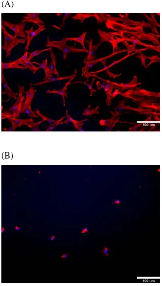
signaling. In this study, after 3 days of incubation, distributions of cytoskeletal actin proteins were examined. The immunocytochemistry with rhodamine staining of actin showed that the spanning F-actin fibers presented in the ACL cells cultured on PCL and the cytoskeletal actin network was very clear and extensive; while cultured on chitosan, the actin organization was small, aggregated, and circular (Fig. 3). In addition, cells cultured on PCL exhibited spindle-like shape but cells cultured on the chitosan did not extend their cytoploasm. Therefore, ACL cells plated on PCL had a much more extensive actin network organization than on chitosan.
Flow cytometry
In this study, the expression of α5 and β1 integrin subunits on the surface of ACL cells, after cultured on the PCL and chitosan membranes for 3 days, were evaluated by flow cytometric analysis. Fig. 4A shows it was difficult to distinguish the fluorescence intensity of α5 integrin subunit between the ACL cells cultured on PCL and chitosan, but the number of cells with the expression of α5 integrin subunit on PCL was higher than that on chitosan. In contrast, Fig. 4B shows the fluorescence intensity of β1 integrin subunit in the ACL cells cultured on PCL is considerably higher than that on chitosan. Therefore, after cultured on PCL or chitosan, the
expression of α5 and β1 integrin subunits on the surface of ACL cells exhibited different behaviors.
Western-blot
Because the FN is abounded in serum, the FN adsorbed on material surface was
examined in this study. This protein is also an important ECM that could mediate cells adhering on biomaterials. Briefly, the PCL and chitosan substrates coated on TCPS were exported to medium containing 10% FBS. After incubated overnight, the serum containing culture medium was discarded and the FN adsorbed on the material surface was examined by western-blot. Each of bands was converted into integrated density value (IDV) by AlphaEaseFC 4.0 analysis software and the IDV of TCPS is a calibrator to convert each of IDV into ratio. Fig. 5, the ratio of blots, illustrated the ratios of FN adsorbed on the PCL and chitosan were 0.86±0.05 and 0.395±0.025, respectively. This result demonstrated that the adsorbed FN on PCL was more than that on chitosan (p<0.05).
mRNA expression
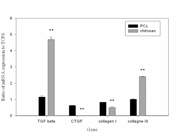
The ruptured human knee ACL explants were received from patients who were range from 20 to 40 years. Their ACL cells were incubated with TCPS, PCL and chitosan 3 days and detected the mRNA expression by real-time PCR. The mRNA expression of ACL cells on TCPS was a calibrator sample in this study. The trend of the mRNA expression was the same in all of patients in this study. Fig. 6 showed the expression of TGF β1 mRNA and type III collagen of ACL cells cultured on chitosan was 4-fold and 2.36-fold more than that of ACL cells cultured on PCL (p<0.01). On the contrary, the expressions of CTGF mRNA and type I collagen mRNA of ACL cells cultured on chitosan were 90-fold (p<0.01) and 1.46-fold (p<0.05) lower than that of ACL cells cultured on PCL, respectively.
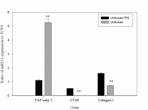
In the additional experiment, the ACL cells were seeded onto two materials that were chitosan-FN and chitosan. As following the above real-time PCR method, the data of ACL cells cultured on chitosan-FN showed that the expression of TGF β1 mRNA was reducing and 4.7-fold lower than that of ACL cells cultured on chitosan. Moreover, the trend of others mRNA expression in ACL cells cultured on chitosan-FN was as same as the ACL cells cultured on PCL did (Fig. 7).
DISCUSSION
Because of the different cell types presented different cellular behaviors, a material
could not be suitable for any kinds of cell. Therefore, in tissue engineering, to choose a suitable material for cell growing is the first step [18]. From the molecular level to
detect the behaviors of cell-material interaction is a reasonable way, which could totally sketch what responses the cells have from inner the cell to outside of the cell.
The results of this in vitro study demonstrated that the PCL is a more suitable material for ACL cells than chitosan did. The ACL cells could attach well, construct their cytoskeleton and proliferate on PCL. However, ACL cells aggregated and suspended in the culture medium instead of anchoring on the surface of chitosan. It is well known that the material surface characteristics, surface topography, surface wettability and surface charge, could affect the cell-material interaction [19]. Moreover, the ECM adsorbed on material play the pivot role to mediate cells attaching on materials. Therefore, in this study, we narrowed down the effected factors to the Fn, because the FN is abounded in serum. Fn is a disulfide-bonded dimmer of 220- to 250-kDa subunits and each subunit is fold into five or six rodlike domains. Through the tripeptide sequence Arg-Gly-Asp (RGD), the cell surface receptor, integrin α5β1, could bind to the Fn [9, 20, 21]. With the integrins, the anchorage-dependent cells could receive the information from the outside environment and do some biochemical reaction to adapt the environment [10, 11].
The western-blotting showed more adsorbed FN presented on PCL than that did on chitosan and the flow cytometry also revealed that ACL cells expressed more α5β1 integrin on PCL. These subtle changes between the interface of ACL cell and material influenced the signal cascades because ACL cells received the extracellular information through the integrin. Without the good integrin-ECM interaction, cells could not spread well on material and could not construct their cytoskeleton well. Hence, our SEM and immunocytochemistry results indeed illustrated that the ACL cells still presented round shape and filamentary global actins on the chitosan, after 3 days incubation.
Furthermore, in our additional experiment of ACL cells incubated with Fn coated on chitosan, the expression of TGF β1 mRNA was decreasing. Unlike the behaviors of ACL cells seeded on chitosan, the ACL cells seeded on the chitosan-FN flatten well and the trend of mRNA expression was same as ACL cells seeded on PCL. Base on these results, we found the critical factor to affect these cellular behaviors was FN. The reason we supposed was because of the ligament cells had FN and laminin receptors [22] on their cellular membrane and only do the FN present in the cell-cultured serum.
The function of TGF β1 was stimulating the expression of FN and collagen and it also had an effect on the cell-FN binding interaction [23, 24]. In this study, ACL cells expressed lots of TGF β1 mRNA when the ACL cells were seeded on the chitosasn. It seemed that the ACL cells could not find the FN on the chitosan surface; therefore the ACL cells expressed more TGF β1 mRNA in order to get more strong interaction with
FN. This observation could be demonstrated by the less TGF β1 mRNA expression in ACL cells seeded on the chitosan-FN system.
Type I and Type III collagen were the typical markers of the ligament cells [25]. Type III collagen is much more prevalent in the fetal ACL and is sometimes referred to as embryonic or fetal collagen [26]. In this study, ACL cells expressed more type III collagen mRNA on chitosan than that on PCL. To combine the results of the TGF β1 mRNA and the type III collagen expression, it is reasonable to assume that ACL cells expressed two kinds of these mRNA to exert them to establish their ECM on the surface of chitosan.
In the Ott and his coworkers’ research, modulation of the cytoskeleton architecture was shown to regulate the expression of CTGF. In this presented data, the ACL cells cultured on the chitosan showed that the cytoskeleton was short and aggregation. We assumed it was the reason that ACL cells expressed less CTGF mRNA while ACL cells cultured on the chitosan. On the contrary, whether ACL cells cultured on PCL or ACL cells cultured on chitosan-FN, the expression of CTGF mRNA almost equals to that of ACL cells cultured on the TCPS.
Conclusion
In this study, the contact angle of PCL and chitosan were range from 70°~75° and it was no significant difference between these biomaterials. Consequently, it was not the critical factor to affect the FN adsorbed on the materials and it needs the further study to investigate what affecting the FN adsorption. Moreover, we found an interesting relationship among the FN adsorbed on the material surface, the integrin expression of ACL cells and the TGF β1 mRNA expression. Once the FN adsorbed on the material surface enough, ACL cells would express α5β1 integrin to bind to the FN and made them to anchor on the material surface. With this phenomenon, ACL cells could construct their cytoskeleton, F-actin, well and the biochemical reaction could be initiated. After that, the outside information was sent into the ACL cells and ACL cells presented the different mRNA expression to adapt the extra-cellular environment. In our observation, the different mRNA expressions were that the higher expression of TGF β1 mRNA and type III collagen in the lower adsorbed FN ACL cell-chitosan cultured system and the lower expression of TGF β1 mRNA and type III collagen in the higher adsorbed FN ACL cell-PCL cultured system. On the above-mentioned, when the ACL cells cultured on the lower adsorbed FN materials, the ACL cells will express more TGF β1 mRNA and type III collagen to make them to get more cell-FN binding interaction and to establish their ECM.
ACKNOWLEDGMENT
The authors thank the National Science Council of Taiwan, the Republic of China for their financial support of this research.
REFERENCE
1. Amiel D, Nagineni CN, Choi SH, Lee J. Intrinsic properties of ACL and MCL cells and their responses to growth factors. Med Sci Sports Exerc. 1995; 27(6): 844-851.
2. Murry MM, Martin SD, Spector M. Histological changes in the human anterior cruciate ligament after rupture. J Bone Joint Surg 2000; 82: 1387-1402.
3. Altman GH, Horan RL, Lu HH, Moreau J, Martin I, Kaplan DL, et al. Silk matrix for tissue engineering anterior cruciate ligaments. Biomaterials 2002; 23: 4131-41.
4. Eijk FV, Saris DBF, Riesle J, Willems WJ, Blitterswijk CV, Dhert WJA, et al. Tissue engineering of ligament: a comparison of bone marrow stromal cells, anterior cruciate ligament, and skin fibroblasts as cell source. Tissue engineering 2004; 10 (5/6): 893-903.
5. Lin VS, Lee MC, O’neal S, Mckean J, Sung K-L P. Ligament tissue engineering using synthetic biodegradable fiber scaffolds. Tissue engineering 1999; 5 (5): 443-51.
6. Lu HH, Cooper Jr. JA, Manuel S, Freeman JW, Attawia MA, Laurencia CT et al. Anterior cruciate ligament regeneration using braided biodegradable scaffolds: in vitro optimization studies. Biomaterials 2005; 26: 4805-16.
7. Mosmann T. Rapid colorimetric assay for cellular growth and survival: application of proliferation and cytotoxicity assays. J Immunol Methods 1983; 65: 55-63.
8. Brakebusch C, Fässler R. The integrin-actin connection, an eternal love affair.
The EMBO journal 2003; 22(10): 2324-33.
9. García AJ. Get a grip: integrins in cell-biomaterial interactions. Biomaterials 2005; 26: 7525-29.
10. Clark EA, Brugge JS. Integrin and signal transduction pathways: the road taken.
Science 1995; 268 (5208): 233-39.
11. Schwartz MA. Transmembrane signaling by integrins. Trends Cell Biol 1992; 2:
304-08.
12. Ueno H, Nakamura F, Murakami M, Okumura M, Kadosawa T, Fujinaga T.
Evaluation effects of chitosan for the extracellular matrix production by fibroblasts and the growth factors production by macrophages. Biomaterials 2001; 22: 2125-30.
13. Fakhry A, Schneider GB, Zaharias R. Senel S. Chitosan supports the initial attachment and spreading of osteoblasts preferentially over fibroblasts.
Biomaterials 2004; 25: 2075-79.
Bone cell attachment and growth on well-characterized chitosan films. Polym Int 2007; 56: 641-47.
15. Young TH, Yao CH, Sun JS, Lai CP, Chen LW. The effect of morphology variety of EVAL membranes on the behavior of myoblasts in vitro. Biomaterials 1998; 19: 717-24.
16. Lee IC, Wang JH, Lee YT, Young TH. Development of a useful technique to
discriminate anterior cruciate ligament cells and mesenchymal stem cells-the application of cell electrophoresis. Biochem Biophys Res Commun 2007; 352(1): 147-52.
17. Rajaraman R, Rounds DE, Rembaum A. A scanning electron microscope study
of cell adhension and spreading in vitro. Exp Cell Res 1974; 88: 327-39.
18. Rosso F, Giordano A, Barbarisi M, Barbarisi A. From cell-ECM interactions to
tissue engineering. J Cell Physiol 2004; 199: 174-80.
19. Wilson C J, Clegg R E, Leavesley D I, Pearcy M J. Mediation of biomaterial-cell interactions by adsorbed proteins: a review. Tissue Eng 2005; 11 (1/2): 1-17. 20. Keselowsky BG, García AJ. Quantitative methods for analysis of integrin binding
and focal adhension formation on biomaterial surfaces. Biomaterials 2005; 26: 413-18.
21. Vogel V, Baneyx G. The tissue engineering puzzle: a molecular perspective. Annu
Rev Biomed Eng 2003; 5: 441-63.
22. Sung K-L P, Kwan MK, Maldonado F, Akeson WH. Adhesion strength of human ligament fibroblasts. J Biomech Eng 1994; 116: 237-42.
23. Ignotz RA, Massagué J. Transforming growth factor-β stimulates the expression of fibronectin and collagen and their incorporation into the extracellular matrix. J
Biol Chem 1986; 261 (9): 4337-45.
24. Molloy T, Wang Y, Murrell G AC. The role of growth factors in tendon and ligament healing. Sports Med 2003; 33 (5): 381-94.
25. Altman G, Horan R, Martin I, Farhadi J, Stark P, Volloch V, Kaplan D I. Cell differentiation by mechanical stress. FASEB J 2002; 16: 270-72.
26. Jackson D W, Heinrich J T, Simon T M. Current concepts biologic and synthetic implants to replace the anterior cruciate ligament. Arthroscope 1994, 10 (4): 442-52.
27. Ott C, Iwanciw D, Graness A, Giehl K, Goppelt-struebe M. Modulation of the expression of connective tissue growth factor by alterations of the cytoskeleton. J
LEGENDS
Figure 1. (A) The relative MTT conversion of cells attached on the PCL and chitosan to that of cells attached on TCPS 4 h after cell seeding, n=4. Data are presented as the mean ± standard error. (B) SEM photographs of ACL cells cultured on PCL, after 4 h incubation (1,500Χ). (C) SEM photographs of ACL cells cultured on chitosan, after 4 h incubation (1,500Χ).
Figure 2. (A) The relative MTT conversion of cells attached on the PCL and chitosan to that of cells attached on TCPS 3days after cell seeding, n=4. Data are presented as the mean ± standard error. Asterisk denotes significant differences (** p < 0.01) of relative MTT conversion compared to the chitosan. (B) SEM photographs of ACL cells cultured on PCL, after 3days incubation (1,500Χ). (C) SEM photographs of ACL cells cultured on chitosan, after 3days incubation (1,500Χ).
Figure 3. Immuncytochemistrical staining of F-actin. F-actin filaments were stained with rhodamine phalloidin (red staining) and nuclei were stained with DAPI (blue staining). (A) ACL cells cultured on the PCL. (B) ACL cells cultured on the chitosan. Scale bar = 100 μm.
Figure 4. Flow cytometric analysis of the ACL cell-surface integrins. For all graphs, isotype control staining was indicated by the red histogram and specific markers staining were indicated by the white histogram. (A) The green curve revealed the expression of α5 integrin in the ACL cell-PCL cultured system. The black curve revealed the expression of α5 integrin in the ACL cell-chitosan cultured system. (B) The green curve revealed the expression of β1 integrin in the ACL cell-PCL cultured system. The black curve revealed the expression of β1 integrin in the ACL cell-chitosan cultured system.
Figure 5. Western blotting of fibronrctin adsorbed onto PCL (gray) and chitosan (striped). Blots were analyzed by AlphaEaseFC 4.0 analysis software and the integated density value (IDV) was converted to related ratio to TCPS. Data (n=2) are presented as the mean ± standard error. Asterisk denotes significant differences (* p < 0.05) of IDV ratio compared to the chitosan.
Figure 6. Real-time PCR results illustrated the ratio of mRNA expression of ACL cells cultured on the PCL (black) and the chitosan (striped). Asterisks denotes significant differences (* P <0.05, ** P <0.01) of mRNA ratio compared to chitosan.
Figure 7. Real-time PCR results illustrated the ratio of mRNA expression of ACL cells cultured on the chitosan-FN (black) and the chitosan (striped). Asterisks denotes static significant differences (* P <0.05, ** P <0.01) of mRNA ratio compared to chitosan.
(A) (B) Materials PCL chitosan Ratio of cell n u m b er to TCPS 0 1 2 (C)
Figure 1. (A) The relative MTT conversion of cells attached on the PCL and chitosan to that of cells attached on TCPS 4 h after cell seeding, n=4. Data are presented as the mean ± standard error. (B) SEM photographs of ACL cells cultured on PCL, after 4 h incubation (1,500Χ). (C) SEM photographs of ACL cells cultured on chitosan, after 4 h incubation (1,500Χ).
(A) (B)
(C)
Figure 2. (A) The relative MTT conversion of cells attached on the PCL and chitosan to that of cells attached on TCPS 3days after cell seeding, n=4. Data are presented as the mean ± standard error. Asterisk denotes significant differences (** p < 0.01) of relative MTT conversion compared to the chitosan. (B) SEM photographs of ACL cells cultured on PCL, after 3days incubation (1,500Χ). (C) SEM photographs of ACL cells cultured on chitosan, after 3days incubation (1,500Χ).
(A)
(B)
Figure 3. Immuncytochemistrical staining of F-actin. F-actin filaments were stained with rhodamine phalloidin (red staining) and nuclei were stained with DAPI (blue staining). (A) ACL cells cultured on PCL. (B) ACL cells cultured on chitosan. Scale bar = 100 μm.
(A)
(B)
Figure 4. Flow cytometric analysis of the ACL cell-surface integrins. For all graphs, isotype control staining was indicated by the red histogram and specific markers staining were indicated by the white histogram. (A) The green curve revealed the expression of α5 integrin in the ACL cell-PCL cultured system. The black curve revealed the expression of α5 integrin in the ACL cell-chitosan cultured system. (B) The green curve revealed the expression of β1 integrin in the ACL cell-PCL cultured system. The black curve revealed the expression of β1 integrin in the ACL cell-chitosan cultured system.
Figure 5. Western blotting of fibronrctin adsorbed onto PCL (gray) and chitosan (striped). Blots were analyzed by AlphaEaseFC 4.0 analysis software and the integrated density value (IDV) was converted to related ratio to TCPS. Data (n=2) are presented as the mean ± standard error. Asterisk denotes significant differences (* p < 0.05) of IDV ratio compared to the chitosan.
Figure 6. Real-time PCR results illustrated the ratio of mRNA expression of ACL cells cultured on the PCL (black) and the chitosan (striped). Asterisks denotes significant differences (* P <0.05, ** P <0.01) of mRNA ratio compared to chitosan.
Figure 7. Real-time PCR results illustrated the ratio of mRNA expression of ACL cells cultured on the chitosan-FN (black) and the chitosan (striped). Asterisks denotes static significant differences (* P <0.05, ** P <0.01) of mRNA ratio compared to chitosan.