行政院國家科學委員會專題研究計畫 成果報告
核磁共振儀對人類軟骨細胞的影響
計畫類別: 個別型計畫 計畫編號: NSC93-2314-B-002-078- 執行期間: 93 年 08 月 01 日至 94 年 07 月 31 日 執行單位: 國立臺灣大學醫學院骨科 計畫主持人: 江清泉 計畫參與人員: 謝昌訓 報告類型: 精簡報告 處理方式: 本計畫可公開查詢中 華 民 國 94 年 10 月 24 日
中文摘要
由於核磁共振儀為非入侵的醫療評估方式,目前在臨床醫學的使用極為普遍,包括 腦部斷層掃瞄、腫瘤追蹤及外科復原評估等等。然而,在近幾年的文獻中不斷探討高磁 場環境與無線電電波對細胞、生理和心裡的影響;包括 c-jun、c-fos 基因的表現,鈣離子 濃度的增加以及幽閉空間恐懼症等等,顯示磁場環境與無線電電波會對細胞有某種程度 的影響,而這方面的研究一直未有明確的結果。在豬隻軟骨修復與評估的計畫中,相同 的實驗條件下,利用核磁共振評估與沒有利用核磁共振評估的豬隻相較下發現,使用核 磁共振之豬隻軟骨修復情況極為不好,修復位置(即原造成損傷位置)有一嚴重的凹陷 損傷。推測原因可能在軟骨修復過程中,因為暴露在核磁共振環境下造成細胞死亡或細 胞轉性所造成。 實驗方式:利用人類軟骨細胞為實驗材料,將人類軟骨細胞暴露在核磁共振儀環境 中,利用暴露時間的長短、是否使用無線電電波掃瞄的條件下,及暴露後細胞恢復時間 點之不同,分別採用細胞染色以及毒性測試實驗對細胞的增生與死亡做探討。另外利用 西方點墨轉漬法與北方點墨轉漬法來探討細胞程式化死亡過程中,p53、-jun、c-fos 等蛋 白質與 RNA 的表現量與表現時間點。測量細胞之間與細胞內的鈣離子濃度的變化,檢測 鈣離子是否擔任造成細胞程式化死亡的訊息傳遞者。 實驗結果:(1)暴露過 3 T 的高磁場環境下,人類軟骨細胞的生長數目會比未暴露 過高磁場環境下的少,其原因為高磁場環境下會造成軟骨細胞的程式化死亡。(2)在核磁 共振的影響下,其軟骨細胞內部的基礎鈣離子濃度明顯升高(3)在核磁共振的影響下, 其細胞程式化死亡的機制是經由 p21 蛋白質表現路徑。 這個實驗結果顯示核磁共振會影響軟骨細胞的生長,軟骨修復手術術後不宜用核磁共 振做為評估的工具。 關鍵字:核磁共振、軟骨細胞、鈣離子、細胞程式化死亡和 p53Abstract
As the MRI is an uninvading physical assessment method, it is prevailing in clinic, including brain CT,tumortracing,and assessmentofsurgery recovery…etc.In recentyears, many papers discuss about the influence of high magnetic field or radiation on the cell, physiology and psychology, including expression of c-jun, c-fos gene、 influencing of Ca2+ fluxes, particularly Ca2+ entry from the extracellular environment through the plasma membrane, and inducing the syndrome of cloustrophobia…etc.Studieswith cellularsystems using different exposure setups, exposure durations, amplitudes, frequencies and wave forms indicate that biological effects of magnetic fields on cellular systems are at hand. But there is no definite conclusion in this field. In the plan of pig cartilage regeneration, the experiment group of pig cartilages that assess with MRI repair is not well as control group of pig cartilages that assessment without MRI. The possible reason is chondrocytes exposing to the high magnetic field inducing cell apoptosis or cell transformation in the cartilage repair process.
Experiment method: for tests the material using the human chondrocytes, exposes the human chondrocytes in the nuclear magnetic resonance, using detection time length, whether uses the radio-frequence scan under the condition, after and the exposition the cell restores time to be different, separately uses the cell staining and cellular toxic test experiment make the research to the cell proliferation and cell apoptosis. Moreover by Western blot and Northern blot to discuss in the cell program death process and time spot, the expression of p53, c-jun, and c-fos. To measure the Ca2+ of intracellular and intercellular compartment, examination Ca2+ whether does hold the post of the news transmission which causes the cell apoptosis.
Results: (1) 3T magnetic field and radio frequency influence cell proliferation and induce
cell apoptosis. (2)Exposing 3T magnetic field and radio frequency, the basis of intracellular
calcium concentration would be raised. (3) Exposing 3T magnetic field and radio frequency,
the cell apoptosis via p21 protein expression pathway.
The chondrocytes are affected by the magnetic resonance. MRI is not a choice for postop evaluation of cartilage repair.
報告內容:
Background and Significance
Background:
MRI provides a high magnetic field environment and is an un-invading physical assessment method in clinic. It has been shown that high magnetic field and radio frequency affect gene regulation (1, 2), cell proliferation (3, 4), calcium signaling (5, 6) and apoptosis (7, 8). In this study, we investigated the effect of 3T (1 Tesla = 10000 gauss) static magnetic field and radio frequency on chondrocytes.
Specific Aims 暴露於磁場環境的細胞,因為其外在環境的改變,為了維護細胞內部的恆定性,會 有一堆細胞內部生理的連鎖反應以因應環境的改變。當環境改變的太劇烈,細胞無法提 供保持細胞內部恆定機制的時候(例如:DNA 修補機制),細胞會選擇走向細胞程式化 死亡以保護其它未受傷害的細胞,這是一個在生物個體中非常重要的受環境影響之保護 機制。核磁共振儀提供了約一般地球磁場的數十萬倍的磁場環境配合化學劑藥與水分子 的震動以供我們進行影像的分析與探索。這個技術在近幾年來更經常性的應用於基礎科 學研究與臨床醫學探討(Hakumaki JM. and Brindle KM., 2003)。在臨床醫學方面,軟骨 外科修復後的追蹤經常使用核磁共振儀來評估軟骨恢復的情形。我們想探討在核磁共振 的高磁場環境下,對軟骨細胞生長的影響,提出下列三個實驗目標: (1) 核磁共振儀對人類軟骨細胞的影響,在不同暴露的時間點上,是否會隨著暴 露時間的增長而造成細胞的程式化死亡情形? (2) 探討在核磁共振儀的影響下,細胞之間與細胞內部的鈣離子濃度的變化量? (3) 探討在核磁共振儀的影響下,其細胞程式化死亡的機制是否經由 p53 蛋白質 表現路徑,如果鈣離子濃度有變化的話,是否與鈣離子濃度改變有關?
Methods:
We isolated human chondrocytes from surgical tissues. One day before magnetic fields exposure, 1000 human chondrocytes were seeded in 96 wells plate. The medium was replaced with fresh medium one hour before the 3T static magnetic fields exposure. Parts of the human chondrocytes were unexposed as control group. To investigate the effects of magnetic field on cell proliferation in human chondrocytes, we counted the cell numbers at recovery time points including 8-hr, 1-, 2-, 3- and 4-days by the MTS assay kit (Promega). To examine the cell death caused by 3T static magnetic exposure, we seeded human chondrocytes in 25T flasks to about 80% confluence on the day of exposure. The cells were exposed to 3T static magnetic fields for one hour (MRI group) or exposed to 3T static magnetic fields plus 60 MHz radio frequency for one hour (RF group). Apoptosis of non-exposed control group, MRI group and RF group were compared at different recovery time points by staining with Annexin V-fluorescein and propidium iodide (Boehringer Mannheim) according the manufacturer’sprotocol. The percentage of apoptotic cells was determined by flow cytometry. To investigate the apoptosis-related gene expression such as p53 and p21 genes, we seeded human chondrocytes in 25T flasks to about 80% confluence and exposed the cells to 3T static magnetic fields for one hour, then recovered at 37℃ under 5% carbon dioxide. At various recovery time points, we prepared the total cell proteins for Western blotting. 50g of total proteins per time point were resolved on a sodium dodecyl sulfate-10% polyacrylamide gel and transferred to Immobilon-P polyvinylidene difluoride membrane (Millipore). The expression of p53 and p21 were detected by Western blotting using specific monoclonal antibodies (Santa Cruz Biotechnology). The blot was visualized with an ECL kit (Amersham-Pharmacia Biotech). To examine whether the high magnetic fields affect chondrocytes regeneration in vivo, we used 3-month-old Lee-Sung strain pig as animal model. To mimic human cartilage regeneration and application of MRI in clinical assessment, we created a defect of 7 mm in diameter on the knee of hind leg in six Lee-Sung strain pigs. After three months, three Lee-Sung strain pigs were assessed in 3T MRI as experimental group and the others were not exposed as control group. These pigs were sacrificed 3 months latter and the effects of MRI and radio-frequency on cartilage recovery were compared between the experiment and control groups.
Results:
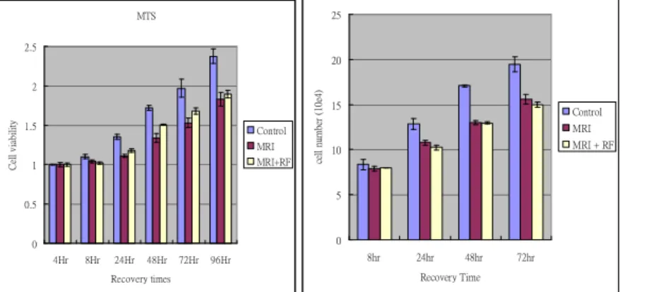
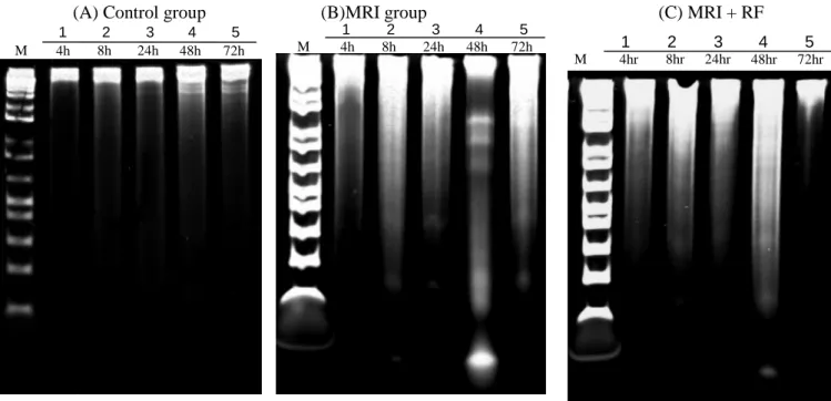
Human chondrocytes were isolated and were treated with 3T MRI for 1 hr. The cells were then recovered at 37℃ under 5% carbon dioxide in cellular incubator and the cell proliferation were assayed by MTS assay at 8 -, 24 -, 48 -, 72 and 96 - hr recovery. The cells of control group proliferated faster than the ones of MRI group, especially between 24- to 48-hour recovery, such as fig 1. The human chondrocytes might be damaged by MRI therefore slow down growth for repairing. In addition, we exposed the isolated chondrocytes to MRI for 1 hour followed by 60MHz radio-frequency for another one hour (RF group). At the indicated time points, e.g. 8 -, 24 -, 48 -, 72 - and 96 - hr, the proliferation of control was better than MRI group and RF group at all indicated time points. The chondrocytes of MRI group had the intermediate growth and ones in RF group had slowest growth among these three groups. To evalutate the influence of 3T MRI and RF on human chondrocytes, the DNA was examined. The DNA laddering was shown in fig 1, the MRI group and RF group had DNA damage situation at 48 hour. The cells from these three groups were stained with annexin V and PI to investigate the effect of MRI and radio-frequency on cell death. After recovery for 24 hour, the Annexin V - PI - stained cells of MRI group and RF group increased more than untreated control group, such as fig 2. At 48 and 72 hour time points, the cells of RF group were Annexin V –PI - stained more than these of MRI group and control group. There were no staining difference between the MRI group and RF group at 72 hr. By the way, the number of Annexin V –PI –stained cells had no difference between control group, MRI group, and RF group at 96 hours. To evaluate the influence of 3T magnetic field and radio frequency on human chondrocytes, the DNA was examined. The DNA laddering is commonly used to establish if a decrease in cell viability to due to apoptosis rather than necrosis. As shown in Fig. 3, polyacrylamide gel electrophoresis of DNA of 3T magnetic field and magnetic field plus radio frequency treated human chondrocytes revealed DNA fragmentation, characteristic of apoptotic cells (DNA ladder). The results indicated that the exposure 3T magnetic field and 3T magnetic field plus radio frequency of human chondrocytes to lead to DNA fragmentation in recovery 24 and 48 hours, suggesting that high magnetic field induced cell death by apoptosis. In addition, the radio frequency did not enhance degree of cell apopsotis comparing with only exposing high magnetic field. The control group had no DNA fragmentation in recovery different times (fig 3a). The results are consistent with observations of cell apoptosis of human chondrocytes by MRI and MRI + RF treatment. To explore the basis change of intracellular calcium concentration after exposing in 3T magnetic field and radio frequency on human chondrocytes, the intracellular calcium concentration was measured by Fura-2AM stained. The basis of intracellular calcium concentration had no difference between control, MRI, and MRI + RF group after recovery 1.5 and 3 hours (fig.4). In MRI and MRI + RF group, the basis of intracellular calcium concentration increased a little than control group after recovery 4.5 hours. The MRI + RF group of basis intracellular calcium concentration a bit higher than MRI group
after recovery 4.5 hours. After recovery 6 hours, the MRI and MRI + RF group of basis intracellular calcium concentration increased more than control group. At the same time, the MRI group of basis intracellular calcium concentration increased more than MRI + RF group. However, exposing normal environment, 3T static magnetic field, and 3T static magnetic field pulse radio frequency, the basis of intracellular calcium concentration could increase during short-term recovery time (~6 hours). But the basis of intracellular calcium concentration could decrease and no difference between control, MRI, and MRI + RF group after recovery 72 hours.
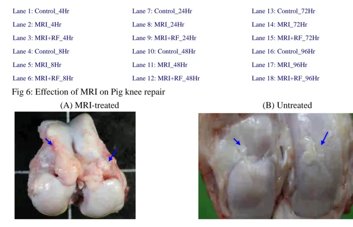
To explore whether high static magnetic fields and radio-frequency causes a change in the expression of the apoptosis-related genes such as p21 and p53, the chondrocytes were treated with 3T MRI for 1 hr, or 3T MRI plus 60 MHz radio frequency for 1 hr. The protein level was detected by Western blotting. p53 expression clearly increased at 24 hrs in MRI treatment and MRI plus 60 MHz radio-frequency treatment. p21 expression increased at 24 hrs and 48 hrs in MRI treatment and MRI pulse 60 MHz radio-frequency treatment, such as fig 5. To examine whether overdose of MRI assessment affect cartilage repair in clinic, we used Lee-Sung strain pig as animal model. We evaluated the knee cartilage regeneration with or without MRI assessment. The detail was described briefly in methods. During the 6-month, we found the defects still existed in the cartilage with MRI exposure (fig. 6A). However, in the cartilage without MRI exposure, the defects were well repaired (fig. 6B).
Fig 1: The human chondrocytes were exposed in normal environment, 3T magnetic field (MRI) and 3T magntic field plus radio frequency (MRI + RF) one hour. (A) On a series of recovery time points, the cells were monitored cell viability by MTS assay, F(10, 54) = 2.193, p < 0.05. (B) At the beginning stage, the same number of cells was seeded to 6 cm-culture dish, F(6, 36) = 16.566, p < 0.01.
MTS 0 0.5 1 1.5 2 2.5 4Hr 8Hr 24Hr 48Hr 72Hr 96Hr Recovery times C el lv ia b il it y Control MRI MRI+RF 0 5 10 15 20 25 8hr 24hr 48hr 72hr Recovery Time ce ll nu m be r (1 0e 4) Control MRI MRI + RF
Fig2: Effect of a 3T static magnetic field (MRI) and 3T static magnetic field plus radio frequency (MRI + RF) induced apoptosis in human chondrocytes as measured by flow cytometric bivariate analysis. (a) Recovery 48 hours
(b) Recovery 72 hours
(B)MRI group (A) Control group
Intracellular calcium concentration
0 0.5 1 1.5 2 2.5 3 1.5 3 4.5 6 72
recovery time (hours)
C a2 + (1 00 nM / 2 * 10 e5 ce ll s) Control MRI MRI + RF
Fig 3: DNA fragmentation detected in human chondrocytes after exposing 3T magnetic field (MRI) and 3T magnetic field plus radio frequency (MRI + RF) one hour.
Fig4: Magnetic field and magnetic field plus radio frequency increase capacitative Ca2+influx in human chondrocytes.
Fig5: MRI induce p53 and p21 expression
M 4hr 8hr 24hr 48hr 72hr 1 2 3 4 5 M 4h 8h 24h 48h 72h 1 2 3 4 5 M 4h 8h 24h 48h 72h 1 2 3 4 5
p21
p53
1 2 3 4 5 6 7 8 9 10 11 12 13 14 15 16 17 18Col II
(C) MRI + RF
-actin
Lane 1: Control_4Hr Lane 2: MRI_4Hr Lane 3: MRI+RF_4Hr Lane 4: Control_8Hr Lane 5: MRI_8Hr Lane 6: MRI+RF_8Hr Lane 7: Control_24Hr Lane 8: MRI_24Hr Lane 9: MRI+RF_24Hr Lane 10: Control_48Hr Lane 11: MRI_48Hr Lane 12: MRI+RF_48Hr Lane 13: Control_72Hr Lane 14: MRI_72Hr Lane 15: MRI+RF_72Hr Lane 16: Control_96Hr Lane 17: MRI_96Hr Lane 18: MRI+RF_96Hr
Fig 6: Effection of MRI on Pig knee repair
(A) MRI-treated (B) Untreated
Discussion:
Our results showed that the 3T static magnetic fields and radio frequency could suppress cell growth and increase cell apoptosis through p21 and p53 protein expression. We found that pig cartilage without MRI assessment repaired better than the one with MRI assessment using pig as model. It is possible that exposure to the high magnetic field induces chondrocytes apoptosis or transformation during the cartilage repair process. Our data suggests a low dosage and short exposure time for the use of MRI in clinical assessment.
Conclusion:
MRI is a high magnetic field environment. Because MRI could made some stimulation for chondocytes, human chondrocytes grow slowly to recovery after treating 3T MRI environment. The MRI stimulation may induce human chondrocytes apoptosis. It hit the assessment of cartilage in clinic, safely short time and low doses
Reference:
1. Colman MS., et al. Mutat. Res. 2000;462: 179-188. 2. Lin H., et al., J Cell Biochem. 1994 ; 54: 281-288. 3. Buemi M., et al., Nephron. 2001 ; 87: 269-273. 4. Olsson G., et al., Mut Res. 2001 ; 493: 55-66.
5. Walleczek J., et al., Bioelectromagnetics 1994;16: 129-145. 6. Denis F., et al., J Toxic. Environ. Health part A 1998 ; 54 : 53-76. 7. Fanelli C., et al., FEBS L. 1999 ; 13 : 95-102.