行政院國家科學委員會專題研究計畫成果報告
糖尿病足底皮膚之剪力波速的測量
Measur ement of the Shear Wave Velocity of
the Sole Skin in Diabetic Patients
計畫編號:NSC 89-2320-B-002-047
執行期限:88 年 8 月 1 日至 89 年 7 月 31 日
主持人:王亭貴 醫師 台大醫院復健科
協同主持人:邵耀華 副教授 台大應用力學研究所
Abstr act 糖尿病足潰瘍與腳底軟組織病變有密切的 關係。由於皮膚增厚、硬化或足底產生胼 胝,因此腳掌皮膚機械特性的改變對糖尿 病足潰瘍的發展是一個重要指標。 本研究 發展一套皮膚剪力波速量測裝置可安全地 應用於較脆或易裂的皮膚。 透過剪力波 速在足底的分佈,我們可以探討糖尿病足 潰瘍易發生的部位。 初步結果顯示手臂 皮膚剪力波速與文獻資料相符,與皮膚的 硬度成線性關係,腳底皮膚剪力波速似乎 相對地比較慢,而皮膚厚度、濕度的影響 也不容忽視。Diabetic foot ulcer is often accompanied by cutaneous changes, including skin thickening, callus formation and increased stiffness at the callus areas. Therefore, variation in the mechanical properties of the sole skin plays an important role in the development of diabetic foot ulceration. In this study, a novel device is developed for the measurements of shear wave propagation velocity (SWV) on the fragile skin. It is applicable for the evaluation of diabetic foots properties to find the regions that are vulnerable for ulceration. The preliminary results of forearm SWV showed good agreement with the literature. The SWV for the sole skin of the foot appears to be slower and the influence due to skin thickness and the moisture level is substantial.
一、背景與目的 (Intr oduction)
The skin is a heterogeneous tissue that composed of three superimposed layers; epidermis, papillary dermis and reticular dermis. They have distinct mechanical characteristics, in which the collagen fibres provide the main source of strength and stiffness for the skin and the elastin fibres are responsible for the recoiling mechanism during deformation (Reihner et al 1995).
Diabetic foot ulcers are the end points of a cascade initiated by hyperglycemia. The initial event in the process of ulceration relates to glycation and its effects upon the collagen. These lead to many cutaneous changes, including thickened and indurated skin, callus formation and altered elasticity, which may predispose the foot, especially at the callus areas, to ulceration. Therefore, variation in the mechanical properties of the sole skin plays an important role in the development of diabetic foot ulceration.
In literature, several instruments (i.e., cutometer, extensometer, impact hammer) have been developed to measure the skin mechanical properties directly or indirectly. Vogel (1995) conducted an overview of the experimental methods for the measurement of skin mechanical properties. The changes in mechanical properties (i.e., stress-strain curves) of the skin were often associated with biological aging process (Oikarinen 1994,
Diridollou et al. 1999), sun exposure, epidermal hydration (Jemec and Serup 1989), thickening (Takema et al 1994), or the fibril matrix (Reihsner et al. 1995, Sanders et al. 1997).
The response of skin to suction (i.e., cutometer) has been widely used in the qualitative study of skin mechanical properties in normal tissues and in the patients with diabetes (Nikkels-Tassoudji et al 1996), burn (Enomoto et al. 1996) and connective tissue disease (Ishikawa et al. 1995; Dobrev 1998). However, the indexes derived for skin elasticity and relaxation have the drawbacks of being relative scales and unable to differentiate the properties between the epidermis and dermis skin layers. In these studies, relatively large strain was applied to the skin and the works were focused mostly on the facial skin or the forearm. The contact (i.e., normal stress, slippage) between the sensors and the skin surface played an important role in the accuracy of these measurements. Therefore, the dynamic behaviors of the skin were found to vary with the contact area (Pierard et al 1995), or the distance between the measurement sites.
Potts et al. (1983) developed a device to determine the changes of skin mechanical properties by measuring the shear wave propagation velocity under low amplitude (i.e., 10 µm) disturbance. Since the transit time between the driving stylus and the receiving stylus was used in the calculation the wave velocity, the magnitude of skin response to the action of shearing force became an irrelevant issue. With the minimum disturbance to the skin, the results would reflect mostly on the stiffness of non-collagen contents of the superficial skin. Also, by using the sinusoidal perturbation at a frequency range of 10-2000 Hz, the shear wave velocity and the damping length were found to correlate linearly with the frequency applied. The shear wave was found propagate markedly faster in surface with greater stiffness. However, the effects of physical
and anatomical changes in skin properties on the wave velocity were not resolved since the variation in skin thickness was not considered. Davis et al. (1989) adopted the same design for the evaluation of age-related change of forearm skin properties of a larger study group. McHugh et al. (1997) and Clark et al. (1996) found that the SWV was markedly higher in scar. Nevertheless, very limited studies addressed the potential of using their instrumentation in the pathologic-related changes of skin or in the documentation of the skin on the foot.
The main purpose of this study is to develop a novel device that applicable to the evaluation of skin mechanical properties, especially for the fragile skin in diabetic foots or patients with burns. The current concept originated basically from Potts’ design, however, major changes were implemented since the record cutting heads had the risk of damaging the thin skin.
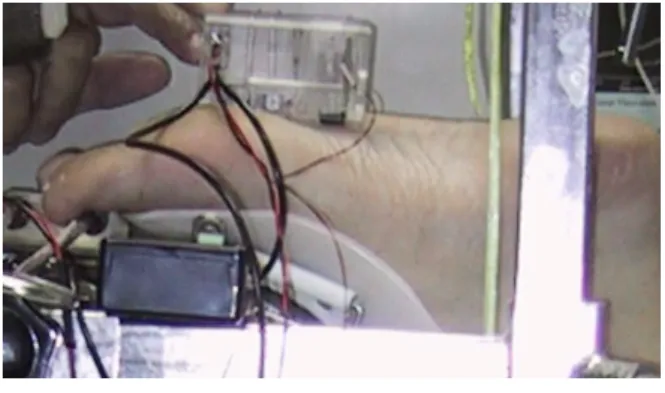
三、研究方法 (Materials and Methods) The skin shear-wave measurement system developed in this study consists of an oscillatory displacement generator that contacts with the skin 0.5-1.0 cm away from the nearby receiver. For safety, the shearing displacement is kept on the order of 20-50
µm at a contact area of 0.6 cm2. Two receiving pins of mass of 0.4g that separated at a distance of 5-8 mm were placed on the skin surface to sense the shear-wave arrivals. The movements of the skin were detected by LED sensor that has superior frequency response and excellent sensitivity (Figure 1).
Five subjects with healthy skin aged between 22-45 were randomly selected in this study to verify the capability of the instrumentation. Both the forearm skin and the skin under the foot were tested. Such a process was considered important for finding the potential problems before the device being practiced on the skin of the patients. A phantom of pork skin was also used to verify the SWV measurement system.
The time-delay of the shear wave sensed at the two receivers was measured using a digital real-time oscilloscope (100 MHz, TDS-224, Tektronix). The signals pickup by the receivers were highly coherent, although the displacement measured at the transducer closer to the actuator was about three-four times higher in amplitude than the other one. (Figure 2) This indicated that the skin also contained somewhat viscoelasticity (damping). Also, the skin thickness was measured using a high-resolution ultrasound system with 12 MHz linear array transducer (HDI-5000, L12-5 38mm, ATL). The oscillating frequency of the driving shear displacement can be altered from 10 Hz to 200 Hz by adjusting the supply voltage of the vibrating motor (from 2 to 9 V).
For the measurement of the skin under the foot, each subject was placed prone with the ankle in neutral position and the knee in 90° flexion, thus, the skin of the foot was relaxed and faced up as shown in Figure 1.
Fig. 2 Measurement of time-delay for skin SWV response Case Thickness (mm) Dx (mm) Dt (msec) SWV (m/s) A 1.4 8 8.5 9.4 B 1.5 8 9.2 8.70 C 1.4 8 7.8 10.26 D 1.6 8 7.5 10.67 E 1.5 8 8.6 9.30 Table 1. Forearm skin SWV and thickness
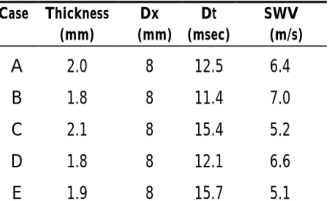
Case Thickness (mm) Dx (mm) Dt (msec) SWV (m/s) A 2.0 8 12.5 6.4 B 1.8 8 11.4 7.0 C 2.1 8 15.4 5.2 D 1.8 8 12.1 6.6 E 1.9 8 15.7 5.1 Table 2. SWV and thickness of the sole skin
of the foot
四、結果與討論(Results and Discussion) The results of skin SWV and thickness for the forearm and the sole skin at the first metatarsal head of five subjects are listed in Table 1 and Table 2 respectively (with the driving oscillatory frequency, f =35 Hz).
The skin of the forearm for healthy subjects is about 1.48 mm in thickness and the shear wave propagates at a velocity of about 9.6 m/s. The current results agree very well with Davis et al (1989). The older subjects had a higher SWV as observed. With respect to the sole skin of the foot, the averaged skin thickness was about 1.92mm, while the epidermis along was about 0.7-0.8 mm. The SWV was about 6.1 m/s.
The skin covers the entire external surface of the body and is fundamentally composed of epidermis and dermis. The epidermis is a compound tissue containing self-renewing and self-repairing stratified keratinized squamous epithelium. It is a primary site of immunosurveillance against
the entry of antigens, and protect human body against mechanical, chemical, osmotic, thermal and photic damage (Bannister 1995). The dermis tissues are composed of collagen fibers in the form of organized macromolecules in bundles. The ground substances in which polysaccharides and protein linked to form the macromolecules with a remarkable capacity for holding water (Gibson et al. 1970).
The geometry and interaction of collagen and elastin networks in the dermis govern the mechanical properties of skin, while the ground substance contributes mainly to the rheological behavior of skin (Lanir 1979). The skin behaves elastically only at low stress levels. Beyond the ‘toe region’ the effect of elastin on the biomechanics decreases resulted in a viscoelastic behavior induced by the straightened collagen fibers. The changes in skin mechanical behavior can be related to the increased cross-linking of collagen fibers and the degradation of elastin network (Braverman & Fonferko 1982).
The diabetic foot conditions include neuropathy, structural deformity, callus, skin and nail changes, foot ulcers, infection, and vascular disease. Foot ulcers are the most common complication of diabetes leading to amputation. The skin distensibility showed marked variations with the location of the measurement. Two diabetic patients were enrolled in this study. Since their foot-skin had decreased extensibility in response to traction, the shear-wave propagated with a higher velocity (5-15%) than the normal soft skin as expected. However, we find the moisture level and the skin thickness have non-negligible effects on the SWV results. The ultrasound couplant that applied on the skin for thickness measurement may slow down the SWV by 5-10%. A more detail investigation of skin thickness (higher frequency ultrasound 20-50MHz), skin hydration (electric resistance and
capacitance), and larger study group is needed to draw a precise conclusion.
五、成果自評
This research program complete the design and verification of skin shear wave velocity (SWV) measurement system to evaluate the corresponding skin mechanical properties. It was demonstrated to be useful in the evaluation of biomechanical properties of sole skin in DM foots to find the regions that are impending or at risk to ulceration.
六、參考文獻
1. Bannister LH. Integumental system: skin and breasts. In: Williams PL, editor. Gray’s anatomy, 38th ed. New York: Churchill Livingstone, 1995, p.375-424. 2. Braverman IM, Fonferko E. Studies in
cutaneous aging: The elastin fiber network. J Invest Dermatol 1982;78:434-443.
3. Clark JA, Change JCY, Leung KS. Mechanical properties of normal skin and hypertrophic scar. Burns 1996;22:443-446.
4. Davis BD, Bahniuk E, Young JK, Barnard CM and Mansour JM, Age-dependent changes in the shear wave propagation through human skin, Experimental Gerontology 1989; 24:201-210.
5. Dobrev H. In vivo study of skin mechanical properties in scleroderma of buschke. Acta Derm Venereol 1998;78:103-106.
6. Enomoto DNH, Mekkes JR, Bossuyt PMM, Hoekzema R, Bos JD. Quantification of cutaneous sclerosis with a skin elasticity meter in patients with generalized scleroderma. J Am Acad Dermatol 1996;35:381-387.
7. Gibbons G, Eliopolos G. Infection of the diabetic foot. In: Kozak G, editor. Management of diabetic foot problems. Philadelphia: WB Saunders; 1984, pp.
97-102.
8. Gibson T, Kenedi RM. In: Montagna W, Bentley JP, Dobson RL, editors. The dermis. New York: Meredith Corp.; 1970, pp. 19-38.
9. Hanna W, Friesen D, Bombardier C, Gladman D, Hanna A. Pathologic features of diabetic thick skin. J Am Acad Dermatol 1987;16:546-533.
10. Irie T, Oka H, Yamamoto T. Measurement of hardness of human skin with impact force. Med Biol Eng Comput 1994;32:231-233.
11. Ishikawa T, Ishikawa O, Miyachi Y. Measurement of skin elastic properties with a new suction device (I): relationship to age, sex, and the degree of obesity in normal individuals. J Dermatology 1995; 22:713-717.
12. Jemec GB and Serup J, Epidermal hydration and skin mechanics, Acta Derm Venereol (Stockh) 1990; 70:245-247. 13. Lanir Y. The rheological behavior of skin.
Biorheology 1979; 16:191-202.
14. McHugh AA, Fowlkes BJ, Maevsky EI, Smith DJ Jr., Rodriguez JL. Garner WL. Biomechanical alterations in normal skin and hypertrophic scar after thermal injury. J Burn Care Rehabil 1997; 18:104-108. 15. Nikkels-Tassoudji N, Henry F, Letawe C,
Piérard-Franchimont C, Lefébvre P, Piérard GE. Mechanical properties of the diabetic waxy skin. Dermatology 1996; 192:19-22.
16. Oikarinen A, Aging of the skin connective tissue: how to measure the biochemical and mechanical properties of aging dermis, Photodermatol, Photoimmunol & Photomed 1994; 10:47-52.
17. Palumbo PJ, Melton LJ. Peripheral vascular disease and diabetes. In: Harris MI, Hamman RF, editors. Diabetes in America. NIH pub no 851468. Bethesda Md; 1985, P. XV 1-21.
18. Piérard GE, Nikkels-Tassoudji N, Piérard-Franchimont C. Influence of the test area on the mechanical properties of skin. Dermatology 1995;191:9-15.
19. Potts RO, Chrisman DA, Buras EM, Jr., The dynamic mechanical properties of human skin in vivo, J Biomech 1983; 16:365-372.
20. Reihner R, Balogh B, Menzel EJ. Two-dimensional elastic properties of human skin in terms of an incremental model at the in vivo configuration. Med Eng Phys 1995;17:304-313.
21. Takema Y, Yorimoto Y, Kawai M and Imokawa G, Age-related changes in the elastic properties and thickness of human facial skin, Brit J Dermat 1994; 131:641-648.
22. Vogel H, Mechanical measurements of skin, Acta Derm Venereol (Stockh) 1994; Suppl. 185:39-43.