行政院國家科學委員會專題研究計畫 成果報告
研究利用新型微脂體 (LPPC) 包覆藥物及吸附導向分子以
治療犬乳癌
研究成果報告(精簡版)
計 畫 類 別 : 個別型 計 畫 編 號 : NSC 97-2313-B-009-002- 執 行 期 間 : 97 年 08 月 01 日至 98 年 07 月 31 日 執 行 單 位 : 國立交通大學生物科技學系(所) 計 畫 主 持 人 : 廖光文 共 同 主 持 人 : 朱瑞民 計畫參與人員: 碩士班研究生-兼任助理人員:徐維瞳 博士班研究生-兼任助理人員:林于鈴 報 告 附 件 : 出席國際會議研究心得報告及發表論文 處 理 方 式 : 本計畫可公開查詢中 華 民 國 98 年 12 月 08 日
行政院國家科學委員會補助專題研究計畫
■ 成 果 報 告
□期中進度報告
研究利用新型微脂體 (LPPC) 包覆藥物及吸附導向分子以治療犬乳癌
計畫類別:■ 個別型計畫
□ 整合型計畫
計畫編號:NSC
97-2313-B-009-002
執行期間:
2008 年
8 月
1 日至
2009 年
7 月
31 日
計畫主持人:
廖光文
共同主持人:
朱瑞民
計畫參與人員:
成果報告類型(依經費核定清單規定繳交):■精簡報告
□完整報告
本成果報告包括以下應繳交之附件:
□赴國外出差或研習心得報告一份
□赴大陸地區出差或研習心得報告一份
■出席國際學術會議心得報告及發表之論文各一份
□國際合作研究計畫國外研究報告書一份
處理方式:除產學合作研究計畫、提升產業技術及人才培育研究計畫、列
管計畫及下列情形者外,得立即公開查詢
■涉及專利或其他智慧財產權,□一年■二年後可公開查詢
執行單位: 國立交通大學生物科技系(所)
中
華
民
國
2009
年
9 月
14
日
目錄
1. 前言與文獻探討...3
2. 目的...4
3. 研究材料與方法...4
3-1. Materials...4
3-2. Preparation of the Lipo-PEI-PEG complex (LPPC)...4
3-3. Microstructure of liposome (LPPC) ...4
3-4. Particle size and charge of liposome (LPPC) ...4
3-5. Stability of liposome (LPPC) ...5
3-6. Adsorptive tests of liposome (LPPC) with proteins...5
3-7. Competition test of LPPC...5
3-8. ITC analysis of LPPC...5
3-9. 0.2M glycine test of LPPC...6
3-10. Activities of adsorbed proteins on LPPC...6
3-11. Statistical Analysis...6
4. 結果...6
5. 討論...8
6. 參考文獻...11
7. 結果圖表...13
Figure 1. The microstructure of the LPPC. ...13
Figure 2. Stabilities of the LPPCs. ...14
Figure 3. Adsorptive ability of LPPC with BSA-Au. ...16
Figure 4. Protein adsorptive abilities of different liposomes. ...17
Figure 5. Adsorption capacity and timing test of LPPC. ...18
Figure 6. Competition test of the LPPC. ...20
Figure 7. ITC analysis of liposomes...21
Figure 8. Effect of 0.2 M glycine solution to adsorptive ability. ...22
Figure 9. Activities of adsorbed proteins on LPPC in serum contained medium. ...23
Figure 10. Activities of adsorbed antibodies on LPPC. ...24
Table 1. Substances adsorbed with LPPC. ...25
1. 前言與文獻探討
Composed of biocompatible and biodegradable phospholipids, liposome is one of the earliest established strategies for delivering genes or drugs into the cells. Several strategies have been developed to modify the liposome to increase the transfection efficiency (Kovacs, Karasz et al. 2009; Obata, Ciofani et al. 2009; Zidovska, Evans et al. 2009) or drug delivery, such as doxorubicin (Wu, Lee et al. 2007). Currently, cationic lipids are incorporated into the liposome to enhance the efficiency of delivery, because they preferentially bind to the cell surface with the negative charge to cause the increase in the endocytosis efficiency (Kovacs, Karasz et al. 2009).
Combination of liposome with cationic polymers to increase delivery efficiency has also been developed recently. Among the polymers, cationic polymer polyethyleneimine (PEI) has been shown to be a carrier to transfer genes into the cells (Boussif, Lezoualch et al. 1995; Boussif, Zanta et al. 1996; Huh, Do et al. 2007). Because of proton sponge effect, PEI causes the endosome rupture by inducing osmotic swelling, and carried DNA could escape into the cytoplasm (Boussif, Lezoualch et al. 1995; Sonawane, Szoka et al. 2003). Therefore, lipid encapsulation of polyplex plasmid DNA (Heyes, Palmer et al. 2007) or modification of cationic polymers PEI (Yamazaki, Nango et al. 2000) is developed for enhancing the transferring efficacy.
In order to utilize the polycationic liposome in vivo, specificity is another important issue for enhancing drug efficacy and reducing the side effect of toxic drug. Therefore, several studies have investigated targeting ability of the liposome decorated with peptides, such as RGD peptides binding with v3 integrins (Temming, Schiffelers et al. 2005; Zhao, Wang et al. 2009), GE11 peptides
binding with EGFR (Song, Liu et al. 2008), and mBAFF motif peptides binding to TNF receptor (Zhang, Gao et al. 2008). In the other hand, immunoliposome are also developed for improving the specificity of liposome by combining with the targeting antibodies, including anti-VCAM antibodies (Gosk, Moos et al. 2008) and anti-MT1-MMP antibodies (Hatakeyama, Akita et al. 2007). However, most of targetable liposome are manufactured by conjugating the targeting molecules on the components of liposome, such as cholesterols or side chain of polymer-modified lipids. In the process of conjugation, activities of targeting molecules would dramatically decrease, because the linkage of functional domain or degradation of peptides within the reaction; thus, functions of targeting molecules on liposome will be affected (Nobs, Buchegger et al. 2004; Kocbek, Obermajer et al. 2007). Additionally, the process of conjugation is complicated and time-exhausted, which would influence the activities of the targeting molecules on liposome.
In this study, we report a new polycationic liposome, Lipo/PEI/PEG complex (LPPC), to provide an alternative method to combine with proteins by non-covalent conjugation. The polymer offers electric forces and hydrogen bonds to adsorb with proteins, and may also provide structural barriers to strongly anchor them. Surprisingly, compared with other non-covalent conjugations which have weak interaction between the proteins and carriers (Nobs, Buchegger et al. 2004), LPPC could adsorb considerable quantities of proteins with stable interaction even competed by 10-fold of BSA. Applicable potential was also proven by adsorption with the antibodies in the serum-contained
medium.
2. 目的
For specifically transfer the drug-encapsulated carrier to specific site, we will develop a biodegradable and biocompatible liposome which is capable of stably combining with proteins on theliposome’ssurfaceand maintaining the activities of them. Therefore, we could further transfer the drug-encapsulated carrier to tumor site by combining the specific antibodies.
3. 研究材料與方法 3-1. Materials
1,2-Dioleoyl-sn-Glycero-3-Phosphocholine (DOPC) and 1,2-Dilauroyl-sn-Glycero-3-Phosphocholine (DLPC) were purchased from Avanti Polar Lipids (Alabaster, AL). Polyethylene glycol (PEG, MW 8,000) and polyethyleneimine (branched PEI, MW 25,000) were purchased from Sigma chemicals (Sigma-Aldrich, St. Louis, MO). DMEM and fetal bovine serum were purchased from Invitrogen (Gaithersburg, MD, USA). Penicillin/streptomycin/amphotericin (PSA) was purchased from Biological industries (Beithaemek, Israel).
3-2. Preparation of the Lipo-PEI-PEG complex (LPPC)
The 50 mg DOPC and 50 mg DLPC were mixed and coated onto a round bottom flask by a rotary evaporator (EYELA, N-1000S, Japan) in order to yield a thin lipid film. The lipid films were hydrated by steam for 2~3 hours and then 5 ml of PEG-PEI solution (44 mg/ml PEG and 133 mg/ml PEI) were added to the container. The lipid films were vigorously resuspended for 10 min and then the suspension was extruded through a LiposoFast extruder (Avestin Inc., Ottawa, Canada) via a 200 nm mesh nine times to give the correct particle size. The suspensions were diluted in H2O 50 fold
and centrifuged at 5,900 g for 5 min to remove any unincorporated substances. Finally, the pellets were resuspended with 5 ml of ddH2O and were stored at 4oC until needed. Before use, liposomes
were warmed to room temperature.
3-3. Microstructure of liposome (LPPC)
The morphology of the LPPC particles were observed by transmission electron microscopy (TEM, JEM 1230; JEOL, Tokyo, Japan) at 100 kV and screening electron microscopy (SEM, Hitachi S-4700)
3-4. Particle size and charge of liposome (LPPC)
The particle size of the LPPC were observed by Dynamic Light Scattering (BI-200SM Goniometer, Brookhaven, New York, USA), and the particle size were determined by Zetasizer
(Nano ZS90, Malvern Instruments Ltd.). The data were analysis and drew by the program (Windows 9KDLSW versions 1.25, Brookhaven).
3-5. Stability of liposome (LPPC)
The 200 g LPPC were stored at 4℃ and 37℃ for different time, respectively, and were diluted in H2O 50 fold and centrifuged at 5,900 g for 5 min to remove any separated substance.
Then, pellets were resuspended with 200l H2O, and transferred 20l resuspended LPPC into the
96-well plate for detecting the quantities of PEI by Coomassie Plus Protein Assay Reagent (PIERCE, USA), and transferred 100l resuspended LPPC into cuvette for measuring the turbidity by spectrophotometer (Eppendorf AG 22331, Hamburg, Germany).
3-6. Adsorptive tests of liposome (LPPC) with proteins
The 40 g LPPC were suspended in 1 ml of H2O and incubated with several proteins. After
incubation for 20 min, the samples were centrifuged at 5,900g for 5 min to remove free proteins. Therefore, the bovine serum albumin (BSA, Invitrogen, Gaithersburg, MD, USA), beta-glucuronidase or HRP conjugated antibodies on LPPC were determined by mixing with Coomassie Plus Protein Assay Reagent (PIERCE, USA), PNPG or TMB(Pierce Chemical Company, Rockford, IL), and detected by ELISA reader (TECAN, Austria). The BSA-FITC were detected by spectrophotometer (Ultrospec 3100 pro, Amersham Biosciences, Sweden) at Ex=488nm and Em=515nm. And urease B and heat shock protein of H. pylori were detected by spectrophotometer (Eppendorf AG 22331, Hamburg, Germany) at OD 280nm. But in the timing test, 40g LPPC were incubated with 200g BSA forvarious time as mentioned in legend.
The 40 g different liposome were also manufactured and incubated with HRP conjugated antibodies for 20 min. After centrifuging at 5,900g for 5 min, resuspended pellets were mixed with TMB (Pierce Chemical Company, Rockford, IL) and detected by ELISA reader (TECAN, Austria)
3-7. Competition test of LPPC
The 40 g LPPC were previous incubated with 10 ng HRP conjugated antibodies (R1356HRP, Acris), 2.2 g beta-glucuronidase, or 42 g BSA-FITC for 20 min. Then, different mounts of BSA were added in and competed for 20 min at 37℃. After centrifuging at 5,900g for 5 min, resuspended pellets were added with substrates and detected with ELISA reader (TECAN, Austria) or spectrophotometer (Ultrospec 3100 pro, Amersham Biosciences, Sweden).
3-8. ITC analysis of LPPC
The 200 g of LPPC or LipofectamineTM 2000 (Invitrogen, Gaithersburg, MD, USA) were suspended in 2.8 ml H2O, and 10l BSA (10 g/l) were drop into the container of ITC (2277
tam , Thermometric, Jarfalla, Sweden) each time. The data were analysis and drew by the program (Digitam v4.1, Thermometric).
3-9. 0.2M glycine test of LPPC
The 40 g LPPC were adsorbed with 200 g BSA as described above, and transferred the pellet into the 0.2M glycine solution for 20 min. After centrifuging to remove the unbound proteins, BSA adsorbed on LPPC were measure by Coomassie Plus Protein Assay Reagent (PIERCE, USA), PNPG or TMB (Pierce Chemical Company, Rockford, IL) and detected with ELISA reader (TECAN, Austria). Another experiment proceeded by adsorbing 200 g BSA with 40 g LPPC in 0.2M glycine solution for 20 min, and the quantities of BSA on LPPC were determined as describe above.
3-10. Activities of adsorbed proteins on LPPC
The 40 g LPPC were adsorbed with 4.4 g beta-glucuronidase and 100 g BSA for 20 min. After centrifuging at 5,900 g for 5 min, resuspended pellets of LPPC were mixed with serum-contained DMEM and transferred into the 96-well plate, blocked with BSA, for 1 hr. Because the adsorptive site on the surface of LPPC have not saturated, LPPC/G complex could bind with the BSA coated on well. Washing the plate for 3 times, and added 10% PNPG of PBS into the well and detected with ELISA reader (TECAN, Austria).
In this study, anti-CD3 monoclonal antibody (2C11 or OKT3) was utilized as first signal for activation of T cell, and the other monoclonal antibody, anti-CD28 as second signal was for optimal activation of T cell. PBMC (1×10⁵cells per well) or were respectively dispensed into 96-well culture plates and then except for control treated with different condition. 1 μg LPPC with 60 ng anti-CD3 mAb or with 60 ng anti-CD3 and 60 ng anti-CD28 mAb treated PBMCs for 72hrs. By using MTT assay, and then the cell proliferation rate was calculated as O.D. value of sample divide into O.D. value of PBMC alone.
3-11. Statistical Analysis
The results were analyzed using the GraphPad statistical software package (GraphPad Software, Inc., San Diego, CA, USA). The results were expressed as mean ± SD. The t-test was used when comparing two independent samples and the ANOVA test was used when comparing multiple samples. Differences where p<0.05 were considered statistically significant.
4. 結果
1. Structure of LPPC
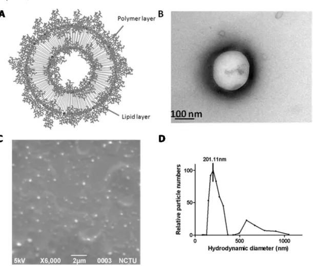
LPPC which combined the hydrophilic PEI and PEG with hydrophobic lipid layer was manufactured through modified procedure as described before. In this study, we expected that polymers would intertwine and surround the surface of lipid layer through the hydrophobic portion stubbed into lipid layer and the hydrophilic portion exposed out of membrane (Fig.1A). Figure 1B,
resembled with the schematic diagram, reveals that a round contour which formed the LPPC surface was sphered with dark shadow that expected the polymers existed. In addition, the image of SEM conspicuously shows the sphere shape with diameter at about 200nm (Fig 1C), was coincided with results measured by DLS (Fig 1D). The major particle sizes range from 150 to 270nm but few of them are over 500 nm (the average of size is 186.8 ± 34.6 nm). It is also notable that the Z potential value of LPPC is 41.4 ± 1.2 mV (data not shown). Since stubbed polymers increased the density of LPPC, they could be centrifuged at 6900g within 5 mins.
2. Functions of LPPC
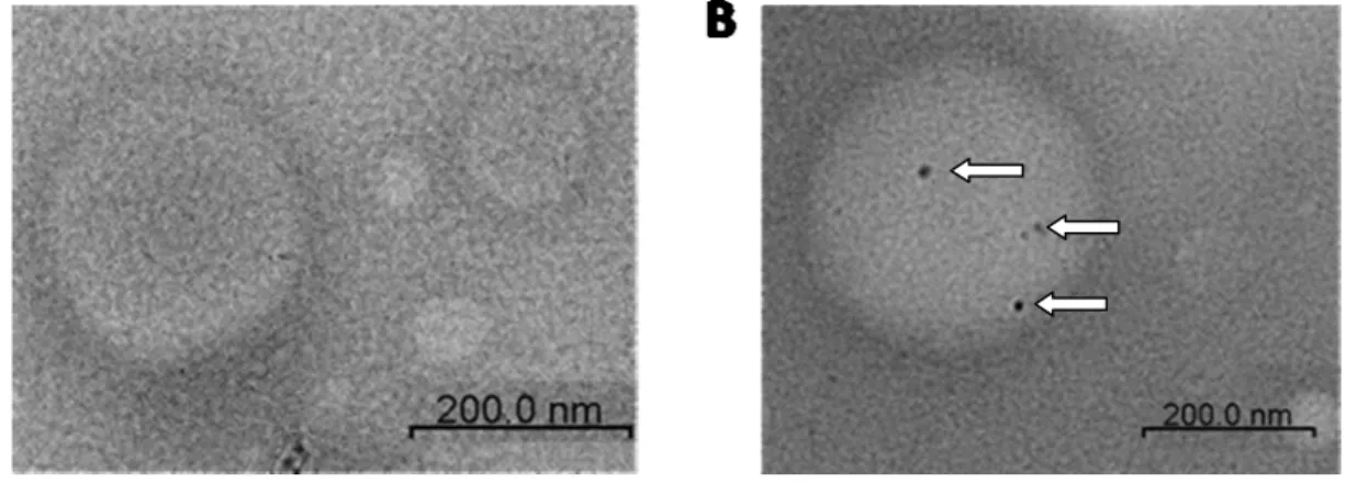
We first measured the turbidity of LPPC to verify whether the liposome particles did alter. As result showed, no significant differences were detected on different incubation times at 4℃ or 37℃ separately. It is respected that the LPPC could stably exist (Fig. 2A). Then, for bio-applications, we tested the interaction between the LPPC and proteins. Table 1 shows that proteins such as bovine serum albumin, FITC-labeled albumin, beta-glucunonidase, HRP-conjugated antibodies, NS-1 protein of Dengue virus, urease B and heat shock protein of H. pylori could be adsorbed with LPPC. Furthermore, photograph of electron microscopy confirmed that BSA-Au was certainly attached with LPPC (Fig. 3).
In order to testify that the adsorptive ability was contributed to branched PEI with positive charge, various liposomes were manufactured and experimented. Figure 4 shows that liposomes with PEI could retain activities of protein adsorption, but others without PEI lost or had slight activities. That is to say, PEI plays the key role in adsorptive ability of LPPC. Therefore, stability in combination of PEI and lipid layer was also detected. We found that no significant decrease of PEI was detected, in other word, PEI could steadily stubbed on LPPC at 4℃ or 37℃ even for 72 hours (Fig. 2B).
3. Capacity of protein adsorption on LPPC
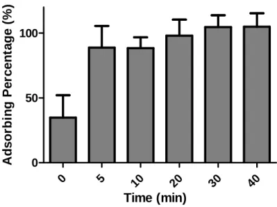
Next, we measured the capacity of LPPC in protein adsorption. As shown in figure 5A, LPPC could not adsorb proteins at environment of low BSA concentration, whereas LPPC revealed activities when BSA over 40ug/ml and the mounts of adsorbed BSA was correlated to concentration of BSA, and adsorptive capacity of LPPC was 169.3 ± 11.9ug. Moreover, as illustrated in figure 5B, LPPC could adsorb with BSA over 80% of adsorptive capacity of LPPC within 5 mins.
4. Irreplaceable of pre-adsorbed functional proteins on LPPC
To clarify the interaction between the pre-adsorbed functional proteins on LPPC and environmental proteins, competitive experiment was performed. The result reveals that pre-adsorbed HRP conjugated antibodies or beta-glucunonidases were not replaced by BSA even 500ug (Figure 6A and 6B). In addition, as illustrated in figure 6c, pre-adsorbed BSA-FITC, a derivative of BSA, was irreplaceable, even 600ug BSA was added. Overall, extra-addition of proteins could not displace
pre-adsorbed proteins on LPPC.
5. Energy patterns of interaction between LPPC and adsorbed proteins
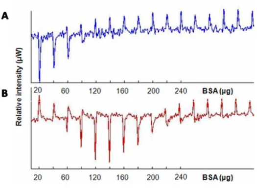
Energy patterns of interaction between LPPC or other cationic liposome, lipofectamine, with proteins was investigated by ITC (Fig.7). The energy pattern of lipofectamine revealed that signals could be observed from 20 ug to 80 ug of the addition of BSA, but reaction terminated when over 80ug BSA was dropped. However, no reaction occurred when BSA less than 40ug, but energy release had been monitored form 40 to 200 ug BSA. In other word, either total mount or pattern of enthalpy for LPPC was distinct from general cationic liposome.
6. Effect of 0.2M glycine solution to adsorptive ability
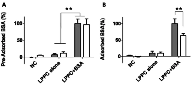
Since irreplaceable ability and particular energy pattern of LPPC had been observed before, we further examined the effect of electric charge on amine group of PEI to adsorptive ability. The result showed that pre-adsorbed proteins on LPPC was not affected by transference of environment into 0.2M glycine (pH=5.4) (Fig. 8A), whereas interaction between the non- adsorbed proteins and LPPC was affected by environment variation but LPPC retained over 60% adsorptive capacity (Fig. 8B). Hence, we speculated that the uncompetitable ability of LPPC was not merely supported by positive charge.
7. Activities of adsorbed proteins on LPPC in serum contained medium
In order to apply LPPC in bio-environment, the influent of serum contained medium to activities of adsorbed functional proteins on LPPC was further investigated. As shown in figure 9, adsorbed beta-glucunonidases still existed on LPPC and retained their activities in serum contained DMEM. On the other hand, as shown in figure 10, antibodies adsorbed on LPPC could stimulate the proliferation of PBMC in serum contained medium, and secretion of INF-γwith 2303±57 pg/ml and IL-2 with 204±29 pg/ml. In short, adsorbed proteins could retain their activities even in serum contained medium.
5. 討論
In this study, the cationic LPPC was manufactured from hydrophobic lipid and hydrophilic polymers without covalent bond formation and had the great capacity for protein adsorption. Interestingly, prior adsorbed proteins on LPPC would not be replaced by the posterior additional proteins. As our knowledge, we provided the first report to this property that the bound protein is irreplaceable.
Without the chemical linker, the sphere shaped LPPC was produced by hydrating the lipid film with polymers-contained solution, which means the hydrophilic polymers PEI and PEG could complex with hydrophobic lipid. As our results, ninhydrin test indicated that the primary amines which only existed in PEI were detected in LPPC (result not shown) and the figure 4 also showed
that the liposome lost the ability to adsorb proteins without PEI incorporation. Thus, PEI should be existed in LPPC. Another component, PEG, was a negatively charged polymer, which would theoretically neutralize the PEI’s positivecharges. As expected, figure 4 indeed showed PEG incorporation suppress the binding ability of LPPC, which indicated that PEG should be incorporated into LPPC. Moreover, hair-liked structure on TEM was demonstrated that polymers indeed stocked on the surface of the LPPC as we predicted.
In the aspect of functions, as the results mentioned, LPPC could stably adsorb with the proteins within 20 mins, and its maximal capacity was over 160μg BSA (per40 g LPPC),the measured values was coincided with the values measured from the ITC (Fig. 7). Compared to the commercial cationic liposome, lipofectamine (80 g pe r 40 g l iposome), LPPC’s adsorptive capacity is great higher than this cationic liposome. The adsorptions might be caused by the formation of more electrostatic interactions between the amine groups of the PEI on LPPC with the basic residues in adsorbed proteins. In addition, a lot of the primary amine groups provided by PEI could also lead to the formation of more hydrogen bonds with the bound proteins (Khoo, Teh et al. 2009; Ong, Leong et al. 2009).
Interestingly, pre-adsorbed proteins, such as beta-glucunonidase and HRP-conjugated antibodies on LPPC could not be competitively replaced by 10-fold of BSA and the same as pre-adsorbed BSA-FITC, the analog of the BSA (Fig. 6C). Consequently, there are several lines of possibilities to explain the mechanism of the irreplaceable adsorption. First, the prior bound individual protein was captured by hundred of the electrostatic forces and hydrogen bonds provided by PEI (Khoo, Teh et al. 2009; Ong, Leong et al. 2009), and it is difficult to simultaneously remove these hundred of the forces to dissociate the pre-adsorbed proteins from the binding site. Secondary, the binding of the pre-adsorbed proteins on LPPC would result in a steric barrier for the posterior additional proteins. Third, a large number of tiny branches of PEI might stab and interact with the microstructure of an individual protein by “Velcro”-like hook side, resembling the interaction between some proteins (Creze, Castang et al. 2008; Durrieu, Lavery et al. 2008). Such simultaneously mechanical interactions may cause the pre-adsorbed proteins to hardly dissociate from the binding site. Thus, we proposed that the irreplaceable adsorption should not only be completely caused by electrostatic forces but the other interactions also involved. This hypothesis was further supported by the Figure 8A, the protein-adsorbed on LPPC would not be dissociated from the surface when it was transferred into the low pH glycine buffer (0.2 M); nevertheless, the low pH glycine buffer indeed lowered the interactions of protein and LPPC. Together these results demonstrated that the electrostatic forces might be not the only factor for the interactions of the pre-adsorbed protein and LPPC, but electrostatic forces should be an essential factor involved in protein adsorption.
Currently, the targeting molecules were covalently coupled with the liposome, which might influence theprotein’sstructure;thus,themaintenance fortheactivitiesofthetargeting molecules usually was important for the therapeutic efficacy (Nobs, Buchegger et al. 2004; Kocbek, Obermajer
et al. 2007). To overcome this effect, LPPC did not only adsorb the proteins on surface but also remain their activities, even in the serum-contained medium (Fig.9 and Fig. 10). In our unpublished results, peptide-loaded HLA-A2 and anti-CD28 antibodies were adsorbed on LPPCs and the immuno-complexes could trigger specific T-cell responses but the unbound peptide-loaded HLA-A2 and anti-CD28 antibodies completely loss the ability.
In summary, we provided a novel lipocomplex, LPPC which could adsorb with proteins, such as enzymes or antibodies and retained their activities. Although the exact mechanisms for the irreplaceable adsorption of LPPC are not clear yet, this adsorptive function of LPPC could possibly provide more convenient and flexible applications. To combine with targeting molecules or functionalmoleculeson LPPC’ssurfacewillallow to specifically deliverdrug orgeneinto specific tissues or cells.
6. 參考文獻
Boussif, O., F. Lezoualch, et al. (1995). "A VERSATILE VECTOR FOR GENE AND OLIGONUCLEOTIDE TRANSFER INTO CELLS IN CULTURE AND INVIVO -POLYETHYLENIMINE." Proceedings of the National Academy of Sciences of the United States of America 92(16): 7297-7301.
Boussif, O., M. A. Zanta, et al. (1996). "Optimized galenics improve in vitro gene transfer with cationic molecules up to 1000-fold." Gene Therapy 3(12): 1074-1080.
Chen, S. T., Y. L. Lin, et al. (2008). "CLEC5A is critical for dengue-virus-induced lethal disease." Nature 453(7195): 672-U12.
Creze, C., S. Castang, et al. (2008). "The crystal structure of pectate lyase PelI from soft rot pathogen Erwinia chrysanthemi in complex with its substrate." Journal of Biological Chemistry
283(26): 18260-18268.
Durrieu, M. P., R. Lavery, et al. (2008). "Interactions between neuronal fusion proteins explored by molecular dynamics." Biophysical Journal 94(9): 3436-3446.
Gosk, S., T. Moos, et al. (2008). "VCAM-1 directed immunoliposomes selectively target tumor vasculature in vivo." Biochimica Et Biophysica Acta-Biomembranes 1778(4): 854-863. Hatakeyama, H., H. Akita, et al. (2007). "Tumor targeting of doxorubicin by anti-MT1-MMP
antibody-modified PEG liposomes." International Journal of Pharmaceutics 342(1-2): 194-200.
Heyes, J., L. Palmer, et al. (2007). "Lipid encapsulation enables the effective systemic delivery of polyplex plasmid DNA." Molecular Therapy 15(4): 713-720.
Huh, S. H., H. J. Do, et al. (2007). "Optimization of 25 kDa linear polyethylenimine for efficient gene delivery." Biologicals 35(3): 165-171.
Khoo, K. S., E. J. Teh, et al. (2009). "Hydrogen Bonding and Interparticle Forces in Platelet alpha-Al2O3 Dispersions: Yield Stress and Zeta Potential." Langmuir 25(6): 3418-3424. Kocbek, P., N. Obermajer, et al. (2007). "Targeting cancer cells using PLGA nanoparticles surface
modified with monoclonal antibody." Journal of Controlled Release 120(1-2): 18-26.
Kovacs, T., A. Karasz, et al. (2009). "The density of GM1-enriched lipid rafts correlates inversely with the efficiency of transfection mediated by cationic liposomes." Cytometry A.
Leserman, L. D., J. N. Weinstein, et al. (1980). "SPECIFIC INTERACTION OF MYELOMA TUMOR-CELLS WITH HAPTEN-BEARING LIPOSOMES CONTAINING METHOTREXATE AND CARBOXYFLUORESCEIN." Cancer Research 40(12):
4768-4774.
Li Yan, Q. and B. You Han (2007). "Self-assembled polyethylenimine-graft-poly( epsiv -caprolactone) micelles as potential dual carriers of genes and anti cancer drugs." Biomaterials 28(28): 4132-4142.
Nobs, L., F. Buchegger, et al. (2004). "Current methods for attaching targeting ligands to liposomes and nanoparticles." Journal of Pharmaceutical Sciences 93(8): 1980-1992.
Obata, Y., G. Ciofani, et al. (2009). "Evaluation of cationic liposomes composed of an amino-acid-based lipid for neuronal transfection." Nanomedicine.
Ong, B. C., Y. K. Leong, et al. (2009). "Interparticle forces in spherical monodispersed silica dispersions: Effects of branched polyethylenimine and molecular weight." J Colloid Interface Sci.
Skjorringe, T., T. Gjetting, et al. (2009). "A modified protocol for efficient DNA encapsulation into pegylated immunoliposomes (PILs)." J Control Release.
Sonawane, N. D., F. C. Szoka, et al. (2003). "Chloride accumulation and swelling in endosomes enhances DNA transfer by polyamine-DNA polyplexes." Journal of Biological Chemistry
278(45): 44826-44831.
Song, S. X., D. Liu, et al. (2008). "Peptide ligand-mediated liposome distribution and targeting to EGFR expressing tumor in vivo." International Journal of Pharmaceutics 363(1-2): 155-161. Temming, K., R. M. Schiffelers, et al. (2005). "RGD-based strategies for selective delivery of
therapeutics and imaging agents to the tumour vasculature." Drug Resistance Updates 8(6): 381-402.
Weissmann, G., A. Brand, et al. (1974). "Interaction of immunoglobulins with liposomes." J Clin Invest 53(2): 536-43.
Wu, J., A. Lee, et al. (2007). "Vascular targeting of doxorubicin using cationic liposomes." International Journal of Pharmaceutics 337(1-2): 329-335.
Yamazaki, Y., M. Nango, et al. (2000). "Polycation liposomes, a novel nonviral gene transfer system, constructed from cetylated polyethylenimine." Gene Therapy 7(13): 1148-1155.
Yang, J. M., Y. F. Chen, et al. (2007). "Combinatorial computational approaches to identify tetracycline derivatives as flavivirus inhibitors." PLoS One 2(5): e428.
Zhang, L., H. G. Gao, et al. (2008). "Tumor targeting of vincristine by mBAFF-modified PEG liposomes in B lymphoma cells." Cancer Letters 269(1): 26-36.
Zhao, H., J. C. Wang, et al. (2009). "RGD-based strategies for improving antitumor activity of paclitaxel-loaded liposomes in nude mice xenografted with human ovarian cancer." Journal of Drug Targeting 17(1): 10-18.
Zidovska, A., H. M. Evans, et al. (2009). "The role of cholesterol and structurally related molecules in enhancing transfection of cationic liposome-DNA complexes." J Phys Chem B 113(15): 5208-16.
7. 結果圖表
Figure 1. The microstructure of the LPPC. (A) Schematic diagram, (B) TEM and (C) SEM
images showed the microstructure of the LPPC, which had dense circle regions and hair-like surface. (D) The particle sizes of LPPCs were measured by DLS, and the major particle size is about 201nm.
(A)
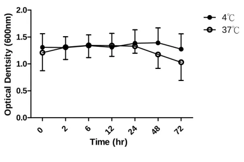
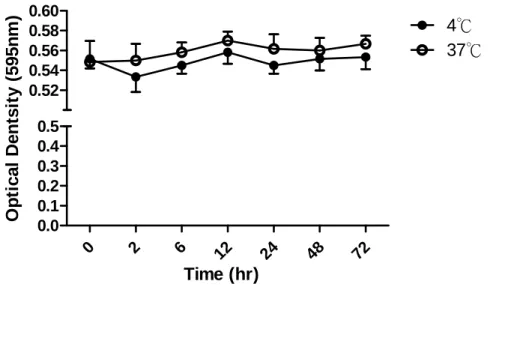
Figure 2. Stabilities of the LPPCs. (A) The 40 g of LPPC were stored at 4℃ or 37℃ environments separately for different times. The pellets were collected and measured the turbidities of LPPC by OD 600 to indicate the stabilities of particle sizes and numbers. (means ± SD, n=6)
0 2 6 12 24 48 72 0.0 0.5 1.0 1.5 2.0 4℃ 37℃ Time (hr) O p ti c a l D e n ts it y (6 0 0 n m )
(B)
Figure 2. Stabilities of the LPPCs. (B) The 40 g of LPPC were stored at 4℃ or 37℃ environments separately for different times. The pellets were collected and measured the amounts of PEI on the LPPC by Coomassie Plus reagent to indicate the stabilities ofparticle’scomponents. (means ± SD, n=6) 72 48 24 12 6 2 0 0.0 0.1 0.2 0.3 0.4 0.5 0.52 0.54 0.56 0.58 0.60 4℃ 37℃ Time (hr) O p ti c a l D e n ts it y (5 9 5 n m )
Figure 3. Adsorptive ability of LPPC with BSA-Au. TEM images of (A) LPPC alone and (B)
LPPC adsorbed with BSA-Au. The block spot (arrows indicated) in the center of the LPPC were nano-gold conjugated BSA (BSA-Au).
Figure 4. Protein adsorptive abilities of different liposomes. Liposome were manufactured with
different components for investigating the effects on adsorptive abilities. Liposome could adsorb proteins when PEI was existed. (means ± SD, n=6)
(A)
Figure 5. Adsorption capacity and timing test of LPPC. (A) LPPC (40 g) was incubated with different amounts of BSA for 20min, and detected the quantities of BSA adsorbed on LPPC by Coomassie Plus Protein Assay Reagent. The capacity of LPPC is 169 ± 11.9 (means ± SD, n=6)g.
50 100 150 200 250 -50 0 50 100 150 200 250 Added BSA (g) T o ta l o f D e te c te d B S A ( g )
(B)
Figure 5. Adsorption capacity and timing test of LPPC. (B) LPPC (40 ) was incubated withg 200 BSA for different reaction time, and measured the quantities of BSA adsorbed on LPPC byg Coomassie Plus Protein Assay Reagent. LPPC could nearly complete the adsorptive reaction at 20 min. (means ± SD, n=6) 0 5 10 20 30 40 0 50 100 Time (min) A d s o rb in g P e rc e n ta g e (% )
Figure 6. Competition test of the LPPC. (A) HRP-conjugated antibodies, (B) beta-glucuronidases
and (C) BSA-FITCs, which prior incubated with the 40 μg LPPC, were competed by different amounts of BSA. Percentages of adsorbed proteins on LPPCs were normalized with positive control, which adsorbed the same amount of proteins without BSA competition. NC was protein alone following the same procedure; LPPC indicate the LPPC alone without the protein adsorption. (means ± SD, n=6)
Figure 7. ITC analysis of liposomes. The 40 μg of (A) Lipofectamine and (B) LPPC were dropped with continued BSA titration, and the thermo releases were detected by ITC.
Figure 8. Effect of 0.2 M glycine solution to adsorptive ability. LPPC was incubated with BSA (A)
before or (B) after transferring into the 0.2 M glycine solution. The quantities of BSA adsorbed on LPPC were measured by Coomassie Plus Protein Assay Reagent. (closed bar is the environment of H2O, and open bar is the environment of 0.2M glycine solution. means ± SD, n=6)
Figure 9. Activities of adsorbed proteins on LPPC in serum contained medium. LPPC/G complex in serum contained DMEM were transferred into the 96-well, which prior blocked with BSA on the bottom, and measured by ELISA. NC is background of medium alone; LPPC is LPPC without G adsorbed; G is beta-glucuronidase alone following following the same procedure (means ± SD, n=6) NC LPP C βG LPPC +βG 0.0 0.5 1.0 1.5 2.0 O p ti c a l D e n s it y (4 0 5 n m )
Figure 10. Activities of adsorbed antibodies on LPPC. LPPC was incubated with anti-CD3 and/or
anti-CD28 monoclonal antibodies for stimulating (A) cell proliferation, (B) IL-2 or (C) INF- release of PBMC. The results revealed that the monoclonal antibodies could retained their activities to stimulate the cells. (means ± SD, n=6)
8. 計畫成果自評 本次計畫內容如預期目標所示,提供(1)最佳化之微脂體 LPPC 製備方式及成分比例,(2)LPPC 基本性質之測試,如吸附蛋白質能力、吸附力穩定性測試、組成成份穩定性測試等,(3)LPPC 穩定吸附蛋白質之特性探討,如 ITC 及 pH test,(4)所吸附蛋白質之活性探討,分別量測含血 清培養液及不含血清培養液中之功能性測試,以上結果皆表示出本計畫所研究之 LPPC 其獨特 性及新穎性。 基於此計畫所開發之微脂體特性,未來於犬類疫苗之開發,如包覆藥物並吸附導向分子以 治療腫瘤,甚至是人類疫苗方面皆可有效利用。其製備時間短且產量大,再加上其穩定吸附之 特性,可以避免未完全之化學反應對活體之傷害及所結合蛋白活性之喪失,因此在生醫產業之 應用性將非常地廣,甚至在生物材料及生物偵測實驗中皆可有效利用。 感謝國科會提供之研究經費,本計畫中之研究已撰寫並準備投稿至國際知名期刊,而其特 性分別在台灣及美國兩地申請專利中(專利申請程序進行中)。
出國報告(出國類別: C 類國際會議)
題目:International Conference on Materials
for Advanced Technology 2009
服務機關:生化工程研究所
姓名職稱:廖光文 副教授
前往國家:新加坡 Suntech
出國期間:2009/06/28~07/03
報告日期:2009/06/30
參與國際型研討會 (ICMAT 2009)
出國報告書
98 年 7 月 10 日 報告內容:
一、參加經過
6/28 Day1 出發至新加坡,到研討會會場(Suntech city mall)報到和拿取相關資料 6/29 Day2 參與大會邀請學者演講 6/30 Day3 海報展覽及口頭講說 7/01 Day4 參與大會邀請學者演講 7/02 Day5 參與大會邀請學者演講 7/03 Day6 參與大會邀請學者演講,返國 二、心得(可含照片) 建立實驗室至今以發展腫瘤免疫治療方向努力,研究領域包含胃幽門螺旋桿菌之熱休克 蛋白致病機制和利用微脂體複合體於免疫治療平台開發,而對於微脂體在生醫材料的應 用發展已有初步研究成果,因此選擇在新加坡舉辦生醫材料相關技術的研討會,此次研 討會之行,除了分享目前實驗室的研究成果外,也藉此了解國際間於生醫材料上的發展。 參與研討會邀請的學者演講這幾天中,其中有學者利用 DNA 組成不同的結構,建立新 穎的傳送平台,擁有專一性且生物可分解性之特性,有趣的是學者利用此平台搭配 hydrogel 於生物體外製造蛋白質,節省時間及增加效率,有別於傳統式利用細菌或是細 胞,利用此新穎方法,激發我和學生們需要利用新方向去思考自我身旁之資源,而另外 有學者是利用親水疏水性質不同之聚合物合成複合體成功增加藥物在免疫治療上的療 效。 於海報展覽當天,和學生於會場分享本實驗室成果,研討會成員們對於本實驗室建 立的微脂體複合體的新穎吸附能力相關特性應用和搭配抗癌藥物之實驗成果,感到很有 興趣,且熱烈討論報告中有趣之發現和對於實驗結果表示肯定,此外比較其他國家實驗 室的成果分享,發現大多海報的展覽多著重於實驗目的和設計概念,但在實驗結果發表, 普遍都著重於建立生醫材料平台上基本特性的測試,並未有進一步應用之成果,不過本 實驗室開發之微脂體複合體此平台於生醫方面應用都較有卓越的成效及進展,突顯出交 通大學在生物科技領域發展之努力和進步,此外,研討會後感覺本實驗室和其他各國比 較仍具有一定之競爭力和潛力。
(a) 海報前合影之一(我與學生劉彥谷) (b) 海報前合影之二(我與學生陳家弘)
(c) 研討會報到地點 (d) 海報展覽區
三、建議 a. 此次研討會之行,發現交通大學本校與其他學校於生物科技的競爭力並無相差太多, 但是在外語能力方面如聽力或演說較略顯不足,建議校方於外語能力的培養課程中可 以多著重於聽力和演說。 b. 建議贊助費用額度提高,因為與生物科技相關之比較大型國際型研討會大都集中於美 國、加拿大、德國等國,但除了該研討會註冊費較高外,餐旅費都相對來得比亞洲國 家更高。 四、攜回資料名稱及內容 a. 研討會摘要手冊 b. 研討會會議流程手冊 c. 研討會摘要內容光碟 五、其他 無
出國報告(出國類別: C 類國際會議)
題目:International Conference on Materials
for Advanced Technology 2009
服務機關:生化工程研究所
姓名職稱:劉彥谷
博班學生
前往國家:新加坡 Suntech
出國期間:2009/06/28~07/03
報告日期:2009/06/30
出國會議報告書
感謝國科會給予計畫經費之補助,使學生得以參加 2009 年 ICMAT & IUMRS-ICA (International Conference on Materials for Advanced Technologies 2009 & International Union of Materials Research Societies-International Conference in Asia 2009),此次會議為第五屆 ICMAT 研討會,6/28~7/3 日於新加坡會議中心舉辦。經由國科會審核之計畫經費,使得學生能夠完 成 Development of Novel Liposome with Stable Protein Adsorption Ability for Bio-Application 之 研究內容,並於 2009/6/30 於會議之壁報中展示成果,會議中不乏各國研究學者前來詢問及討 論研究之內容,並在討論其中各國學者皆對此研究充滿高度興趣,甚至更深入的詢問研究之 做法及源起之動機,而學生亦在討論中發現此研究仍然充滿許多有趣的問題及題目可以深入 探討或應用,如可應用此可吸附蛋白質之微脂體吸附螢光蛋白及標靶性分子,以建構新型之 偵測試劑,並結合流式細胞儀將可快速且有效的分析細胞表面之標誌分子。 與會期間,學生亦與指導教授參與不少口頭報告會議,其中另人印象最為深刻的莫過於 曾發表於 Nature 之作者 Dan LUO,所報告之 DNA-based Hydrogels for Drug Delivery and P-gels for Protein Production without Any Living Cells,此篇作者利用 DNA 做為一種攜帶藥物進入到 細胞內部之載體,此構想源自於現今載體幾乎都是利用其帶電的特性吸附特定物質進而送入 至細胞內部,但往往這些載體都會具有些許的毒性,因此作者想利用細胞本身的物質來運送 藥物進入到細胞內部,此做法將可有效的降低載體對於細胞的相容性及毒性上的問題。如此 新穎的構想,的確令人眼睛為之一亮,並且提供藥物載體建構之另一種思維。但於壁報之展 示中,我們發現國內研究載體之內容相較起來較有新穎性,且提供之數據內容也較為完整, 因此,國科會所提供之載體相關之研究成果皆相當豐碩,但國外研究仍有我們該學習的地方, 譬如說其對照組較為完整,且使用之技術及儀器亦較為高級及方便,這些皆是我們國內研究 需要加強的地方。
出國報告(出國類別: C 類國際會議)
題目:International Conference on Materials
for Advanced Technology 2009
服務機關:生化工程研究所
姓名職稱:陳家弘 碩班學生
前往國家:新加坡 Suntech
出國期間:2009/06/28~07/03
報告日期:2009/06/30
出國會議報告書
感謝老師和學姊給予我這個機會讓我出國學習,由此次準備新加坡之旅,學習如何從網 路上找尋適合領域的研討會,及事後註冊報名、辦理機票護照、訂購房間等等前置作業,到 後來於研討會會場中和學者們分享實驗結果,在討論切磋中了解自己本身實驗設計和分析結 果之優缺點,並且給我們一個很好的思考方向來改進。而且出國參加研討會對自己來說可以 了解其他領域或是相關領域的學者們的看法,也可以比較其他各國實驗室在生物科技的發 展,除此之外由研討會邀請的演講者吸收到許多新穎的想法,這樣的激盪下所激發出的科技 火花,讓自己更加成長茁壯。 此外英語的聽說能力對於這次研討會非常重要,因為在研討會中我們大多利用英語分享 及解說,常常在討論中會遇到口音不同或是使用詞彙不恰當,有時會有雞同鴨講的狀況發生, 所以我認為舉凡參加國際型的研討會都需要擁有,這樣在涉獵汲取新知外,和世界各國的先 進們都可以盡情的切磋討論。 綜合來說,參加這次新加坡研討會之旅,除了讓自己了解語言能力尚待加強外,也看到 其他相關領域學者的實驗架構想法和實驗設計進步,未來如果有機會的話,我會建議實驗室 同仁多多參加。Development of Novel Liposome with Stable Protein
Adsorption Ability for Bio‐Application
College of Biological Science and Technology, National Chiao Tung University, Taiwan, ROC Yen‐Ku Liu, Yu‐Ling Lin, Chia‐Hung Chen, Chia‐Ching Chang and Kuang‐Wen Liao Abstract L i p o s o m e h a v e b e e n a p p l i e d i n d r u g encapsulation and delivery; they can entrap both hydrophilic and hydrophobic drugs. Liposome combined with targeting molecule, such as antibodies can deliver drug to specific site. Therefore, retaining activities of targeting molecules or protein-drugs by convenient process would be an important issue to manufacture drug carriers. The novel liposome, Lipo-PEI-PEG C o m pl e x (L P P C ) , w a s c o m p o s e d o f l i p i d , polyethyleneimine and polyethylene glycol. With our procedure, TEM showed that polymers were dis per sed o n t he l ipi d l a yer of lip o so me. Interestingly, LPPC could stably adsorb various proteins, such as antibodies, beta-glucuronidase etc., even 10-fold competitive protein could not replace pre-adsorbed proteins on LPPC, and adsorptive reaction was accomplishable within 20 min. The results showed that PEI played an important role in proteins adsorption and conspicuously different enthalpy pattern of LPPC was observed. Moreover, the proteins adsorbed on LPPC would retain their activities in serum contained medium. That is to say, LPPC could be applied to bio-application, such as adsorbed anti-VEGFR antibodies for specific target to cancer cell. This kind of quick and stable adsorptive ability could improve medical material and vaccines.Strategy
We developed a novel liposome-polymer complex, LPPC, which was a liposome modified by PEG and PEI without covalent conjugation. The LPPC was used to combine with proteins for investigating the bio-functions of proteins on the liposome.
Results
Figure.1. The microstructure of LPPC.
(A) Schematic diagram, (B) TEM and (C) SEM images showed the microstructure of LPPC, which had dense circle regions and hair-like surface.
A B C
100 nm
Substance Assay Method
Bovine serum albumin(BSA) Coomassie Plus Reagent FITC-conjugated BSA Spectrofluorometer Beta-glucuronidase ELISA
HRP-conjugated Antibody ELISA Hp Hsp60 OD 280 Ureas B OD 280 DNA Transfection
Table 1. Substances adsorbed with LPPC.
Figure 2. Protein adsorption ability of different liposome.
Liposome were manufactured with different components for investigating the effects on adsorptive abilities. Liposome could adsorb proteins when PEI was existed. (mean ± SD, n=6)
0.0 0.5 1.0 O p ti cal D ent si ty ( 595nm ) Ab-HRP + + + + - - - - + PEI + + - - + + - - -PEG + - + - + - + - -Lipid + + + + + + + + -* * 0 5 10 20 30 40 0 50 100 Time (min) Ad so rb in g Pe rc en ta g e (%) 50 100 150 200 250 -50 0 50 100 150 200 250 Added BSA (μg) T o tal o f De tect ed B S A ( μg)
Figure 3. Adsorption capacity and timing test of LPPC (40 μg).
(A) LPPC was incubated with different amounts of BSA for 20min, and detected the quantities of BSA adsorbed on LPPC by ELISA. (B) LPPC was incubated with certain quantities of BSA-FITC for di fferent reacti on time, a nd a nal yze d by spectrofluorometer. (mean ± SD, n=6)
(A) (B)
Figure 4. Competition test of LPPC (40 μg)
( A) HRP-conj ugated antibodies, (B) beta-glucuronidases and (C) BSA-FITCs, which prior incubated with the LPPC, were competed by BSA. The results showed that the pre-adsorbed proteins
NC LPPC alone LPPC +BSA 0 50 100 P re-A d so rb ed B S A ( % ) NC LPPC alo ne LPPC +BSA 0 50 100 ddH2O 0.2M Glycine Solution Ad so rb ed BS A( % ) ** ** (pH=5.4) NC LPPC βG LPPC +βG 0.0 0.5 1.0 1.5 2.0 O p ti cal Den si ty (4 05 n m ) PBMC alon e anti-C D3(free) anti-CD3/28( free) LPPC LPPC +a nti-CD3 LPPC +a nti-CD3/2 8 0 100 200 300 IL -2 (p g /m l) PBM C alo ne anti-CD3( free) anti-CD3/ 28(free) LPPC LPPC +an ti-CD3 LPPC+ an ti-CD3/ 28 0 1000 2000 3000 INF-γ (p g /ml ) (A) (B) (C)
Figure 5. Effect of 0.2 M glycine solution to adsorptive ability.
LPPC was incubated with BSA before or after transferring into the 0.2 M glycine solution. The results showed that the protein-adsorbed on LPPC would not be dissociated from the surface when it was transferred into the low pH glycine buffer (0.2 M); nevertheless, the low pH glycine buffer indeed lowered the interactions of protein and LPPC. (mean ± SD, n=6)
Figure 6.Activities of adsorbed proteins on
LPPC in serum contained medium.
LPPC was incubated with beta-glucuronidase for 20mins. And the results showed that the beta-glucuronidase still existed on LPPC and retained their activities in serum contained DMEM. (mean ± SD, n=6)
Figure 7.Activities of adsorbed antibodies
on LPPC.
LPPC was incubated with anti-CD3 and/or a nt i- CD28 monoc lonal a ntibodies f or stimulating cell proliferation and cytokine release of PBMC. The results revealed that the monoclonal antibodies could retained their activities to stimulate the cells. (mean ± SD, n=6)
1.LPPC could adsorb with several substance, such as proteins, antibodies and DNA, by PEI and this interaction might not be only
provided by electric force.
2.Pre-adsorbed proteins on LPPC could retain their activities, and would not be replaced by other proteins; thus, LPPC is a
Conclusion DMSOPBM C anti -CD3 (free ) anti-CD3/2 8(free)LPPC LPPC+ anti-CD3 LPPC+a nti-CD3 /28 0.0 0.5 1.0 1.5 2.0 O D val u e (5 95n m ) NC LPPC 20 0 300 400 500 0 (PC ) 0 50 100
Total of competing BSA (μg)
P re -Ad so rb ed HRP -A b (%) NC LPPC 20 0 300 400 500 0 (PC ) 0 50 100
Total of competing BSA (μg)
P re-A d so rb ed β G ( % ) 200 300 400 500 0 (PC) 0 50 100
Total of competing BSA (μg)
P re-A d so rb ed BS A-F IT C ( % )
A Novel Liposome Complex for Encapsulating Curcumin to
Enhance the Antitumor Effect of Curcumin
College of Biological Science and Technology, National Chiao Tung University
Yu-Ling Lin, Yen-Ku Liu, Chia-Hung Chen and Kuang-Wen Liao
T h e l i p o s o m e - b a s e d e n c a p s u l a t i o n o f
chemotherapeutic drugs has been developed well for
the increases in drug delivery and efficacy, and the
decrease in side effect. Several natural extracts have
been reported that they have the selective toxicities to
n o r m a l a n d t u m o r c e l l s , H o w e v e r, t h e y a r e
hydrophobic components and limited in their poor
bioavailability. We firstly developed a novel cationic
l i p o s o m e - P E G - P E I c o m p l e x ( L P P C ) w h i c h
e n c a p s u l a t e d t h e h y d r o p h o b i c c u r c u m i n
(curcumin/LPPC) and converted the curcumin to
a v a i l a b l e h y d r o p h i l e . Tr a n s m i s s i o n e l e c t r o n
microscopy image showed that the curcumin/LPPC
had roughly spherical shape and hairlike surface. In
addition, the
curcumin/LPPC’s encapsulating rate of
curcumin were 45-55% for different packaged formulae,
and 90% of encased curcumin would stably release
from curcumin/LPPC at 37℃ for 72 h. Surprisingly, the
cytotoxic activities of the encapsulated curcumin were
higher to 490% than free curcumin, even more 20-fold
t o c u r c u m i n - r e s i s t a n c e c a n c e r c e l l l i n e .
Curcumin/LPPC could increase the cell cycle arrest at
G2/M phase and rapidly result in apoptosis. These
great cytotoxic activities of curcumin/LPPC were
contributed to rapidly transport across cell membrane.
In vivo, administration with low dose of curcumin had
slight or even no effects on inhibiting tumor growth in
the subcutaneous CT26 or B16F10 tumor model.
However, the same dose of curcumin/LPPC was more
effective for tumor inhibition. Therefore, this drug
encapsulation technology of LPPC can enhance the
antitumor effects and provide a new tool for cancer
therapies.
Abstract
Results
Conclusions
Table 1. Characteristics of LPPC for encapsulating
curcumin.
Characteristics
Empty LPPC
Curcumin/LPPC
10: 3: 3
b
10: 1: 1
Particle size (nm)
258.8 ± 4.6
a
268.9 ± 4.2 269.7 ± 4.1
Zeta potential (mV)
40.7 ± 0.4
40.6 ± 3.1
40.1 ± 2.3
Encapsulate rate (%)
-
45 ± 0.2
55 ± 0.5
a
All values are mean of three independent
experiments and duplicate (n=6).
b
The ratio means the formulae of curcumin/LPPC,
lipids: PEG: PEI = 10: 3: 3 or 10: 1: 1.
0
10
20
30
40
50
60
70
80
90
100
0
6
12
18
24
30
36
42
48
54
60
66
72
Time (hr)
C
u
rc
u
m
in
r
e
le
a
s
e
r
a
te
(
%
)
10: 3: 3 (37℃)
10: 1: 1 (37℃)
10: 3: 3 (4℃)
10: 1: 1 (4℃)
Table 2. Effects of curcuminal LPPC on
proliferation in different cell lines
a
.
Cell lines
Cancer
types
Curcumin
(
M)
Cur LPPC
10: 3: 3
(
M)
Cur LPPC
10: 1: 1
(
M)
Mouse
B16/F10
melanoma
8.2 ± 1.0
1.1 ± 0.1
(6.5) b
1.0 ± 0.1
(7.2)
LL-2
lung carcinoma
10.8 ± 2.3
1.4 ± 0.2
(6.7)
1.5 ± 0.1
(6.2)
CT-26
colorectal
adenocarcinoma
7.9 ± 0.8
1.2 ± 0.1
(5.6)
1.6 ± 0.2
(3.9)
JC
breast
adenocarcinoma
11.0 ± 1.5
1.3 ± 0.1
(7.5)
1.5 ± 0.2
(6.3)
Human
HepG2
hepatocellular
carcinoma
12.2 ± 1.1
1.7 ± 0.2
(6.2)
1.6 ± 0.2
(6.6)
A549
lung carcinoma
30.0 ± 9.5
1.4 ± 0.1
(20.4)
1.4 ± 0.1
(20.4)
HT-29
colorectal
adenocarcinoma
12.9 ± 1.2
1.5 ± 0.1
(7.6)
1.4 ± 0.2
(8.2)
HeLa
cervical cancer
17.7 ± 7.0
1.2 ± 0.2
(13.8)
1.2 ± 0.1
(13.8)
a
Inhibition of cells exposed to IC
50
levels of curcumin.
b
Enhanced fold of curcumin cytotoxicity = ([Free from
curcumin]
IC50
/[ Encapsulation from curcumin]
IC50
)-1
0
10
20
30
40
50
60
70
SubG1
G0/G1
S
G2/M
Phase of cell cycle
% o
f t
otal
cel
l
PBS Curcumin_e Empty LPPC Cur LPPCSub-G1
G2/M
DNA content
*
*
*
*
DNA content Sub-G1 G2/M
0 10 20 30 40 50 60 SubG1 G0/G1 S G2/M