Correlation between Imaging Characteristics and Microbiology in Patients with Deep Neck Infections: A Retrospective Review of 161 Cases
Short Title: Deep neck infections in Taiwan
Ryh-Hsin Lin, MD1, Chia-Chang Huang, MD1, Yung-An Tsou, MD1,2, Chia-Der Lin, MD1,2, Ming-Hsui Tsai, MD1,2, Jin-Hua Chen, PhD3, Chuan-Mu Chen, PhD4,5
, Yi-Tzone Shiao, MPH3
1Department of Otolaryngology-Head and Neck Surgery, China Medical University Hospital, 2Graduate Institute of Clinical Medical Science, 3Biostatistics Center, China Medical University, Taichung, Taiwan, and 4Embryology&DNA Methylation Lab.Department of Life Sciences, Agricultural Biotechnology Center, National Chung Hsing University, and 5
Rong Hsing Research Center for Translational Medicine, and the iEGG Center, National Chung Hsing University, Taichung, Taichung, Taiwan
Corresponding author: Yung-An Tsou
Address: Yuh-Der Rd. No. 2, Taichung, Taiwan
Neck Surgery, China Medical University Hospital E-mail: address: [email protected] Abstract
Background: This study is to review our recent experience with deep neck infections in order to propose recommendations in selecting presumptiveempiric antibiotics according to
imaging characteristics and identifying predisposing factors of of life-threatening complications.
Materials and Methods: The records of 161 patients treated for deep neck infections at the Department of Otolaryngology-Head and Neck Surgery, China Medical
University Hospital from 2002 to 2012 were retrospectively reviewed. The
demographic data, co-morbidities, source of infections, complications, duration of hospital stay, imaging characteristics, and bacteriological studies were evaluated. The involved neck space was determined by computed tomogram (CT) scan with contrast. Complications included mortality and life threatening conditions
Results: The most common cause of deep neck infections in our study was odontogenic infection (20.5%), followed by pharyngo-tonsillitis (18.6%) and lymphadenitis (10.5%). The most commonly involved neck space was the submandibular space (40.9%), followed by the carotid space (37.2%), and the para-pharyngeal space (33.5%). Gas formation was detected in 31 (19.3%) cases. Infections of the different neck spaces and patients with gas formation noted on
cComputed tTomogram (CT) scan showed a specific distribution of common microorganisms. Streptococcus spp. was the most common pathogen in
submandibular/ sublingual space infections. Klebsiella pneumonia infection accounts for 53.1% of peri-tonsillar/para-pharyngeal space infections and 40% of carotid space infections. When gas formation noted on CT imaging, anaerobic infection was the most common pathogen. Chronic kidney disease, diabetes mellitus (DM), multiple space infection, and gas formation present on CT scan were independent predictors of complications (P < 0.05).
Conclusion: The imaging characteristics and microbiology of patients with deep neck infections are correlated and can facilitate the optimal selection of antibiotics. We can administerrated more precise empiric presumptive antibiotics according to the
identified involved neck space on CT scan. Patients with predisposing factors of life-threatening
complications require early aggressive multi-disciplinary management to prevent severe sequelae.
INTRODUCTION
Deep neck infection is defined as a suppurative infectious process of the neck that often starts as soft tissue cellulitis and eventually leads to abscess. Preceding infection of the upper aerodigestive tract, such as pharyngo-tonsillitis, lymphadenitis, dental caries, post-dental procedure infection, trauma, surgery, or foreign body, spreads into the neck spaces. If untreated, life-threatening complications may arise, including airway compromise, sepsis, and mediastinal abscess. Necrotizing fascitis may develop quickly and especially in immuno-compromised individuals.
The clinical consequence depends on the virulence of the causative pathogen, use of appropriate antibiotics, prompt surgical intervention, and individual immunity. The microbiology of deep neck infections is usually polymicrobial in natureinfections, including both aerobic and anaerobic microorganisms. Appropriate empiric antibiotic treatment contributes to successful disease control Empirical antibiotic administration as initial treatment before isolation of the pathogenic bacteria contributes to disease control. Imaging also plays a crucial role in confirming clinical diagnosis, locating extent of the disease, identifying complications, and monitoring disease progression.
This study aimed to review the imaging characteristics, microbiology, and clinical behavior of deep neck infections in order to propose recommendations in selecting presumptiveempiric antibiotics and identifying predisposing factors of
life-threatening complications.
MATERIALS AND METHODS
This was a retrospective study of patients who underwent surgical drainage for deep neck infection at the Department of Otolaryngology - Head and Neck Surgery of China Medical University Hospital between June 2002 and August 2012. Patients with head and neck cancer, superficial neck abscess, traumatic neck wound or iatrogenic neck wound infection, and peri-tonsillar abscess only treated with non-surgical treatment were excluded. Twenty patients who did not have sufficient data to complete the study were also excluded.
One hundred and sixty-one patients were enrolled in this study. All patients underwent contrast-enhanced computed tomography. 5 mm slides from skull base to the upper mediastinum was obtained. CT scan was extended to include the chest in cases of suspected mediastinitis. Their demographic data, co-morbidities, source of infections, complications, duration of hospital stay, imaging characteristics, and bacteriological studies were reviewed. The involved neck spaces were categorized according to Levitt [1]: peri-tonsillar space, para-pharyngeal space, submandibular space, parotid space, retropharyngeal space, masticator space, pre-vertebralterygo-maxillary space, pre-tracheal space, sublingual space, carotid space, temporal space,
and dangerposterior neck space. Patients with involvement of two or more spaces were classified as multiple space infections.
The associated facts with complications were analysed statistically using Chi-square test with 0.05 significant level. Another, Fisher’s exact test was applied if the large sample size assumption was not heold. Analyses were performed using SAS version 9.2. (SAS Institute, Cary, NC, USA).
Ethical considerations
The retrospective review of data was performed with strict discretion and confidentiality. All data was anonymised without any personally identifiable information. This study has been reviewed and approved by the Institutional Review Board (IRB) of China Medical University Hospital.
RESULTS
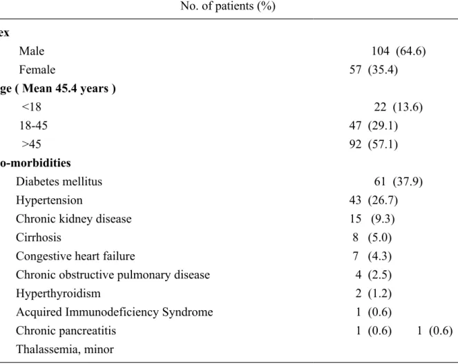
The 161 patients in this study included 104 males (64.6%) and 57 females (35.4%), with mean age of 45.4 years (range, 3 months to 90 years). The age distribution is shown on Table 1. There were 83 patients (51.5%) with underlying systemic diseases, the most common of which was diabetes mellitus (DM) (61 cases, 37.9%). The demographic data and the associated systemic diseases are shown in Table 1. The duration of hospital stay ranged from 2 to 120 days. The mean duration of hospital stay was 13.96 days.
The most common cause was odontogenic infection (33 cases, 20.5%), followed by pharyngo-tonsillitis (30 cases, 18.6%), lymphadenitis (17 cases, 10.5%), sialoadenitis (9 cases, 5.6%), foreign body (3 cases, 1.9%), thyroglossal duct cyst (1 case, 0.6%), brachial cleft cyst (1 case, 0.6%), and tuberculosis (1 case, 0.6%).
Computed tomography (CT) scans performed on all cases helped identify the involved neck space and the extent of infection. The most commonly involved neck space was the submandibular space (66 cases, 40.9%), followed by the carotid space (60 cases, 37.2%) and the para-pharyngeal space (54 cases, 33.5%). Eighty-six (86, 53.4%) cases presented with multiple neck space infection. Gas formation was detected in 31 (19.3%) cases.
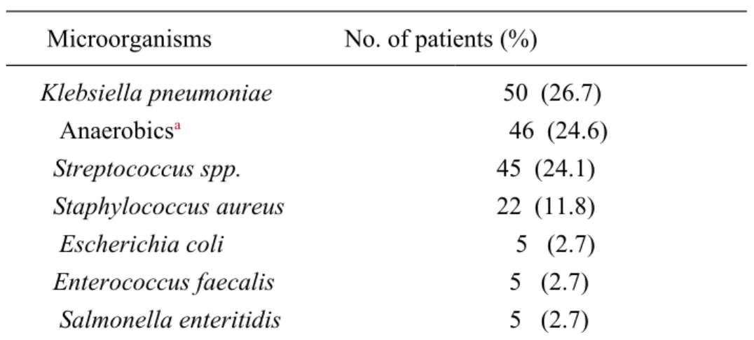
All patients underwent surgical drainage of their neck abscesses. Pus samples were sent for aerobic and anaerobic cultures. The bacterial growth in the pus was 139 patients (86.3%), including 47 (29.2%) with polymicrobial infections. The isolated pathogens and their incidences are shown in Table 2. The distribution of isolated pathogens in three most commonly involved spaces and patients with gas formation on CT scan are shown in Figures 1 and 2. Klebsiella pneumoniae was the most commonly cultured pathogen in the patients with DM. The cultureal rate of Klebsiella pneumoniae in patients with DM was significantly higher than that in those without DMhealth control group (52.46% vs. 18%, p<0.0001).
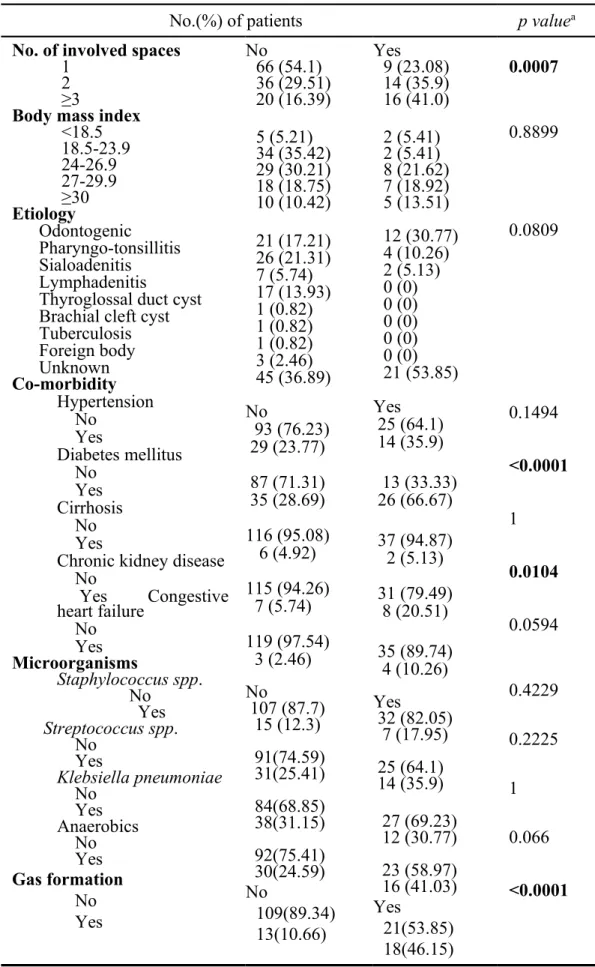
Of the 161 patients, 39 (24.2%) had complications. Airway distress was the most frequent complication (20 cases, 12.4%), followed by pleural effusion (18 cases, 11.3%), pneumonia (16 cases, 9.9%), septic shock (15 cases, 9.3%), mediastinal involvement (13 cases, 8.1%), and necrotizing fasciitis (9 cases, 5.6%). Four patients died from septic shock (mortality rate, 2.5%). All of them had multiple systemic diseases, including DM and hypertension in all patients, chronic kidney disease in one patient, chronic obstructive pulmonary disease in one patient, and congestive heart failure and liver cirrhosis in one patient. Factors associated with complications were analyzed and variables significantly associated with complications are shown in Table 3. There were no statistically significant differences in complication rates among different body mass index values, etiologies, and isolated pathogens.
DISCUSSION
In this study, odontogenic infection is the most common cause of deep neck infection, followed by pharyngo-tonsillitis. This result is consistent with available literature [2,3]. Odontogenic infection usually spreads continuously from the mandible into the submandibular/ sublingual space. This space lies between the mucosa of the floor of the mouth and the superficial layer of deep cervical fascia below. Dental (periapical) infection generally breaks through the lingual cortex of the
mandible and makes its appearance in this space [4].
Odontogenic infection is usually responsible for submandibular/ sublingual space infection, whereas infections originating from the tonsils and pharynx are usually related to para-pharyngeal space infections [545]. These findings explain why the submandibular/sublingual and peri-tonsillar/para-pharyngeal spaces are the most frequently involved spaces in our study.
The carotid space is also a frequently affected space in this study. The carotid space is located posterior to the para-pharyngeal space and is usually responsible for deep neck infection in children, which is commonly related to cervical lymphadenitis. This infection is usually well-controlled by intravenous antibiotic therapy [6]. Para-pharyngeal space infection tends to liquefy fat components and form large quantities ofmuch pus. Due to the lack of definitive boundaries, para-pharyngeal space infections can spread rapidly to the surrounding spaces and may cause fatal complications including airway compromise, jugular vein thrombosis, Lemierre’s syndrome, Horner’s syndrome, mediastinitis, and carotid artery hemorrhage [77-99]. The submandibular space infection is separated by the mylohyoid muscle. If the infection extends to the bilateral supra- and infra-mylohyoid spaces, it is so-called Ludwig’s angina, which can lead to subsequent rapidly progressive airway obstruction [1010].
In consideration of the relatively common involvement of the three above-mentioned spaces and risk of severe sequelae, the distribution of common microorganisms in these spaces was analyzed. To avoid confusing results and interpretation difficulties, two or more space involvement were excluded from analysis, for example, submandibular/sublingual space involvement were patients with abscess formation at submandibular and/or sublingual space only without other space involvement. Infections of the different neck spaces show a specific distribution of common microorganisms, similar to the findings by Lee [11]. Since few studies described this finding, this study provides a guide to facilitating optimal selection of empiric presumptive antibiotics.
Empiric antibiotics are administrated before the definitive results of pus or blood cultures become available. Thus, antibiotics should cover against most of the potential microorganisms in deep neck infections. Early start of appropriate antibiotics at the stage of cellulitis can prevent abscess formation [12Refs]. Initial broad-spectrum antibiotics will be replaced by an antibiotic based on the pathogen identified and its antibiotic susceptibility.
The most common empiric antimicrobial regimen prescribed to the patients in this study is intravenous amoxicillin with clavulanic acid (co-amoxiclav). As previously documented, it has coverage against the common pathogens in deep neck
infections since more than two-thirds of etiologies are beta-lactamase producing pathogens [1322,144]. However, Klebsiella pneumonia infection accounts for 53.1% of peri-tonsillar/para-pharyngeal space infections and 40% of carotid space infections in our study. The high prevalence of decreased susceptibility to co-amoxiclav among Klebsiella pneumonia isolates is demonstrated in early studies and most Klebsiella pneumonia are susceptible to narrow-spectrum cephalosporins [155]. This implies that as the selection of presumptiveempiric antibiotics for infections of the peri-tonsillar/para-pharyngeal and carotid spaces should be modified. Prompt CT reading can facilitate the optimal selection of empiric presumptive antibiotics by identifying the involved neck space.
Gas formation is a clue to diagnosis of anaerobic infection [166]. However, few studies have analyzed the bacteriologic pattern of deep neck abscess with gas formation. The present study demonstrates the link between anaerobic isolates and gas formation present on imaging. This information is likely to contribute to prompt identification of potential anaerobic infections and the initiation of appropriate antibiotics.
Treatment of deep neck infections consists of adequate antimicrobial therapy, early surgical drainage of the abscesses, and appropriate management of complications. Surgical drainage is indicated in patients with extensive multiple neck
space abscesses, impending complications, infection in immuno-compromised patients, or clinical deterioration despite adequate antibiotics use. Non-operative Conservative medical treatment can be applied in patients with cellulitis, small amount of abscess (<3cm), no involvement of danger space s( A neck space lies between the alar and prevertebral fascias, the two divisions of the deep layer of deep cervical fascia, and extends from the skull base down into the mediastinum to the level of the diaphragm. Infection here tends to spread inferiorly and resulting in mediastinitis.), no impending complication, and no predisposing factors of life-threatening complications [177, 188]. The study of Mayor et al. reported successful non-operativeconservative medical treatment in 90.32% (28 of 31) [1919], while Boscolo Rizzo et al. reported 226 patients (61.9%) who responded effectively to intravenous antimicrobial therapy alone [200]. Adequate empiric antibiotic selection is more important in patients who can be treated by conservative non-operative treatment due to no pus culture available in this group. Our results may help physicians to apply adequate empiric presumptive antibiotics by identifying involved spaces and gas formation on CT scan. Based on our data and other existing literature, we elaborated an algorithm (Figure 3) for managing deep neck infections.
susceptible to bacterial infections, more likely to develop complications, and have longer hospital stay compared to non-diabetics because of the dysfunction in polymorphonuclear neutrophil bactericidal function, cellular immunity, and complement activation [211,222]. As has been previously reported[11,23], there was alsois a close association between DM and Klebsiella pneumonia in our study [11,23]. The host defense against Klebsiella pneumonia depends on the host’s macrophage function, which is impaired in hyperglycemia [244]. Aggressive control of blood glucose concentration in plasma is imperative in treating diabetic patients with deep neck infection [255].
Several studies have identified DM and multiple space infections as significant risk factor for morbidity and mortality [17,20]. Our study shows that DM, multiple space infection, and gas formation present on CT scan are strong independent predictors of complications. Patients with these variables should be considered to have potentially life-threatening complications and require early aggressive multi-disciplinary management.
The limitations of this study included its retrospective, nonrandomized design. Patients who did not undergo surgical drainage were excluded from this study due to no pus culture available for pathogen analysis and low positive rate of blood culture. This may produce selection bias in terms of disease severity. Our study showed
specific bacteriologic patterns in different neck space infection and gas formation noted on CT scan. The result was partially similar to the Asian study [11]. Other geographic regions could experience different bacteriologic patterns among patients with neck infections, as has been previously reported [11]. Thus, further studies could be warrented to determine the patterns seen in Western patients."
However, previous studies had demonstrated different bacteriological pattern in deep neck infection geographically [11]. Further studies on bacteriologic pattern in different neck space infection with larger sample size should be directed to the Westerners.
CONCLUSIONS
This study corroborates the specific bacteriologic patterns in different neck space infection and gas formation noted on CT scan. The imaging characteristics and microbiology of patients with deep neck infections are correlated and can facilitate the optimal selection of antibiotics.
AUTHOR DISCLOSURE STATEMENT
This work is supported by CMUH-DMR-103-25 and NSC 101-2314-B-039-013 -MY3. .No financial funding was obtained for this study. None of the authors has any conflicts of interest to disclose.
REFERENCES
1. Levitt GW. (1970) Cervical fascia and deep neck infections. Laryngoscope. 80, 409-435.
2. Bakir S., Tanriverdi M.H., Gün R. et al. (2012) Deep neck space infections: a retrospective review of 173 cases. Am J Otolaryngol. 33,56-63.
3. Huang TT, Liu TC, Chen PR et al. (2004) Deep neck infection: analysis of 185 cases. Head Neck. 26, 854-860.
4. Yonetsu K, Izumi M, Nakamura T. (1998) Deep facial infections of odontogenic origin: CT assessment of pathways of space involvement. AJNR Am J
Neuroradiol. 19, 123–128. 5.
6. Blomquist IK, Bayer AS. (1988) Life-threatening deep fascial space infections of the head and neck. Infect Dis Clin North Am. Mar, 2(1), 237-264.
7. Sichel J, Attal P, Hocwald E et al. (2006) Redefining para-pharyngeal space infections. Ann Otol Rhinol Laryngol. 115, 117-23.
8. Gidley PW, Ghorayeb BY, Stiernberg CM. (1997) Contemporary management of deep neck space infections. Otolaryngol Head Neck Surg. 116, 16-22. 9. Koivunen P, Lopponen H. (1999) Internal carotid artery thrombosis and
Horner’s syndrome as complications of para-pharyngeal abscess. Otolaryngol Head Neck Surg. 121, 160-162.
10. Sethi DS, Stanley RE. (1991) Para-pharyngeal abscesses. J Laryngol Otol. 105, 1025-1030.
11. Mosier KM. (2008) Non-oncologic imaging of the oral cavity and jaws. Otolaryngol Clin N Am. 41, 103-137.
12. Lee YQ, Kanagalingam J. (2011) Bacteriology of deep neck abscesses: a retrospective review of 96 consecutive cases. Singapore Med J. 52, 351-5. 13. Brook I. (2012) Anaerobic bacteria in upper respiratory tract and head and neck
infections: Microbiology and treatment. Anaerobe. 18, 214-220.
14. Page C, Biet A, Zaatar R et al. (2008) Para-pharyngeal abscess: diagnosis and treatment. Eur Arch Otorhinolaryngol. 265(6), 681-686.
15.
16. Brook I. (1987) Microbiology of abscesses of the head and neck in children. Ann Otol Rhinol Laryngol. 96, 429-433.
17. Pérez-Moreno MO, Centelles-Serrano MJ, Cortell-Ortolá M, et al. (2011) Molecular epidemiology and resistance mechanisms involved in reduced susceptibility to amoxicillin/clavulanic acid in Klebsiella pneumoniae isolates from a chronic care centre. Int J Antimicrob Agents. 37(5), 462-466.
18. Meyer RD, Finegold SM. (1976) Anaerobic infections: diagnosis and treatment. South Med J. 69(9), 1178-1195.
19. Boscolo-Rizzo P, Marchiori C, Zanetti F, et al. (2006) Conservative management of deep neck abscesses in adults: the importance of CECT findings. Otolaryngol Head Neck Surg. 135, 894-899.
20. Wang LF, Tai CF, Kuo WR, et al. (2010) Predisposing factors of complicated deep neck infections: 12-year experience at a single institution. J Otolaryngol Head Neck Surg. 39, 335-341.
21. Plaza Mayor G, Martinez-San Millan J, Martinez-Vidal A. (2001) Is
conservative treatment of deep neck space infections appropriate? Head Neck. 23, 126-133.
22. Boscolo-Rizzo P, Stellin M, Muzzi E, et al. (2012) Deep neck infections: a study of 365 cases highlighting recommendations for management and treatment. Eur Arch Otorhinolaryngol. 269, 1241-1249.
23. Iwata T, Sekine Y, Shibuya K, et al. (2005) Early open thoracotomy and mediastino-pleural irrigation for severe descending necrotizing mediastinitis. Eur J Cardiothorac Surg. 28, 384-388.
24. Hasegawa J, Hidaka H, Tateda M, et al. (2011) An analysis of clinical risk factors of deep neck infection. Auris Nasus Larynx. 38, 101-107.
25. Chen MK, Wen YS, Chang CC, et al. (2000) Deep neck infections in diabetic patients. Am J Otolaryngol. 21, 169-173.
26. Huang TT, Tseng FY, Yeh TH, et al. (2006) Factors affecting the bacteriology of deep neck infection: a retrospective study of 128 patients. Acta Otolaryngol. 126, 396-401.
27. Zheng L, Yang C, Kim E, et al. (2012) The clinical features of severe multi-space infections of the head and neck in patients with diabetes mellitus compared to non-diabetic patients. Br J Oral Maxillofac Surg. 50(8), 757-761.
Table 1. Patient characteristics (n=161)
No. of patients (%) Sex
Male Female
Age ( Mean 45.4 years ) <18 18-45 >45 Co-morbidities Diabetes mellitus Hypertension
Chronic kidney disease Cirrhosis
Congestive heart failure
Chronic obstructive pulmonary disease Hyperthyroidism Acquired Immunodeficiency Syndrome Chronic pancreatitis Thalassemia, minor 104 (64.6) 57 (35.4) 22 (13.6) 47 (29.1) 92 (57.1) 61 (37.9) 43 (26.7) 15 (9.3) 8 (5.0) 7 (4.3) 4 (2.5) 2 (1.2) 1 (0.6) 1 (0.6) 1 (0.6)
Table 2. Bacterial cultures from pus
Microorganisms No. of patients (%) Klebsiella pneumoniae Anaerobicsa Streptococcus spp. Staphylococcus aureus Escherichia coli Enterococcus faecalis Salmonella enteritidis 50 (26.7) 46 (24.6) 45 (24.1) 22 (11.8) 5 (2.7) 5 (2.7) 5 (2.7) a
Anaerobic organism in our study including
Fusobacterium spp., Peptostreptococcus
spp., Clostridium bifermentans, Bacteroides fragilis, Bifidobacterium spp., Veillonella spp., Prevotella intermedia, Propionibacterium acnes.
Table 3. Factors that were associated with complications
No.(%) of patients p valuea No. of involved spaces
1 2 ≥3
Body mass index <18.5 18.5-23.9 24-26.9 27-29.9 ≥30 Etiology Odontogenic Pharyngo-tonsillitis Sialoadenitis Lymphadenitis
Thyroglossal duct cyst Brachial cleft cyst Tuberculosis Foreign body Unknown Co-morbidity Hypertension No Yes Diabetes mellitus No Yes Cirrhosis No Yes
Chronic kidney disease No Yes Congestive heart failure No Yes Microorganisms Staphylococcus spp. No Yes Streptococcus spp. No Yes Klebsiella pneumoniae No Yes Anaerobics No Yes Gas formation No Yes No 66 (54.1) 36 (29.51) 20 (16.39) 5 (5.21) 34 (35.42) 29 (30.21) 18 (18.75) 10 (10.42) 21 (17.21) 26 (21.31) 7 (5.74) 17 (13.93) 1 (0.82) 1 (0.82) 1 (0.82) 3 (2.46) 45 (36.89) No 93 (76.23) 29 (23.77) 87 (71.31) 35 (28.69) 116 (95.08) 6 (4.92) 115 (94.26) 7 (5.74) 119 (97.54) 3 (2.46) No 107 (87.7) 15 (12.3) 91(74.59) 31(25.41) 84(68.85) 38(31.15) 92(75.41) 30(24.59) No 109(89.34) 13(10.66) Yes 9 (23.08) 14 (35.9) 16 (41.0) 2 (5.41) 2 (5.41) 8 (21.62) 7 (18.92) 5 (13.51) 12 (30.77) 4 (10.26) 2 (5.13) 0 (0) 0 (0) 0 (0) 0 (0) 0 (0) 21 (53.85) Yes 25 (64.1) 14 (35.9) 13 (33.33) 26 (66.67) 37 (94.87) 2 (5.13) 31 (79.49) 8 (20.51) 35 (89.74) 4 (10.26) Yes 32 (82.05) 7 (17.95) 25 (64.1) 14 (35.9) 27 (69.23) 12 (30.77) 23 (58.97) 16 (41.03) Yes 21(53.85) 18(46.15) 0.0007 0.8899 0.0809 0.1494 <0.0001 1 0.0104 0.0594 0.4229 0.2225 1 0.066 <0.0001