Original Article
Safety and efficacy of quadrapeutics
versus chemoradiation in head and
neck carcinoma xenograft model
Ekaterina Y Lukianova-Hleb1*, Yoo-Shin Kim2*, Bhawani Aryasomayajula3, Teni Boulikas4, Jack Phan5, Mien-Chie Hung6,7, Vladimir P Torchilin3, Brian E O’Neill2, Dmitri O Lapotko1
1Department of BioSciences at Rice, Rice University, Houston, TX 77005, USA; 2Department of
Translational Imaging, Houston Methodist Research Institute, Houston, TX 77030, USA; 3Center
for Pharmaceutical Biotechnology and Nanomedicine, Northeastern University, Boston, MA 02115, USA; 4Regulon Inc., Alimos 17455, Greece; 5Department of Radiation Oncology, The
University of Texas MD Anderson Cancer Center, Houston, TX
77030, USA; 6Department of Molecular and Cellular Oncology, The University of Texas M. D.
Anderson Cancer Center, Houston, Texas 77030, USA; 7Center for Molecular Medicine and
Graduate Institute of Cancer Biology, China Medical University, Taichung, Taiwan. *Equal
contributors.
Abstract: Chemoradiation is the strongest anti-tumor therapy but in resistant unresectable cancers it often lacks safety and efficacy. We compared our recently developed cell-level combination approach, quadrapeutics, to chemo- radiation therapy to establish pre-clinical data for its biodistribution, safety and efficacy in head and neck squamous cell carcinoma (HNSCC), as a clinically challenging aggressive and resistant cancer. In vitro and in vivo models of four carcinomas were treated with standard chemoradiation and quadrapeutics using identical drug and radia- tion doses. We applied liposomal cisplatin or doxorubicin, colloidal gold, near-infrared laser pulses and radiation, all at low safe doses. The final evaluation used a xenograft model of HNSCC. Quadrapeutics enhanced standard chemoradiation in vitro by reducing head and neck cancer cell proliferation by 1000-fold, inhibiting tumor growth in vivo by 34-fold and improving animal survival by 5-fold, and reducing the side effects to a negligible level. In quad-rapeutics, we observed an “inversion” of the drug efficacy of two standard drugs: doxorubicin, a low efficacy drug for the cancers studied, was two times more efficient than cisplatin, the first choice drug in clinic for HNSCC. The radical therapeutic gain of quadrapeutics resulted from the intracellular synergy of the four components employed which we administered in a specific sequence, while the reduction in the toxicity was due to the low doses of all four components. The biodistribution, safety and efficacy data for quadrapeutics in HNSCC ensure its high translational potential and justify the possibility of clinical trials.
Keywords: Carcinoma, drug resistance, laser, plasmonic nanobubble, quadrapeutics, chemoradiation, nanomedi- cine
Introducti on
Chemoradiation is generally considered as the strongest therapy for unresectable tumors but even so, it often fails to safely and efficiently treat resistant and aggressive cancers [1-3]. To overcome this challenge, we recently converted current standard macro treatments-surgery, chemo- and radiation therapies-into one can- cer cell-specific intracellular micro-modality named quadrapeutics [4]. To achieve
such a macro-to-micro conversion of standard thera- pies, we employed non-stationary mechanical
intracellular nanoevents, plasmonic nanobub- bles (PNBs) [4, 5] in a four-component treat- ment (Figure 1). Firstly, systemically adminis- tered antibody-targeted gold colloid and a liposo- mal drug are aggregated by the cancer cell into an intracellular mixed cluster [4, 6]. Secondly, a low-energy near-infrared (NIR) laser pulse is applied locally and is instantly converted by the gold cluster into an expanding and collapsing vapor nanobubble named a PNB [7]. A PNB is not a particle, but a nano-explosion, with a mechani- cal, non-thermal, intracellular impact. The high cancer cell specificity of a PNB is due to its
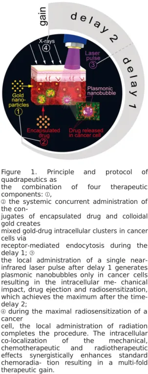
Figure 1. Principle and protocol of quadrapeutics as
the combination of four therapeutic components: ①,
② the systemic concurrent administration of the
con-jugates of encapsulated drug and colloidal gold creates
mixed gold-drug intracellular clusters in cancer cells via
receptor-mediated endocytosis during the delay 1; ③
the local administration of a single near-infrared laser pulse after delay 1 generates plasmonic nanobubbles only in cancer cells resulting in the intracellular me- chanical impact, drug ejection and radiosensitization, which achieves the maximum after the time-delay 2;
④ during the maximal radiosensitization of a cancer
cell, the local administration of radiation completes the procedure. The intracellular co-localization of the mechanical, chemotherapeutic and radiotherapeutic effects synergistically enhances standard chemoradia- tion resulting in a multi-fold therapeutic gain.
threshold generation mechanism: the threshold energy (fluence) of the laser pulse decreases with the cluster size [7] and the cluster size is the largest in cancer cells [4, 8]. This cluster-threshold mechanism results in PNB generation only in cancer cells even despite the non-specific uptake of some gold nanoparticles by adjacent normal cells (which cannot produce a PNB at the same laser fluence [4, 9]). An intracellular gold cluster and a PNB
deliver three therapeutic me- chanisms by using four components: a mechani-cal, non-thermal impact destroys a cancer cell [4,
5, 10-12], or disrupts the co-localized drug-bear- ing liposomes thus ejecting the drug into the cytoplasm [4, 6, 13], and radiosensitizing the cell [4]. The intracellular synergy of these three mech- anisms is the foundation of quadrapeutics and significantly amplifies chemoradiation in resis- tant cancer cells and tumors [4].
In order to determine the translational potential of quadrapeutics and to prepare for future clini- cal trials, firstly for head and neck squamous cell carcinoma (HNSCC), we continue our previous work here and report the in-depth study of this novel technology focusing on the biodistribution, safety and efficacy of quadrapeutics and its com- ponents and on its applicability to other can- cers. We also optimize the quadrapeutics proto- col (because the time sequence of the compo- nent administration is the key to quadrapeutics), and compare it to standard chemoradiation in several resistant carcinomas, with an in-depth evaluation in HNSCC. Compared to our previous works, the focus here is on the key preclinical data for HNSCC: the biodistribution and toxicity of the employed gold and drugs, the influence of the drug type on the therapeutic efficacy, and the analysis of the key therapeutic metric-the overall animal survival rate-for chemoradiation versus quadrapeutics.
Materials and methods
Cancer models
In vitro, we used cell lines of four
epithelial car- cinomas associated with high aggressiveness and drug resistance, and poor therapeutic
out-comes for chemo- and
chemoradiation treat- ments: head and neck squamous cell carcino- ma, HN31, triple-negative breast adenocarci- noma, MDA-MB-468, ovarian adenocarcinoma, SKOV3 IP1, and colon adenocarcinoma, SW48. The cells were obtained from UT MD Anderson Cancer Center (Houston, TX). All these carcino- mas overexpress Epidermal Growth Factor Re- ceptor (EGFR) which was targeted in our study to deliver the gold and drugs by using two clini- cal antibodies, cetuximab (C225, ImClone Sys- tems Inc., Branchburg, NJ) and panitumumab (Vectibix, Amgen Inc., Thousand Oaks, CA). Epithelial cancers with a tumor depth up to sev- eral millimeters can be reliably and safely
(for adjacent normal tissues) accessed with laser radiation in the NIR spectral range and there- fore are a good translational model. To evaluate specificity, we used normal epithelial cell line NOM9 (from J. Myers’s Lab, UT MD Anderson Cancer Center, Houston, TX).
In vivo, a HNSCC xenograft tumor was
raised on a flank of mice (athymic nude, strain CRL-490) to 5 mm diameter by s.c. injection of HN31 cells. We chose HNSCC as a model due to its
high aggressiveness and drug resistance which has not allowed an improvement in patient sur- vival time over the last 30 to 35 years [2, 3, 15]. Two standard metrics of therapeutic efficacy were obtained: tumor volume (within 2 to 3 weeks after the treatment) and the animal overall survival time (measured until the ani-mal reaches the moribund condition which was defined as a tumor size of 10 mm). Six animals were treated in each group. The ani- mals were treated according to the IACUC pro-tocols approved by the Rice University and the Methodist Hospital Research Institute.
Drug and gold targeting and analysis in vivo
Gold spheres of 60 nm diameter (VanPelt Bio- sciences LLC (Ijamsville, MD) and nanoCom- posix Inc (San Diego, CA) were covalently con-jugated to the anti-EGFR antibody clinically- approved for HNSCC, Panitumumab, by using a proprietary conjugation method (performed by VanPelt Biosciences LLC (Ijamsville, MD)). In vitro, gold conjugates were incubated with cells for 24 hours under physiological conditions at the concentration of gold conjugate suspen- sion corresponding to the optical density of
0.08 at the wavelength of 540 nm, the maxi- mum of the optical absorption spectrum. This corresponds approximately to a dose of 0.7 μg/ ml.
In vivo, gold conjugates were administer- ed intravenously in the low dose of 4 mg/kg of body weight and a certain time-delay was allowed in order to form intracellular clusters of gold and drug nanoparticles [4-6, 13]. Two standard liposomal drugs were used: Doxil (Ben Venue Laboratories Inc, Bedford, OH) for doxorubicin and Lipoplatin (Regulon Inc, Alimos, Greece) for cisplatin (the first drug of choice in chemoradiation therapy of HNSCC and other studied carcinomas since the platinum in this drug is known to radio-sensitize the cells). For targeting,
these 100 nm drug liposomes were covalently conjugated to the same antibodies as GNPs by using our previously established and validated methods that show high stability, low toxicity and the long shelf life (> 6 months) of such liposomal conjugates [4, 6, 13].
In vitro, GNPs and drug liposomes were
mixed and incubated with the cells under physiologi- cal conditions for 24 hours at specific concen- trations of gold and drugs. This incubation time was previously shown to provide the maximal
efficacy and specificity of cluster formation in cancer cells via receptor-mediated endocytosis [10, 13]. In vivo, GNPs and drug liposome con- jugates were administered systemically and concurrently. We previously verified the high efficacy and tumor specificity of the formation of gold conjugate clusters in HNSCC tumors in vivo under low doses of colloidal gold conju-gate through the mechanism of receptor-medi- ated endocytosis [4, 6]. The influence of can- cer aggressiveness on the cluster size was also observed previously [4]. Therefore, the described protocol provides the safe and reli- able formation of mixed gold-drug clusters
in vitro and in vivo.
The efficacy of gold and drug conjugate target- ing in vivo was analyzed by measuring the level of gold and platinum in the tumor and other organs which were harvested 24 hours and 72 hours after the systemic administration of the conjugates. Three animals were studied for each time-point. The level of gold and platinum was measured with the mass-spectroscopy method (Perkin Elmer Nexion 300 ICP-MS, Perkin Elmer, Inc., Waltham, MA). The toxicity of the gold conjugates in vivo was measured short-term (24 and 72 hours after administra- tion) and long-term (over 1 month). To deter- mine the short-term toxicity, the harvested liver, kidney, spleen, and lung were analyzed with a pathological method for necrosis, apoptosis and other standard signs of toxicity. The long-term toxicity was assessed by monitoring the animal weight for one month and longer.
Histolo gy
The harvested organs (kidney, lung, liver, heart,) and the tumor were placed in 10% neutral buffered formalin and fixed for 48 hours. The organs were then processed routinely and sec- tions were stained with hematoxylin and eosin (H&E). Sections were examined by a board cer- tified veterinary pathologist (BCVP). Regions
of tumor and necrosis were delineated with the assistance of the BCVP. For the histological study of the therapeutic effect of standard chemoradiation and PNB-enhanced chemora- diation, animals were sacrificed on Day 12, where Day 0 was the day of treatment initiation. This corresponds to 72 hours following the end of the dual treatment. Following sacrifice, the tumors were collected along with the underly- ing muscle and ribcage, and fixed in 10%
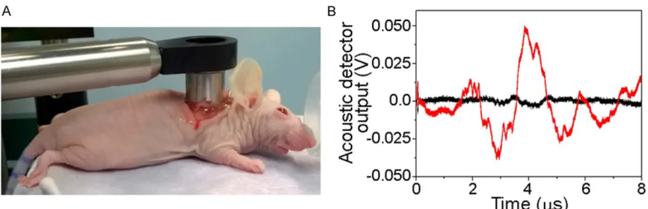
neu-Figure 2. A. The laser pulsed treatment has been applied locally through the custom-designed endoscope with a ring- shape acoustic sensor around the optical exit window (the endoscope was coupled to the wound optically and acousti- cally through transparent ultrasound gel); B. Acoustic time-responses to a single laser pulse (782 nm, 30 ps, 65 mJ/ cm2) of the primary tumor of the animal pretreated with gold conjugates (red) and the tumor without such pretreatment (black).
tral buffered formalin for at least 48 hours. Samples were then processed to HE slides as outlined above.
PNB generation and detection
On-demand intracellular PNBs were generated around clusters of gold colloids with single NIR laser pulses (782 nm, 30 ps, Ekspla PG 500, Ekspla UAB, Lithuania) which were absorbed by the gold spheres and converted into heat. While the stationary optical excitation of gold colloids at 782 nm is not efficient due to their poor opti- cal absorbance (just 6% relative to that in their visible spectral peak of 500-600 nm), our non- stationary excitation method [9] provides an efficient generation of PNBs with a 30 ps laser pulse at the NIR wavelength of 782 nm. The in vivo experiments used our photothermal micro- scope described previously [7]. In the in vivo
experiments, the laser pulse was delivered to the tissue via a custom-made endoscope (Figure 2A). To detect PNBs optically in individ- ual cells in
vitro, we used our established opti- cal
scattering method [7]. In vivo, PNBs were detected with ultrasound sensors installed in the tip of an endoscope via a PNB-specific acoustic signal (Figure 2B).
Radiation treatment
Cells and animals were irradiated with a RS
2000 machine (Rad Source Technologies, Inc., Suwanee, GA). In
vitro, single treatment was used. In vivo, two fractions were administered
locally with a one day interval. The radiation
was administered with a specific time delay
after the laser treatment.
Statistical considerations
We used two-tailed t-tests to compare the cell and animal group metrics. We performed sta- tistical analyses with Origin software (Origin
9.1, OriginLab Corporation, Northampton, MA). P values of < 0.05 were considered statistically significant.
Results and
discussion
This study was aimed at the optimization of the quadrapeutics protocol in several resistant carcinomas to achieve maximal safety and effi-cacy in comparison with the standard of care, chemoradiation therapy.
Optimization of the gold and drug targeting in vivo
Since the therapeutic efficacy of the quad- rapeutics mechanisms depends upon the clus- tering of gold and drugs in the tumor, we first analyzed the safety and efficacy of the systemic targeting of gold and liposomal conjugates in a xenograft model of head and neck squamous cell carcinoma (HNSCC) induced with HN31 cell line. According to our previous observations [4,
9], this is a very resistant and aggressive form of HNSCC. Both conjugates were concurrently i.v. injected with doses of 4 mg/kg (gold) and
12 mg/kg (cisplatin). The systemic administra- tion followed the standard approach in
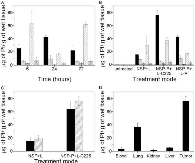
chemo-Figure 3. The levels of drug (platinum) in various organs of nude mice (n = 3) as measured with mass spectros- copy after the concurrent systemic administration of gold colloids and liposomal drug (Lipoplatin). A. Platinum biodistribution measured at specific time-points (Lipoplatin 12 mg/kg, black solid-tumor, light grey solid-liver, dense grey shaded-kidney, shaded white-lung, light solid grey with shading-blood); B. Platinum biodistribution for various combinations of Lipoplatin (12 mg/kg) and gold (4 mg/kg) in 24 hours after their administration (NSP-gold colloids, L-plain Lipoplatin, C225-Cetuximab antibody, P-Panitumumab antibody, antibodies were covalently conjugated to gold or drug liposomes, organs are shown as in “A”; C. Influence of the tumor size and antibody on the biodistribu- tion of platinum 24 hours after their administration: (solid)-the tumors < 3 mm, (shaded)-the tumors > 5 mm; D. The biodistribution of platinum under optimized doses, conjugation of gold (4 mg/kg, conjugated to Panitumumab), Lipoplatin (12 mg/kg, conjugated to C225) and time-delay of 24 hours after their administration.
therapy. In addition, the systemic administra- tion of gold conjugates is more efficient than the local intratumoral injection as we demon-strated recently [12]. We used the concurrent administration of the drug and gold in order to maximize the endocytosis-based intracellular formation of the mixed drug-gold clusters which requires the synchronous internalization of gold and drug liposomes by cancer cells [13].
To reduce the treatment toxicity, mainly associat- ed with the drug, the drug dose was adminis- tered at the level within 15-20% of the human clinical dose equivalent.
The dynamics of the biodistribution of the lipo- somal drug was measured for several organs harvested at 6, 24 and 72 hours in animals with relatively small tumors (within 3 mm). The maximum level of drug in the tumor was achieved in 24 hours (Figure 3A), which coin- cided with the optimal time for the formation of the maximal gold-drug clusters in cells as observed previously in vitro [4, 13]. The effi-cient retention of the drug in a tumor was observed up to 72 h. This relatively long reten- tion time (compared to that for small molecule-based free drugs) should further improve the
radiation treatment by amplifying the radio- sensitization effect of the intracellular drug clusters.
The influence of the drug targeting antibody combination for drug was measured for the drug levels in relatively small (3 mm) tumors harvested 24 hours after the systemic injection of bare gold and drug liposomes versus those conjugated to identical or different antibodies (C225 and Panitumumab). The combination gold-Panitumumab and Lipoplatin-C225 pro- vided the maximal levels of the drug in the tumor (Figure 3B). After optimizing the timing and the antibody combination, we analyzed the influence of the tumor size (early vs mature tumor, Figure 3C). By comparing the drug bio- distribution achieved in 24 hours in animals with early (< 3 mm) and well-developed (> 5 mm) tumors, we concluded that 5 mm tumors provide a much better accumulation of the drug when it is conjugated to the optimal anti- body (Figure 3C). The better-developed vascu- lature in larger tumors improved the systemic delivery of the gold and drug. Never- theless, even small early tumors can be suc-cessfully targeted systemically with low doses of the drug and gold. After optimizing all target- ing conditions, we achieved the relatively high tumor specificity of accumulation of the drug (Figure 3D). In the therapeutic experiments, we used animals with 5 mm tumors. Note that despite using the anti-EGFR antibody, its drug liposome conjugates escaped the so called “liver sink” effect [14] and the liver levels of the drug was relatively low (Figure 3D). Among nor- mal organs, a relatively high amount of the drug was found in the lungs and liver (Figure
3D), although it should not induce any PNB treatment-related toxicity because the laser pulse (which activates the gold and drug) can- not reach either the lungs or the liver.
Coupled with our previous observation of gold biodistribution and gold clusters in tumor cells for a similar targeting of the HNSCC xenograft in mice [6], and a comparison of the intra- tumoral versus the systemic administration of gold conjugates [6], we chose to use the con- current systemic administration of 60 nm gold-Panitumumab and 100 nm drug liposome- C225 conjugates. An advantage of the concur- rent administration is an ability to personalize and independently tune the doses of GNPs and drugs in order to optimize the cluster formation and the therapeutic effect. The advantage of
systemic administration over local intra-tumor- al injection is in using the standard chemother- apy route for drug delivery, whereas the local intratumoral injection of drug liposomoses or gold nanoparticles will have limited delivery and selectivity due to the strong limiting effect of the tissue upon the nanoparticle diffusion. This was directly observed by us previously by comparing the local versus the systemic deliv- ery of the same gold nanoparticles in similar tumor models [6].
Toxicity of systemically administered gold and drug conjugates
Although colloidal gold, drug and antibodies are clinically validated, we studied their short- and long-term toxicity in mice. A histological evalua-tion of organs harvested at 24 hours and 72 hours from intact (Figure 4A-C) and gold/drug- treated mice (Figure 4D-F) revealed no short- term gold-and drug-related toxicity at all (Table 1). This result is in line with (1) the generally
safe nature of gold nanoparticles of this rela- tively large size because they remain chemical- ly and biologically inert (this was also verified in clinic for similar colloidal gold nanoparticles [16-20]), (2) the low doses of gold we used in our experiments which were 5-100 times lower than those reported in the diagnostic and ther- apeutic use of gold nanoparticles in vivo
[21-29], and (3) the significantly reduced dose (25% of the clinical equivalent) of the chemotherapy drug, cisplatin, which is associated with major clinical adverse effects of cisplatin-based che-motherapy and chemoradiation therapy. In addition, we observed the relative changes in the body weight in two groups of intact and quadrapeutics-treated mice for a relatively long term of over one month. We found no toxicity expressed in the weight loss and/or abnormal behavior of the treated animals (Figure 4G). These results reveal that quadrapeutics and its components under the doses employed were safe in vivo and their optimized administration protocol
provided a relatively high accumula-tion of both the gold and drug in the tumor. This systemic pre-treatment was designed to sup- port the next stage of local treatment with a laser and radiation.
Quadrapeutics versus standard therapy effi- cacy in vitro in various carcinomas
We first compared standard chemoradiation,
chemoradiation with targeted liposomal
nano-Figure 4. Evaluation of the safety of quadrapeutics in nude mice (n = 3). (A-F) Histological analysis of organs har- vested from intact (A-C) and treated (D-F) animals 72 hours after the treatment: liver (A, D), lung (B, E), heart (C, F) (scale bar: 100 μm), none of necrosis, apoptosis or inflammations are seen in the treated tissues; (G) body weight of untreated (black) and quadrapeutics-treated mice (red) do not show a long-term adverse effects in the treated group, animal weight is shown in arbitrary units after normalizing by the body weight of untreated group (mean ± SD).
Table 1. Short-term toxicity of treatment
therapeutics and quadrapeutics in cultures of resistant and aggressive head and neck squa- mous cell carcinoma (HNSCC, HN31) and nor-mal epithelial (NOM9) cells by using free and liposomal forms of cisplatin (Lipoplatin), the main clinical choice in HNSCC chemo- and chemoradiation therapies. After optimizing the drug dose to the safest level for normal cells by using the clonogenic test (Figure 5A), both standard and targeted chemoradiation thera- pies revealed the high therapeutic resistance of HNSCC (Figure 6A), which is in line with clini- cal experience [1, 3, 15]. In contrast, quad- rapeutics showed a 1000-fold therapeutic gain
compared with standard
chemoradiation (Fig- ure 6A). At the same time, identically treated normal cells were not damaged (Figure 6A)
because they were unable to generate PNBs unlike cancer cells (Figure 6B).
This 1000-fold therapeutic gain (which exceed- ed a similar previous result by almost ten-fold [4]) was achieved by: -optimizing the timing of
Treatment Time Kidne
y Liver Lung Untreated 24 h 0% 0% 0%
72 h 0% 0% 0% PNB-enhanced
the radiation administration after the laser treatment (Figure 5B)-24-hour delay was found optimal to radiosensitize the cell and to apply a single fraction of the radiation of 4 Gy; -optimizing the antibody conjugation of drug and gold (Figure 5C)-the combina- tion of Lipoplatin-Panitumumab and gold- C225 provided both high efficacy and selectivity of quadrapeutics; -optimizing the
laser pulse administration (Figure 5D). Firstly, a single laser pulse (782 nm, 30 ps, 30 mJ/cm2) was applied 24 hours after administering the drug and gold to ensure their intracellular clus- tering and co-localization. This pulse induced PNBs only in cancer cells (Figure 6B) and there- fore all quadrapeutics mechanisms were trig- gered only in cancer cells, while the identical treatment of normal cells did not trigger the quadrapeutics mechanisms. After optimizing the quadrapeutics sequence timing, targeting antibodies and the number of laser pulses, we studied the influence of the drug type on the therapeutic efficacy in quad-rapeutics and chemoradiation modes. Cisplatin (the first drug of choice in clinic for the cancer studied) was replaced with a similar clinically equivalent dose of liposomal doxorubicin (Doxil) (whose clinical efficacy against HNSCC is con- sidered to be low). The chemoradiation mode revealed the predictably better efficacy of cis- platin (Figure 6C). In contrast, in the
quad-Figure 5. In vitro response of HNSCC HN31 (red dots and bars) and normal epithelial NOM9 (grey dots and bars) cells to the treatments measured as the surviving fraction with a clonogenic test: A. Chemotherapy (24 hour incuba- tion) with liposomal cisplatin (Lipoplatin) in plain (solid circle) and targeted forms (hollow circle-with C225 antibody, hollow triangle-with Panitumumab antibody); B. PNB-enhanced chemoradiation with a variable time-delay between the laser and radiation treatments (Lipoplatin-Panitumumab: 200 ng/ml, gold-C225: 2.4 × 10¹⁰ NPs per ml, laser
782 nm, 30 ps, 45 mJ/cm2, single pulse, radiation: 4 Gy, single fraction), red arrow indicates zero colonies; C. Influ- ence of the intracellular co-localization of gold and drug on the efficacy and selectivity of PNB-enhanced chemora- diation (plain gold colloids, L-plain Lipoplatin, NSP-C225-conjugate of gold colloids with C225, L-P-Lipoplatin conjugated with Panitumumab); D. Influence of the number of laser pulses applied on the efficacy and selectivity of PNB-enhanced chemoradiation.
rapeutics mode, doxorubicin, a “poor” drug against HNSCC, improved the therapeutic effi- cacy by four-fold (Figure 6C) and, most unex- pectedly, outperformed cisplatin by two-fold (Figure 6C). This inversion of drug efficacy (never observed previously) is associated with the PNB-induced mechanical impact and the intracellular drug ejection and radiosensitiza- tion it provided.
Such an inversion may significantly improve current chemoradiation and
therefore it was further studied in three additional drug-resis- tant and aggressive cancers which are current-ly also treated preferabcurrent-ly with cisplatin
(triple- negative breast
ade-nocarcinoma and colon adenocarcinoma). To generate similar PNBs in these different cancer cells, we optimized the level of laser pulse flu-ence for each cell line (Table 2). After identical targeting with gold conjugates, the laser pulse fluence was adjusted to provide the generation of sub-lethal PNBs of a 60-70 ns lifetime as was previously done for HNSCC cells (Table 2). The difference found in the laser fluences is associated with the different efficacy of gold clustering in these cells which was apparently the highest (lowest fluence required to gener- ate similar PNBs) in SW48 cells and the lowest in SKOV3 cells (the highest fluence required to generate similar PNBs). In this experiment, we used the same drug and radiation doses as in
Figure 6. In vitro study of quadrapeutics in cancer and normal cells. A. Surviving fractions of HN31 cancer (red) and NOM9 normal (grey) cells in a clonogenic test after identical treatment with: Chemotherapy (Chem) with liposomal cisplatin (Lipoplatin)-Panitumumab conjugated (200 ng/m), chemoradiation therapy (ChemoRad) with Lipolatin- Panitumumab and a single radiation dose (4 Gy), quadrapeutics (ChemoRad+PNB): NSP-C225 conjugates (2.4
× 1010 NPs per ml) + Lipoplatin-Panitumumab (200 ng/ml), single laser pulse (782 nm, 30 ps, 30 mJ/cm2) and a single dose of the radiation (4 Gy) 6 hours after the laser treatment (data: mean ± SE, n = 3); B. Optically-detected typical time-responses of individual cells to a single laser pulse (782 nm, 30 ps, 30 mJ/cm2): cancer HN31 (red) and normal NOM9 (black), only cancer cells show the PNB-specific optical time-response; C. The ratio of the therapeutic efficacy of liposomal cisplatin (Lipoplatin), 200 ng/ml versus liposomal doxorubicin (Doxil) in standard chemoradia- tion (PNB lifetime = 0) and quadrapeutics (PNB lifetime > 0) in four different carcinoma cell lines (delay between laser treatment and X-ray treatment was 6 hours for Doxil and 24 hours for Lipoplatin): head and neck-black, breast-white, colon-grey and ovarian (shaded) cancer. The ratio < 1 means the higher efficacy of cisplatin, the ratio
> 1means the higher efficacy of doxorubicin ; D. Therapeutic gain delivered by the quadrapeutics (as the ratio of the cell survival for chemoradiation divided by that for quadrapeutics) as the function of the PNB generation efficacy in cancer cells (measured for individual cells as PNB lifetime under identical optical excitation and gold pre-treatment) for Lipoplatin (black) and Doxil (red) for carcinoma cells of the following cancers: head and neck (star), breast (hol- low circle), ovarian (square), colon (solid circle), solid lines show the best polynomial fit, the correlation (Pearson) coefficient is 0.99 for both drugs (data: mean ± SE).
the previous one. In addition to the cell viability measurements, the clonogenic test resulted in zero colonies in the samples of all the addition- al cancer cell lines treated with quadrapeutics (not shown). We therefore used an additional metric-cell viability 24-48 hours after the treat- ment. The
treatment of all three additional can-cer lines with standard chemoradiation and quadrapeutics revealed a similar inversion effect for doxorubicin vs cisplatin (Figure 6C): doxorubicin was more efficient than cisplatin.
Interestingly, in all four carcinoma cell lines, the therapeutic gain for both drugs (quantified through the ratio of cell survival for stand- ard chemoradiation/quadrapeutics)
correlated with the PNB generation efficacy of these cell lines (quantified through the PNB lifetime in individual cells under identical exposure to gold conjugates and laser pulses) (Figure 6D). Therefore, the intracellular mechanical impact of PNBs plays a key role in enhancing chemora- diation in cancer cells in the quadrapeutics
radiation: 34-fold for tumor growth inhibition (Figure 7C, the histological data are shown in Figure 8) and 5.4-fold for the animal survival time (Figure 7D). No short-term (Figure 4A-F; Table 1) or long-short-term (Figure 4G) toxicities were observed after the treatment with quadrapeu- tics, a problem associated with full clinical doses of standard chemoradiation.
mode and explains the observed “inversion” of drug efficacy (for doxorubicin versus cisplatin).
Quadrapeutics versus standard chemoradia- tion in vivo
We further compared standard chemoradia- tion, targeted chemoradiation and quadrapeu- tics in
vivo in a xenograft mouse model of
HNSCC. All three treatments used identical doses of the drug at the level of 18% of its clini- cal equivalent: free cisplatin in the standard mode (12 mg/kg) and Lipoplatin conjugate in targeted chemoradiation and in PNB-enhanced chemoradiation (12 mg/kg). After optimizing targeting antibodies for the drug (C225, Figure
3B), gold (Panitumumab) and their
administra-tion timing (Figure 3A), we achieved the tumor- specific bio-distribution of the gold and drug (Figure 3C). A single NIR laser pulse (782 nm,
30 ps, 65 mJ/cm2) was applied locally via an endoscope (Figure 2A) 24 hours after systemi- cally injecting the drug and gold. The PNB gen- eration in the tumor was confirmed in real time by detecting PNB-specific acoustic signals (Figure 2B). The radiation (two fractions, 6 Gy (day 3) and 3 Gy (day 4)) was administered locally. Each treatment was administered twice with a one-week interval. Compared to the standard mode, chemoradiation with targeted Lipoplatin did not significantly inhibit tumor growth (Figure 7A), although the improvement in the animal survival time was 1.9-fold (Figure
7B). In contrast, the Lipoplatin-based quad- rapeutics provided a 5-fold inhibition of the tumor growth and 3.1-fold improvement in the animal survival
time (Figure 6A, 6B) compared to standard chemoradiation.
Next, after replacing liposomal cisplatin with a similar clinically equivalent dose (18%) of lipo- somal doxorubicin (Doxil), we observed a fur- ther increase in the therapeutic gain in the quadrapeutics mode versus standard
chemo-Table 2. PNBs lifetime on different cells lines
Cell line Laser pulse flu- ence, mJ/cm2 PNB life- time, MDA-MB-468 30 70±6 SW48 22 63±4 SKOV3 IP1 40 62±5 HN31 30 61±4 NOM9 40 0
Thus, our in vitro and in vivo results showed that a drug with a relatively poor clinical effica- cy in HNSCC, doxorubicin, outperformed the first choice drug, cisplatin, in the quadrapeutics mode and provided a more than 5-fold improve- ment in the overall animal survival rate, com-pared to that for standard chemoradiation. Such a radical therapeutic improvement was achieved with a combination of low, safe doses of liposomal doxorubicin and radiation with the cell-level mechanical impact of PNBs (applied locally).
Quadrapeutics mechanisms versus other in- vestigational therapies
Such a radical improvement both in the safety and efficacy of standard chemoradiation was achieved through the intracellular synergy of several PNB-based mechanisms which are spe-cific to quadrapeutics:
The high intracellular concentration of the drug due to its instantaneous ejection by a PNB [4,
6, 7, 13] efficiently sensitizes the target cancer cell. While this mechanism is well-known, PNBs further enhance it by enabling better radiosen- sitization with doxorubicin than with cisplatin (Figure 6C, 6D). This “drug efficacy inversion” is probably the most promising part of radio-sen- sitization because it broadens the drug choice for chemoradiation, while at the same time reducing the efficient therapeutic dose of the drug to a safe level. The intracellular mechani- cal impact of PNBs [4, 5, 10-12] (similar to the mechanical macro-sensitization demonstrated earlier [30-35]. The re-emission of secondary electrons by gold clusters [36-38] both achieve maximal strength in cancer cells with the larg- est gold clusters and PNBs, while sparing adja- cent normal cells (which do not generate PNBs and have much lower levels of non-specifically accumulated gold).
All these mechanisms were not triggered in nor- mal tissue because no PNBs were generated
Figure 7. In vivo study of the quadrapeutics vs standard and targeted chemoradiation in a xenograft mouse model of HNSCC: monitoring of the tumor volume (A, C) and animal survival fractions (in arbitrary units, a.u.) (B, D) after the two treatment cycles: untreated animals (magenta, hollow circles), standard chemoradiation with cisplatin (blue, squares, 12 mg/kg, radiation 9 Gy total), targeted chemoradiation with liposomal cisplatin-Panitumumab (green, crosses, identical doses of the drug and radiation) and quadrapeutics with liposomal cisplatin-Panitumumab (black, stars, identical doses of the drug and radiation, gold colloid-C225 at 4 mg/kg, single laser pulse at 782 nm, 30 ps, 65 mJ/cm2) and liposomal doxorubicin-Panitumumab (red, hollow stars, drug dose 3 mg/kg, other component doses identical to those above for cisplatin-based quadrapeutics). Data: mean ± SE. *P < 0.05.
there under identical laser exposure due to the threshold mechanism of PNB generation (Figure 2B). The high cancer-cell specificity of PNB-enhanced chemoradiation stems from the localized mechanical impact of a PNB and its threshold nature, not only from using antibod- ies to target cancer cells. The PNB generation threshold fluence decreases with the gold clus-ter size, with the lowest PNB threshold in large clusters in cancer cells and the highest PNB threshold in single gold nanoparticles or their small clusters in normal cells [4, 12, 13]. The mechanical non-stationary
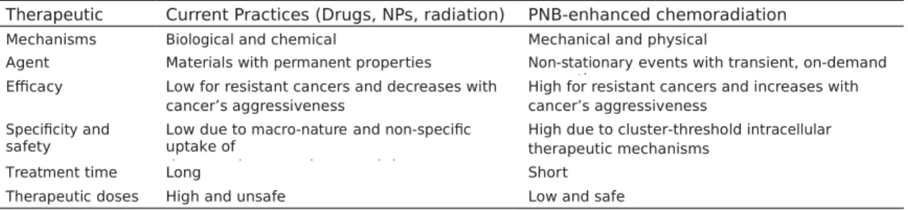
mechanism employed in
quadrapeutics principally differs from
gold nanoparticle-based drug delivery, hyperthermia and radiation therapies which all employ the stationary effects of drugs, heat or secondary radiation (Table 3) [21-29, 36-38].
Such “stationary” nano-therapies suffer from poor cancer cell specificity due to the unavoid- able uptake of nanoparticles by normal cells and their gain is relatively incremental com- pared to that of the quadrapeutics.
Conclusio ns
Our study investigated and optimized the quad- rapeutics protocol for the delivery, safety and efficacy:
1. The delivery of the gold and drugs was veri- fied via a systemic route: no non-specific toxic- ity was observed due to the reduction of the drug dose to 25% of the clinical one and because of using relatively low doses of clini-cally-safe colloidal gold and clinically-approved antibodies (also at low, sub-clinical doses). A
Figure 8. Histological images of the primary HNSCC tumor samples harvested from (A) untreated, (B) chemoradia- tion treated, and (C) quadrapeutics-treated mice (drug: liposomal doxorubicin (Doxil)-Panitumumab, 3 mg/kg, gold colloid-C225: 4 mg/kg, laser: 782 nm, 30 ps, 65 mJ/cm2, radiation: 9 Gy total). All tumors were harvested 12 days following start of the treatment. Scale bar is 2 mm; inset is 10 × magnification of the specified location. Green-border of viable tumor, yellow-boarder of necrotic tumor or edematous tissue associated with the tumor necrosis). All tumors had similar size in the beginning of the treatments.
Table 3. Therapeutic mechanisms and performance of PNB-enhanced chemoradiation vs current practices
Therapeutic
Feature Current Practices (Drugs, NPs, radiation) PNB-enhanced chemoradiation
Mechanisms Biological and chemical Mechanical and physical
Agent Materials with permanent properties Non-stationary events with transient, on-demand properties
Efficacy Low for resistant cancers and decreases with cancer’s aggressiveness
High for resistant cancers and increases with cancer’s aggressiveness
Specificity and safety
Low due to macro-nature and non-specific uptake of
therapeutic agents by normal tissue
High due to cluster-threshold intracellular therapeutic mechanisms
Treatment time Long Short
Therapeutic doses High and unsafe Low and safe
liver “sink effect” did not prevent the efficient targeting of the tumor via EGFR antibodies and the levels of non-specific uptake of drugs and gold were relatively low.
2. After optimizing the quadrapeutics protocol, we achieved a more than 5-fold increase in overall survival (compared to that after chemo-radiation therapy), without increasing either the drug or radiation doses. This gain was achieved mainly due to the conversion of the standard macro-treatment into an intracellular and can-cer cell-specific quadrapeutics micro-treatment which activates the therapeutics mechanisms only in cancer cells.
3. Unlike standard cisplatin-based chemoradia- tion therapy the quadrapeutics mechanisms revealed
the higher efficacy of another drug, doxorubicin, which does not provide high effi- cacy under the standard treatment.
The reported results establish a foundation
for translating quadrapeutics in HNSCC to
clini-cal trials with an unprecedented combination of efficacy and safety compared to standard chemoradiation: a more than five-fold improve- ment in overall animal survival and a significant reduction in side-effects. They also establish the potential of quadrapeutics in other aggres- sive and resistant carcinomas, including triple- negative breast cancer. The high translation potential of quadrapeutics relies upon using only clinically validated components in low, safe doses: 25-30% for the drugs and 15% for radia- tion (of clinical doses). This broadens patient eligibility, especially among those who already failed standard therapies, ensures easy inte- gration with standard protocols and flexibility in choosing the drug type. Quadrapeutics is a non-stationary nanomedicine which can be administered as a stand-alone or adjuvant treatment after standard practices fail. While quadrapeutics is a local treatment by defini- tion, by efficiently destroying a primary or resid-ual epithelial tumor it effectively reduces the probability of both local recurrence and meta- stases.
Acknowledgeme nts
Authors thank Ehab Hanna of UT MD Anderson Cancer Center for the discussion of clinical applications of the method, Andrew Hurrell and Thomas Kelley of Precision Acoustics Ltd (Dorset, UK), Eliberto Batres of Rice University (Houston TX), Aidas Aleknavicius and Rokas Sulcas of EKSPLA UAB (Vilnius, Lithuania) and Tej Pandita of Houston Methodist Research Institute for the help with experimental equip- ment, Ryan Vance, HTL (ASCP), Jennifer Ri- cketts, HTL (ASCP) and Judit Markovits, DVM, PhD, Diplomate ACVP, CMP Pathology, for tech- nical assistance. This work was supported with the grants from Gillson Longenbaugh Foun- dation (Houston, TX), NSF (CBET-1341212), NIH (R01GM094816). Disclosure of conflict of interest None .
Address correspondence to: Dr. Dmitri Lapotko, De- partment of BioSciences at Rice, Rice University,
6100 Main St, MS-140, Houston, TX
77005-18-92, USA. Tel: 348-3708; Fax: 713-348-5154; E-mail: d l 5@ri c e . e du
Referenc es
[1] Haddad RI, Shin DM. Recent advances in head and neck cancer. N Engl J Med 2008; 359:
1143-1154.
[2] Leemans CR, Braakhuis BJ, Brakenhoff RH.
The molecular biology of head and neck can- cer. Nat Rev Cancer 2011; 11: 9-22.
[3] Radosevich JA. Head and Neck cancer: cur- rent perspectives, advances and challenges. Springer; 2013. pp. 1070.
[4] Lukianova-Hleb EY, Ren X, Sawant RR, Wu X, Torchilin VO, Lapotko DO. On-demand intracel- lular amplification of chemoradiation with can- cer-specific plasmonic nanobubbles. Nat Med
2014; 20: 778-784.
[5] Hleb EY, Hafner JH, Myers JN, Hanna EY, Rostro BC, Zhdanok SA, Lapotko DO. LANTCET: elimi- nation of solid tumor cells with photothermal bubbles generated around clusters of gold nanoparticles. Nanomedicine 2008; 3:
647-667.
[6] Lukianova-Hleb EY, Ren X, Townley D, Wu X, Kupferman ME, Lapotko DO. Plasmonic nano- bubbles rapidly detect and destroy drug-resis- tant tumors. Theranostics 2012; 2: 976-987.
[7] Lukianova-Hleb E, Hu Y, Latterini L, Tarpani L, Lee S, Drezek R, Hafner JH, Lapotko DO. Plas- monic nanobubbles as transient vapor nano- bubbles generated around plasmonic nanopar-ticles. ACS Nano 2010; 4: 2109-2123.
[8] Lukianova-Hleb EY, Ren X, Constantinou P, Danysh B, Shenefelt D, Carson D, Farach- Carson M, Kulchitsky V, Wu X, Wagner D, Lapotko DO. Improved cellular specificity of plasmonic nanobubbles versus nanoparticles in heterogeneous cell systems. PLos One
2012; 7: e34537.
[9] Lukianova-Hleb EY, Ren X, Zasadzinski JA, Wu X, Lapotko D. Plasmonic nanobubbles en- hance efficacy and selectivity of chemotherapy against drug-resistant cancer cells. Adv Mater 2012; 24:
3831-3837.
[10] Wagner DS, Delk NA, Lukianova-Hleb EY, Lukianova-Hleb EY, Hafner JH, FarachCarson MC, Lapotko DO. The in vivo performance of plasmonic nanobubbles as cell theranostic agents in zebrafish hosting prostate cancer xe- nografts. Biomaterials 2010; 31: 7567-7574.
[11] Lukianova-Hleb EY, Hanna EY, Hafner JH, Lapotko DO. Tunable plasmonic nanobubbles for cell theranostics. Nanotechnology 2010;
21: 085102.
[12] Lapotko D, Lukianova E, Potapnev M, Aleini- kova O, Oraevsky A. Method of laser activated nanothermolysis for elimination of tumor cells. Cancer Lett 2006; 239: 36-45.
[13] Lukianova-Hleb EY, Belyanin A, Kashinath S, Wu X, Lapotko DO. Plasmonic nanobubble-en- hanced endosomal escape processes for se-lective and guided intracellular delivery of chemotherapy to drug-resistant cancer cells. Biomaterials 2012; 33: 1821-1826.
[14] Goldstein NI, Prewett M, Zuklys K, Rockwell P, Mendelsohn J. Biological efficacy of a chimeric antibody to the epidermal growth factor recep- tor in a human tumor xenograft model. Clin Cancer Res 1995; 1: 1311-1318. [15] Köberle B, Tomicic MT, Usanova S, Kaina B.
Cisplatin resistance: preclinical findings and
clinical implications. Biochim Biophys Acta
2010; 1806: 172-182.
[16] Merchant B. Gold, the noble metal and the paradoxes of its toxicology. Biologicals 1998;
26: 49-59.
[17] Root SW, Andrews GA, Kniseley RM, Tyor MP.
The distribution and radiation effects of intra- venously administered colloidal gold-198 in man. Cancer 1954; 7: 856-866.
[18] Kean WF, Kean IR. Clinical pharmacology of
gold. Inframmopharmacology 2008; 16:
112-125.
[19] Alkilany AM, Murphy CJ. Toxicity and cellular uptake of gold nanoparticles: what we have learned so far? J Nanopart Res 2010; 12:
2313-2333.
[20] Libutti SK, Paciotti GF, Byrnes AA, Alexander HR Jr, Gannon WE, Walker M, Seidel GD, Yulda- sheva N, Tamarkin L. Phase I and pharmacoki- netic studies of CYT-6091, a novel PEGylated colloidal gold-rhTNF nanomedicine. Clin Can- cer Res 2010; 16: 6139-6149.
[21] Ayala-Orozco C, Urban C, Knight MW, Urban AS, Neumann O, Bishnoi SW, Mukherjee S, Good- man AM, Charron H, Mitchell T, Shea M, Roy R, Nanda S, Schiff R, Halas NJ, Joshi A. Au nano-matryoshkas as efficient near-infrared photo- thermal transducers for cancer treatment: benchmarking against nanoshells. ACS Nano
2014; 8: 6372-6381.
[22] Dong X, Mumper RJ. Nanomedicinal strategies to treat multidrug-resistant tumors: current progress. Nanomedicine (Lond) 2010; 5: 597-615.
[23] Koning GA, Eggermont AM, Lindner LH, ten Hagen TL. Hyperthermia and thermosensitive liposomes for improved delivery of chemother-apeutic drugs to solid tumors. Pharm Res
2010; 27: 1750-1754.
[24] Rai P, Mallidi S, Zheng X, Rahmanzadeh R, Mir Y, Elrington S, Khurshid A, Hasan T. Develop- ment and applications of photo-triggered ther- anostic agents. Adv Drug Deliv Rev 2010; 62:
1094-1124.
[25] Carter KA, Shao S, Hoopes MI, Luo D, Ahsan B, Grigoryants VM, Song W, Huang H, Zhang G, Pandey RK, Geng J, Pfeifer BA, Scholes CP, Ortega J, Karttunen M, Lovell JF. Porphyrin-phospholipid liposomes permeabilized by near-infrared light. Nat Commun 2014; 5:
3546.
[26] Troutman TS, Leung SJ, Romanowski M. Light- induced content release from plasmon-reso- nant liposomes. Adv Mater 2009; 21:
2334-2338.
[27] Dreaden EC, Alkilany AM, Huang X, Murphy CJ, El-Sayed MA. The golden age: gold nanoparti- cles for biomedicine. Chem Soc Rev 2012; 41: 2740-2779.
[28] Peer D, Karp JM, Hong S, Farokhzad OC, Matgalit R, Langer R. Nanocarriers as an emerging platform for cancer therapy. Nat Nanotechnol 2007; 2: 751-760.
[29] Wu TT, Zhou SH. Nanoparticle-based targeted therapeutics in head-and-neck cancer. Int J Med Sci 2015; 12: 187-200.
[30] Paliwal S, Mitragotri S. Ultrasound-induced cavitation: applications in drug and gene deliv- ery. Exper Opin Drug Deliv 2006; 3: 713-726.
[31] Czarnota GJ, Karshafian R, Burns PN, Wong S, Al Mahrouki A, Lee JW, Caissie A, Tran W, Kim C, Furukawa M, Wong E, Giles A. Tumor radia- tion response enhancement by acoustical stimulation of the vasculature. Proc Natl Acad Sci U S A 2012; 109: E2033-E2041.
[32] Rapoport N, Nam KH, Gupta R, Gao Z, Mohan P, Payne A, Todd N, Liu X, Kim T, Shea J, Scaife C, Parker DL, Jeong EK, Kennedy AM. Ultra- sound-mediated tumor imaging and nanother-apy using drug loaded, block copolymer stabi- lized perfluorocarbon nanoemulsions. J Control Release 2011; 153: 4-15.
[33] Gao Z, Kennedy AM, Christensen DA, Rapoport NY. Drug-loaded nano/microbubbles for com- bining ultrasonography and targeted chemo-therapy. Ultrasonics 2008; 48: 260-270.
[34] Rapoport NY. Physical stimuli-responsive poly- meric micelles for anti-cancer drug delivery. Prog Polym Sci 2007; 32: 962-990.
[35] Rapoport N, Gao Z, Kamaev P, Christensen DA.
Ultrasound-enhanced localized chemotherapy of drug-sensitive and multidrug resistant tu- mors. Am Inst Physics 2006; 829: 481-483.
[36] Hainfeld JF, Dilmanian FA, Zhong Z, Slatkin DN, Kalef-Ezra JA, Smilowitz HM. Gold nanoparti- cles enhance the radiation therapy of a murine squamous cell carcinoma. Phys Med Biol
2010; 55: 3045-3059.
[37] Hainfeld JF, Slatkin DN, Smilowitz HM. The use of gold nanoparticles to enhance radiotherapy in mice. Phys Med Biol 2004; 49: N309-315.
[38] Zhang XD, Wu D, Shen X, Chen J, Sun YM, Liu PX, Liang XJ. Size-dependent radiosensitiza- tion of PEG-coated gold nanoparticles for can- cer radiation therapy. Biomaterials 2012; 33: