Elsevier Editorial System(tm) for Acta Biomaterialia Manuscript Draft
Manuscript Number:
Title: Development of Novel Nanoparticles Shelled with Heparin for Berberine Delivery to Treat Helicobacter pylori
Article Type: Full Length Article
Keywords: Helicobacter pylori; berberine; heparin; nanoparticles; intercellular spaces Corresponding Author: Dr Yu-Hsin Lin,
Corresponding Author's Institution: China Medical University First Author: Chiung-Hung Chang
Order of Authors: Chiung-Hung Chang; Shu-Fen Peng; Chih-Ho Lai, ; Yi-Hsing Yao; Ting-Yu Chen; Jing-Yan Wu; Wen-Ying Huang; Tsai-Luan Chang; Ming-Ju Liu; Yu-Hsin Lin
Abstract: A variety of approaches have been studied to overcome problems encountered with using antibiotics, which are ineffective in treating Helicobacter pylori infection and the occurrence of unpleasant side effects. This situation has forced researchers to look for alternative strategies to eliminate H. pylori infection. The plant alkyloid berberine has been shown to significantly reduce proliferation of H. pylori. In order to localize berberine to the H. pylori infection site, we developed a novel nanoparticle berberine carrier shelled with heparin. Analysis revealed that our in vitro drug carrier system is able to control berberine release in the simulated gastrointestinal medium, and that the berberine was able to interact specifically with intercellular space at the site of H. pylori infection. Furthermore, the prepared nanoparticles were able to significantly increase the capacity of berberine to inhibit the growth of H. pylori, while efficiently decreasing the cytotoxic effects within the H. pylori infected cells.
1
May 15, 2010
Professor W.R. Wagner
Editor-in-Chief, Acta Biomaterialia
Dear Professor Wagner:
Attached please find a manuscript entitled "Development of novel nanoparticles shelled with heparin for berberine delivery to treat Helicobacter pylori". The manuscript is intended to be published in Acta Biomaterialia. It has been sorely submitted to Acta
Biomaterialia and that it is not concurrently under consideration for publication in any
other journal. We value you and the reviewers' suggestions and comments.
Thank you in advance for arranging the review process for our manuscript. We look forward to hearing from you soon.
Sincerely yours,
Yu-Hsin Lin, PhD Assistant Professor
Department of Biological Science and Technology China Medical University,
Taichung, Taiwan, 40402 Fax: 886-4-2207-1507
E-mail: [email protected]
Suggested reviewers:
1. Prof. C. Perry Chou. Departments of Chemical Engineering and Biology,
Waterloo University, 200 University Avenue West, Waterloo, Ontario, Canada. Email: [email protected]
2. Prof. Tatsuo Yamamoto. Infectious Diseases and International Medicine,
Niigata University, JP. E-mail: [email protected]
3. Prof. Fwu-Long Mi. Department of Biotechnology, Vanung University, Chungli,
Taoyuan, Taiwan, ROC. E-mail: [email protected]
Text only
2
Development of Novel Nanoparticles Shelled with Heparin for Berberine Delivery to Treat Helicobacter pylori
Chiung-Hung Chang1†, Shu-Fen Peng2†, Chih-Ho Lai3, Yi-Hsing Yao2, Ting-Yu Chen2, Jing-Yan Wu2, Wen-Ying Huang4, Tsai-Luan Chang4,
Ming-Ju Liu4, Yu-Hsin Lin2#
1
School of Chinese Medicine, China Medical University, Taichung, Taiwan, ROC.
2
Department of Biological Science and Technology, Center for Inflammation Research, China Medical University, Taichung, Taiwan, ROC.
3
Department of Microbiology, School of Medicine, China Medical University, Taichung, Taiwan, ROC.
4
Department of Applied Cosmetology and Graduate Institute of Cosmetic Science Hungkuang University, Taichung, Taiwan, ROC.
#Correspondence to:
Yu-Hsin Lin, PhD Assistant Professor
Department of Biological Science and Technology China Medical University,
Taichung, Taiwan, 40402 Fax: 886-4-2207-1507
E-mail: [email protected]
†The first two authors (Chiung-Hung Chang and Shu-Fen Peng) contributed equally to this work.
3 Abstract
A variety of approaches have been studied to overcome problems encountered with using antibiotics, which are ineffective in treating Helicobacter pylori infection and the occurrence of unpleasant side effects. This situation has forced researchers to look for alternative strategies to eliminate H. pylori infection. The plant alkyloid berberine has been shown to significantly reduce proliferation of H. pylori. In order to localize berberine to the H. pylori infection site, we developed a novel nanoparticle berberine carrier shelled with heparin. Analysis revealed that our in vitro drug carrier system is able to control berberine release in the simulated gastrointestinal medium, and that the berberine was able to interact specifically with intercellular space at the site of H. pylori infection. Furthermore, the prepared nanoparticles were able to significantly increase the capacity of berberine to inhibit the growth of H. pylori, while efficiently decreasing the cytotoxic effects within the H. pylori infected cells.
4 1. Introduction
Gastric ulcer disease is a global health concern because of the considerable economic burden associated with its high morbidity and mortality rates [1].
Helicobacter pylori infection is considered to be a primary risk factor for
development of gastric ulcers and gastric cancer [2]. The bacterium H. pylori produces the enzyme urease, which is able to hydrolyze urea to ammonia and bicarbonate in order to neutralize the acidic pH of stomach environment. It has been reported that the normal gastric pH of approximately 1 to 3 can be elevated to about 4.5–7.0, thus creating an environment conducive to H. pylori colonization [3]. Moreover, the microorganism mainly exists deep within the gastric mucus layer where it adheres to gastric epithelial cells through a variety of adhesion-like proteins [3,4]. In order to effectively eradicate H. pylori infection, the therapeutic agent must be able to penetrate through the gastric mucus layer and maintain a concentration sufficient for antibacterial activity at the infected site for a suitable length of time. The most widely recommended regimen includes a triple therapy which combines various antibiotics (amoxicillin, clarithromycin, and metronidazole) and a proton pump inhibitor administered over a period of two weeks [5]. However, the occurrence of unpleasant side effects, such as a metallic taste in the mouth, diarrhea and nausea, may cause the patient to interrupt the prescribed course of antibiotics, thus promoting the development of bacterial resistance [6]. This situation has forced researchers to look for alternative strategies to treat and eradicate H. pylori infection.
Several herbal medicines have been tested for their potential antibacterial activity against H. pylori in vitro and in clinical studies as possible candidates for use in modified eradication therapy [2,7]. Berberine (structure shown in Figure 1) is an isoquinoline quaternary alkaloid derived from a number of species of the barberry plant, including Berberis aristate and Coptis chinensis [8]. Berberine has been used for over 2000 years in traditional Eastern medicine to treat gastro–enteritis and secretory diarrhea and is also effective in prevention and treatment of diarrheal illness [9]. Recent pharmacological studies have demonstrated that berberine is able to exert
5
inhibitory effects on the proliferation capacity of H. pylori and the activity of its
N–acetyltransferase [10]. Thus, berberine could act to reduce the growth of H. pylori
in infected individuals, promote anti–neoplastic activity of gastric cancer cells and protect gastric mucosa from damage [11–13]. However, it is unknown whether berberine may be able to readily penetrate the epithelia. Therefore, we sought to develop a site–specific drug delivery system that would facilitate not only penetration of the stomach mucus layer but also deliver a sufficient concentration of berberine to eradicate H pylori infection.
In order to localize berberine at the H. pylori infection site on the gastric epithelium where it may improve the efficacy of concomitantly administered anti–H.
pylori agents, a novel nanoparticle shelled with heparin was developed as berberine
delivery system. Heparin is a polyanionic mucopolysaccharide with a mean molecular weight (MW) of 15 kDa (approximately 45 monosaccharide chains). The polymeric chain is composed of repeating disaccharide units of D–glucosamine and uronic acid linked by 1→4 interglycosidic bonds. The uronic acid residue can be either D–glucuronic acid or L–iduronic acid [14]. Recently, heparin, a well-known anticoagulant, has been reported to have the ability to bind to cell receptors and accelerate gastric ulcer healing, which is associated with mucosal regeneration, proliferation and angiogenesis [15,16]. We found that heparin crosslinked with berberine appears to have a heterogeneous size distribution with an oval donut shape (Figure 1 and Figure 2). To form a more suitable complex, we employed chitosan, a cationic polysaccharide known to be non–toxic, bioabsorbable and biocompatible [17,18]. Chitosan has also been proven to adhere to and open the tight junctions between epithelial cells and to increase the permeability of epithelial cell monolayers [19]. When blended with heparin, the resulting chitosan-heparin complex was determined to be spherical in shape with a relatively homogeneous size distribution (Figure 1).
In this study, we prepared nanoparticles composed of berberine, heparin and chitosan in various weight ratios and examined their physicochemical characteristics
6
with fourier transformed infrared spectroscopy (FT–IR), transmission electron microscopy (TEM), and dynamic light scattering. We also investigated the in vitro release characteristics of berberine from the prepared nanoparticles and examine the
in vitro growth inhibition in H. pylor isolates. In addition, the effect of the
nanoparticles and their mechanism of interaction were investigated in the human gastric mucosal AGS cell line (human gastric adenocarcinoma cell line) [20] with confocal laser scanning microscopy (CLSM).
2. Experiments and protocols 2.1. Materials
Chitosan (MW 50 kDa) with approximately 85% deacetylation was obtained from Koyo Chemical Co. Ltd. (Japan). Heparin (5000 IU/mL, MW 15 kDa, 179 IU/mg) was purchased from Leo Chemical Factory (Ballerup, Denmark). Acetic acid, berberine, phosphate-buffered saline (PBS), 4´,6-diamidino-2-phenylindole (DAPI),
fluoresceinamine isomer I (FA), paraformaldehyde,
3-(4,5-dimethyl-thiazol-yl)-2,5-diphenyltetrazolium bromide (MTT), and Hanks’ balanced salt solution (HBSS) were purchased from Sigma-Aldrich (St Louis, MO). RPMI 1640, fetal bovine serum (FBS), penicillin, streptomycin, and trypsin–EDTA
were from Gibco (Grand Island, NY).
3-(4,5-dimethylthiazol-2-yl)-5-(3-carboxymethoxyphenyl)-2-(4-sulfophenyl)-2H-tetra zolium) (MTS) was purchased from Promega, Madison (WI, USA). DilC18(5)–DS lipophilic dye were purchased from Molecular Probes (Eugene, OR, USA). All other chemicals and reagents were of analytical grade.
2.2. Preparation of berberine/heparin/chitosan nanoparticles
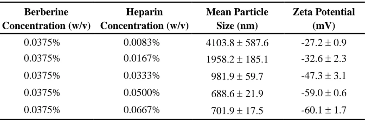
The different composition of nanoparticles (berberine/heparin nanoparticles or berberine/heparin/chitosan nanoparticles) was prepared by a simple ionic gelation method with magnetic stirring at room temperature. Table 1–3 lists the conditions for the nanoparticles preparation, as well as the measured size distribution and zeta
7
potential values for each test sample. First, the berberine/heparin nanoparticles (Table 1) were prepared by aqueous berberine (0.0375% by w/v) was added by flush mixing with a pipette tip into aqueous heparin at various concentrations (0.0083%, 0.0167%, 0.0333%, 0.0500%, 0.0667% by w/v). Second, the berberine/heparin/chitosan nanoparticles (Table 2–3) at distinct compositions (berberine:heparin:chitosan =
0.0375%:0.0500%:0.0200%, 0.0375%:0.0500%:0.0400%,
0.0375%:0.0500%:0.0800%, 0.0200%:0.0500%:0.0400%,
0.0250%:0.0500%:0.0400%, and 0.0300%:0.0500%:0.0400% by w/v) were prepared by dropping aqueous berberine/heparin solution (0.5 mL) into a aqueous chitosan solution (pH 6.0, 0.5 mL).
2.3. Characterization of the prepared nanoparticles
The size distribution and zeta potential of the prepared nanoparticles at DI water were measured using a dynamic light scattering analyzer (Zetasizer ZS90, Malvern Instruments Ltd., Worcestershire, UK). FT–IR spectra of the prepared nanoparticles were recorded with a Fourier transformed infrared spectroscopy (FT–IR, Shimadzu Scientific Instruments, USA). TEM was employed to examine the morphology of the different composition of nanoparticles (berberine/heparin nanoparticles or berberine/heparin/chitosan nanoparticles). The nanoparticle suspension was placed onto a 400 mesh copper grid coated with carbon. About 2 min after deposition, the grid was tapped with a filter paper to remove surface water and positively stained with an alkaline bismuth solution [21].
2.4. Release profiles of berberine
To measurement the berberine loading efficiency of the prepared nanoparticles, the resulting solution containing berberine–loaded nanoparticles was centrifuged at 32,000rpm, 4℃ for 50 min. The amount of free berberine concentration in the supernatant was analyzed using a high–performance liquid chromatography (HPLC) [22]. The berberine loading efficiency of the nanoparticles was calculated from the
8
following equation: [23]
The release profiles of berberine from test nanoparticles were investigated in simulated dissolution medium (pH 1.2 for 120 min, pH 6.0 for 120 min, then pH 7.0 for 240 min, simulating gastric acid, gastric mucosa and the H. pylori survival situation), respectively (n = 5) at 37 °C. At set time intervals, samples were removed and centrifuged, and the supernatants were subjected to HPLC. The percentage of cumulative amount of released berberine was determined by a standard calibration curve. The stability of berberine was determined by analyzing the conformation of the released berberine from nanoparticles using an electrospray ionization–mass spectrometer (ESI–MS) and comparing the spectrum with that of standard berberine. For ESI–MS measurement, berberine released from nanoparticles and standard berberine were separately dissolved at alcohol at the concentrations of 10 ppm directly injected into the electrospray ionization interface of the mass spectrometry. The ionization was on a positive mode with a spray voltage of +4.25 kV [24].
2.5. In Vitro growth inhibition study
H. pylori strain 26695 (ATCC 700392) was obtained from the American Type
Culture Collection (ATCC, Manassas, VA, USA). The bacteria were grown on blood agar plates under microaerophilic conditions for 48–72 hr at 37°C. The colonies were collected and pooled in HBSS solution (pH 6.0) to an optical density of 1.0 at 590 nm (OD590), which corresponded to 106 colony–forming units (CFU)/mL bacteria at 590
nm in a visible spectrophotometer (Biochrom Ltd., England). To characterize the ability of berberine and berberine–loaded nanoparticles to inhibit growth of H. pylori, bacterial suspensions were exposed to berberine solution and berberine/heparin/chitosan nanoparticles with distinct berberine concentrations (0.0, 3.0, 6.0, and 12.0 by mg/L) for 24 hr. Specifically, 5 mL of HBSS solution was inoculated with an equal volume of stock culture; a micropipette was used to aliquot
Loading efficiency = 100%
total amount of berberine
9
appropriate volumes of different formulations to culture vials containing the H. pylori strain. All mixtures were incubated in a candle jar under microaerophilic conditions with humidity at 37°C. The extent of growth inhibition was determined by OD590
measurements at distinct time intervals. Quantification of in vitro antibacterial activity was carried out by calculating percentage growth inhibition as compared to untreated
H. pylori. The percent growth inhibition was determined using the formula [25]:
where control refers to H. pylori broth and sample refers to H. pylori broth incubated with formulations.
2.6. Viability of AGS cells treated with berberine solution and berberine/heparin/chitosna nanoparticles
The AGS cell line (ATCC CRL 1739) was obtained from the American Type Culture Collection (ATCC) and used between passages 40 and 60. The cells were initially grown in 25 cm2 tissue culture flasks with RPMI 1640 medium containing 10% FBS, penicillin (100 U/mL), and streptomycin (100 g/mL) and were kept in an incubator at 37 °C, 95% humidity, and 5% CO2 [3]. The cells were harvested for
subculture every three days with 0.25% trypsin plus 0.05% EDTA solution and were used for the cytotoxicity experiments. The cytotoxicity of the test samples was evaluated in vitro with the MTT assay. The assay is based on mitochondrial dehydrogenase activity as an indicator of cell viability. Briefly, MTT was dissolved in PBS to a concentration of 5 mg/mL as a stock MTT solution and filtered for sterilization. AGS cells were seeded at 5 104 cells/well in 96-well plates and allowed to adhere overnight. The growth medium was replaced with HBSS solution (pH 6.0) that contained various berberine concentrations (0.0, 3.0, 6.0, and 12.0 by mg/L) of berberine solution and berberine/heparin/chitosna nanoparticles formulation for 2 hr. After 2 hr, the test samples were aspirated and the cells were washed twice with 100 L of PBS. The cells were then incubated in growth medium for an
% growth inhibition = OD of control – OD of sample 100% OD of control
10
additional 22 hr. After specific times, the cells were then incubated in growth medium containing 1 mg/mL MTT for an additional 4 h. Dimethyl sulfoxide (DMSO; 100 L) was added to each well to ensure the solubilization of the formazan crystals formed. The optical density was read with a Molecular Devices SpectraMax M2e microplate
spectrofluorometer (Sunnyvale, CA) at a wavelength of 570 nm. All experiments were performed six times with eight replicate wells for every sample and control per assay.
2.7. Association of fluorescent H. pylori and nanoparticles with AGS cells and CLSM visualization
To observe the adhesion of H. pylori to cells, the bacterium was labeled with DilC18(5)-DS fluorescently labeled lipophilic dye to form the DilC18(5)–H. pylori. The bacteria were collected from the agar plates and resuspended in PBS to an optical density at 590 nm (OD590) of 1.0. After centrifugation at 3000 rpm for 10 min, the
bacterial pellet was resuspended in 1.0 mL of diluent A. Then 1.0 mL of diluent A containing 50 L of DilC18(5)–DS dye was added and mixed with the bacterial suspension. The bacterial suspension was mixed again for 5 min at 30°C followed by 15 min incubation on ice. The bacterial pellet was washed three times with ice cold PBS, suspended in PBS, and used to infect cells at a multiplicity of infection (MOI) of 50.
The fluorescent nanoparticles (berberine/FA–heparin/chitosan) were prepared. The synthesis of FA–heparin was based on the reaction between the amine groups of FA and the carboxylic acid groups of heparin [3]. Heparin (3.07 g) and FA (0.0583 g) were dissolved completely in 30 mL of acetonitrile with 0.0408 g of 1-ethyl-3-(3-dimethylaminopropyl)-carbodiimide hydrochloride, and incubated at room temperature for 2 hr. To remove the unconjugated FA, FA–heparin were dialyzed in the dark against 5 L of distilled water, which was replaced on a daily basis until no fluorescence was detected in the supernatant. The resultant FA–heparin was lyophilized in a freeze dryer. The fluorescent nanoparticles were prepared according to the procedure described in preparation of berberine/heparin/chitosan nanoparticles
11
section.
To track the internalization of the test samples, the cells were seeded onto glass coverslips at a density 3 105 cells/cm2 and incubated for two days. The test samples (DilC18(5)–H. pylori and berberine/FA–heparin/chitosan nanoparticles) were then added to the cells for 2 hr. After incubation, the test samples were aspirated. The cells were then washed three times with PBS before they were fixed in 3.7% paraformaldehyde. The cells were examined with excitation at 340 (for berberine), 488 (for FA–heparin), and 633 (for DilC18(5)–H. pylori) under a CLSM [26]. The images were superimposed with the LCS Lite software (version 2.0).
2.8. Evaluating the relationship between H. pylori and nanoparticles
AGS cells were grown to subconfluence and treated with various berberine concentrations (0.0, 3.0, and 6.0 by mg/L) of berberine/heparin/chitosan nanoparticle formulation for 2 hr. The test samples were then aspirated and the cells were washed twice with PBS and then inoculated with H. pylori at a final concentration of 8 108 CFU/mL. After co–culture with H. pylori for 2 hr, the treated AGS cells were washed and incubated in growth medium for an additional 22 hr. The amount of growth inhibition on AGS cells by H. pylori was analyzed using the trypan blue exclusion [27].
The in situ adherence properties of H. pylori are useful for identifying effective nanoparticles. The adherence detection protocol used was carried out as previously described, but with the following modifications [25,28]. The constant berberine 6.0 mg/L of berberine/FA–heparine/chitosan nanoparticles diluted in HBSS solution were added directly to the AGS cells for 2 hr. Meanwhile H. pylori in log phase was inoculated onto glass coverslips; the AGS treated cells were then added to the labeled DilC18(5)–H. pylori at an MOI of 50 and incubated at 37°C for 2 hr. The cells were then washed three times with PBS and fixed in 3.7% paraformaldehyde. Cells were examined by excitation at 340 nm (for berberine), 488 nm (for FA–heparin) and 633 nm (for DilC18(5)–H. pylori) under a CLSM. The images were superimposed by the
12
LCS Lite software (version 2.0).
2.9. Statistical analysis
Statistical analysis of the differences in the measured properties of the groups was performed with one-way analysis of variance and the determination of confidence intervals, with the statistical package Statistical Analysis System, version 6.08 (SAS Institute Inc., Cary, NC). All data are presented as means and standard deviations, indicated as “mean ± SD”. Differences were considered to be statistically significant when the P values were less than 0.05.
3. Results and discussion
3.1. Preparation and characterization of nanoparticles
Berberine/heparin nanoparticles were produced by the ionic gelation of positively charged berberine with negatively charged heparin. As shown in Table 1, berberine:heparin in distinct compositions (0.0375%:0.0083%, 0.0375%:0.0167%, 0.0375%:0.0333, 0.0375%:0.0500%, and 0.0375%:0.0667% by w/v) have a mean size range of 700–4000 nm, with negative zeta potentials, depending on the relative concentrations of berberine and heparin used (Table 1). The particles size distributions, polydispersity indices (PDI) and zeta potentials of the prepared nanoparticles of berberine:heparin at 0.0375%:0.0167% and 0.0375%:0.0500% (w/v) composition in aqueous solution are presented in Figure 2. As shown in Table 1 and Figure 2, the zeta potential value of berberine/heparin nanoparticles (berberine:heparin = 0.0375%:0.0500%) was significantly lower than that of berberine:heparin compositions at 0.0375%:0.0167%, due to the significantly higher amounts of the negatively charged heparin being exposed on their surfaces. Additionally, berberine:heparin compositions at 0.0375%:0.0500% had a smaller particle size (688.6 ± 21.9 nm) with a narrower distribution (PDI 0.51 ± 0.11) than their counterpart berberine:heparin = 0.0375%:0.0167% (Figure 2). Therefore, the berberine:heparin composition of 0.0375%:0.0500% was chosen for use throughout
13
the rest of the study.
Heparin (structure shown in Figure 1) is a polydispersed, highly sulfated polysaccharide composed of repeating 1→4 linked uronic acid and glucosamine residues. There are three types of acidic functional groups (sulfate monoesters, sulfamido groups and carboxylate groups) present in the heparin polymer [3]. Berberine (structure shown in Figure 1) is a yellow–colored quaternary protoberberine alkaloid extracted from the root of the Berberis species shrubs or the bark of the Amur cork tree [29,30]. When prepared in distilled water, berberine, like heparin, is ionized. When prepared in combination the ionized berberine and heparin can form polyelectrolyte complexes (berberine/heparin nanoparticles) by electrostatic interactions between the positively charged (–CN+) on berberine and the negatively charged (–SO4– and –COO–) on heparin. In the Fourier transform infrared (FTIR)
spectra (Figure 3), peaks occur at 1615 cm–1 for the cyano cation (–CN+) on berberine and at 1229 cm–1 and 1635 cm–1 for the sulfate ions (–SO4–) and carboxylic ions
(–COO–), respectively, on heparin [3,31]. As shown, berberine/heparin nanoparticles lose the cyano cation characteristic peak at 1615 cm–1, but gain a new peak at 1619 cm–1; in addition, the characteristic peak of sulfate and carboxylic ions on heparin at 1229 cm–1 and 1635 cm–1 shifted to 1235 cm–1 and 1639 cm–1. The ionized berberine and heparin were, therefore, able to form polyelectrolyte complexes via electrostatic interactions.
However, the results obtained by the TEM examination showed that the morphology of the prepared berberine/heparin nanoparticles was irregular and oval shaped (Figure 1). To improve the complex formed, chitosan was blended with the berberine/heparin complex (Figure 1). The cationic polysaccharide chitosan is derived from chitin by alkaline deacetylation [32]. In our nanoparticles which were prepared with different concentratios of chitosan (0.00%, 0.02%, 0.04%, and 0.08% by w/v), the size distribution and zeta potential in an aqueous environment were investigated by a dynamic light scattering analyzer. As shown in Table 2, as the amount of chitosan incorporated was increased, the size of the resulting nanoparticles decreased
14
appreciably; meanwhile, the zeta potential value increased noticeably as more chitosan was incorporated, with the exception of the chitosan concentration 0.08%. The polydispersity index of nanoparticles measured by a dynamic light scattering analyzer revealed a narrower distribution (PDI 0.39 ± 0.07) than those berberine/heparin nanoparticles not incorporating chitosan (Figure 2). The characteristic FT–IR spectrum peak at 1573 cm-1 that corresponds to the protonated amino group (–NH3+) in aqueous chitosan (pH 6.0) was shifted to 1590 cm-1 in the
prepared berberine/heparin/chitosan nanoparticles (Figure 3). Meanwhile, the characteristic peak for the negatively charged (–SO4– and –COO–) on heparin was
shifted and decreased. These observations can be attributed to the electrostatic interaction between the negatively charged groups on heparin and the positively charged group on chitosan.
The particulate system for translocation permeabilities through gastrointestinal mucin was found to be decreased as the particle size increased [33]. The PDI is a measure of dispersion homogeneity and ranges from 0 to 1. Values close to 0 indicate a homogeneous dispersion while those greater than 0.3 indicate high heterogeneity [34]. As shown in Table 3, the berberine-loaded nanoparticles were obtained instantaneously upon addition of a thoroughly mixed aqueous berberine/heparin to a chitosan solution under magnetic stirring at room temperature. Table 3 and Figure 2 show the particle size, zeta potential, loading efficiency and PDI of the obtained nanoparticles. In the loading process, increasing the amount of berberine led to a larger overall size of the nanoparticles and to a significant increase in their berberine loading efficiency. When the berberine concentration was 0.0250% (w/v), the particle size, loading efficiency and PDI of the nanoparticles was 256.5 ± 2.3 nm, 54.4 ± 3.4% and 0.26 ± 0.02, respectively. Under this specific condition, the berberine-loaded nanoparticles remained spherical (Figure 1). As the berberine concentration was increased, the nanoparticle size became larger, yielding a mean particle size of more than 300 nm. Therefore, the optimal berberine concentration of 0.0250% (w/v) was chosen for subsequent in vitro study.
15 3.2. In vitro drug release
The release profiles of berberine from nanoparticles were investigated in simulated dissolution medium (pH 1.2 for 120 min, pH 6.0 for 120 min, then pH 7.0 for 240 min, simulating gastric acid, gastric mucosa and the H. pylori survival situation), respectively. Figure 4a shows the release profiles of berberine from the prepared nanoparticles at pH 1.2, 6.0, and 7.0, respectively. Heparin is a polydispersed, highly sulfated polysaccharide composed of repeating 1→4–linked uronic acid and glucosamine residues. There are three types of acidic functional groups in the heparin polymer. The sulfate monoesters and sulfamido groups are both highly acidic, with pKa values ranging from 0.5 to 1.5, whereas the extra carboxylate groups are less acidic, with pKa values between 2 to 4 [3]. Chitosan is a weak base, and the amino group on chitosan has a pKa value of around 6.5 [3]. At pH 1.2 (simulating gastric acid), only some of the –COO- groups on heparin became protonated (–COOH), the amino groups (–NH3+) on chitosan and the acidic functional groups (sulfate
ions; –SO4–) on heparin were ionized. Therefore, chitosan and heparin were able to
form polyelectrolyte complexes via electrostatic interaction, but relatively weaker compared with that at prepared nanoparticle situation, resulting in the proportion of berberine released from the nanoparticles in 120 min was 18.98 ± 3.56% (Figure 4a). As shown, at pH 6.0 (simulating the environment of the gastric mucosa or the H.
pylori survival situation), little of the –NH3+ groups on chitosan became deprotonated
and the carboxylic ions (–COO-) and sulfate ions (–SO4–) were ionized on heparin,
this was followed by little release of berberine at pH 6.0, possibly due to the conjugation of berberine and heparin. At pH 7.0 (H. pylori survival situation), most of the –NH3+ groups on chitosan was deprotonated, the nanoparticles became unstable
and broken apart, allowing rapid release of the berberine from the nanoparticles. The HPLC method was developed and validated in order to quantify berberine. Satisfactory retention times and good resolution of berberine were achieved using reverse phase C18 columns and elution with acetonitrile–0.04 M H3PO4 (42:58; v/v)
16
absorbance at 349 nm in the range of 1.0–8.0 mg/L (R2 = 0.999). The linear regression equation for the calibration curve was y = 727528x+33975, where y represented the peak area ratio of berberine and x represented the concentration of berberine (mg/L). Comparative chromatographic studies revealed that berebrine released from nanoparticles with standard berberine solution yielded a pure peak at the same retention time of 6.96 min.
Berberine’s chemical structure contains a quaternary protoberberine alkaloid and it is commercially available in various salt forms, suchas berberine chloride and hemisulfate. ESI–MS experiments were employed to examine the properties of berberine release from nanoparticles in standard berberine solution (Figure 4b, left). Berberine exhibited a base peak at m/z = 336.2 for M+ and all of the alkaloids displayed [M-CH3]+ = 321.4, [M-2CH3]+ = 306.5, [M-C2H4O]+ = 292.5, and
[M-2CH3-CO]+ = 278.4 as the base peaks, no matter what collision energy was used
[35,36]. As evidenced by ESI–MS (Figure 4b), no significant conformational change was observed for the berberine released from nanoparticles, as compared to the standard berberine.
3.3. In vitro growth inhibition study
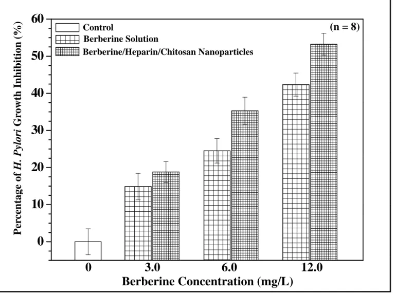
Berberine has been extensively used to treat gastroenteritis and diarrhea caused by bacteria since the 1950s when its clinical safety was experimentally confirmed [37]. Since then, berberine has been characterized as having multiple pharmacologic functions, including antibacterial, antiparasitic, antifungal, hypotensive, antitumoral, anti–inflammatory and inhibition of lymphocyte transformation [30,37]. Moreover, recent pharmacological studies have demonstrated that berberine can inhibit the arylamine N-acetyltransferase activity in strains of H. pylori collected from peptic ulcer patients [11]. N–acetyltransferase–catalyzed reactions are known to activate or detoxify arylamine carcinogens. A study on H. pylori demonstrated that berberine elicited a dose–dependent growth inhibition in the H. pylori cultures, and inhibited the growth of the H. pylori strains with a minimum inhibitory concentration (MIC) range
17
of 0.78 to 25.00 mg/L [12]. Figure 5 shows the percentage of H. pylori growth inhibition effected by treatment with various berberine concentrations (0.0, 3.0, 6.0, 12.0 by mg/L) for 24 hr. The evident growth inhibition observed at 6.0 mg/L was found to be 24.5 ± 3.3 (only berberine solution) and 35.3 ± 3.7 (berberine/heparin/chitosan nanoparticles). Berberine concentration of 12.0 mg/L was 42.4 ± 3.2 (only berberine solution) and 53.2 ± 2.9 (berberine/heparin/chitosan nanoparticles), compared to control (without sample). Our prepared berberine/heparin/chitosan nanoparticles with a negative surface charge (where heparin dominated the surface, berberine:heparin:chitosan = 0.0250%:0.0500%:0.0400% by w/v) were able to increase the values of H. pylori growth inhibition significantly (p < 0.05), compared to berberine solution. Heparin is found primarily in the liver, lungs and gut tissues, while its structural relative, heparin sulfate, resides in the extracellular cell matrix at the surface of most animal cells [38]. Heparin is normally produced by mast cells and is stored in intracellular granules until needed. Recent studies indicate that vacuolating cytotoxin A (VacA) is able to recruit and activate mast cells [39]. Moreover, the C-terminal domain of the 58 kDa subunit of H. pylori VacA can bind both heparin and heparan sulfate [40]. Therefore, when the negatively charged nanoparticles (berberine/heparin/chitosan) specifically interact with H. pylori, the drug is likely released from the nanoparticle to act locally.
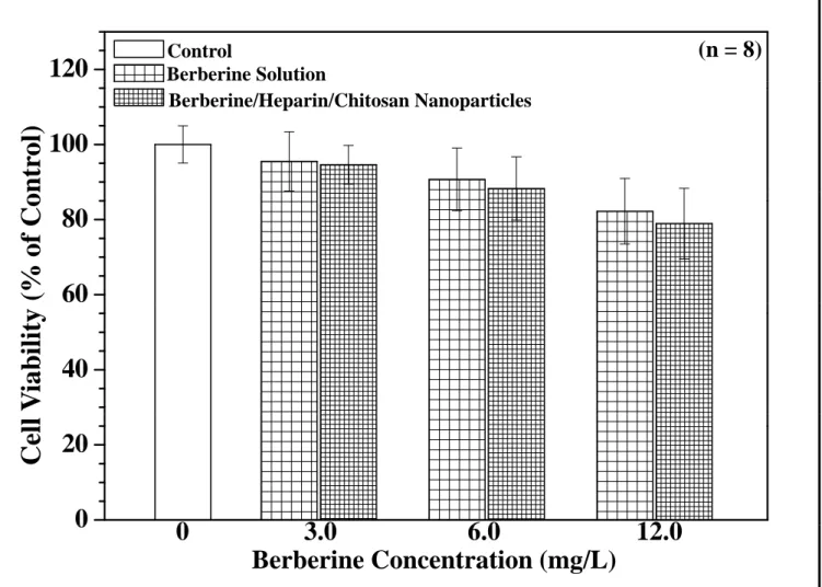
3.4. cytotoxicity of berberine solution and berberine/heparin/chitosan nanoparticles
The anti–neoplastic effects of berberine have drawn extensive attention of researchers hoping to exploit its therapeutic properties. Berberine has since been shown to suppress the growth of a wide variety of tumor cells, including breast cancer, epidermoid carcinoma, oral carcinoma, prostate carcinoma, and gastric carcinoma [41–43]. Lin et al. sought to determine the molecular mechanism of berberine that underlies its effects in human gastric cancer cells. Berberine (8.4–672.4 mg/L) was found to be cytotoxic to human gastric cancer cells in a dose-dependent manner, at an IC50 of 16.1 mg/L [13]. In our study, the cytotoxicity of berberine solution and
18
berberine/neparin/chitosan nanoparticles were evaluated using human gastric adenocarcinoma cells by using an MTT assay (Figure 6). The reduction of cell viability after treatment was significantly higher with 12 mg/L of berberine for all test samples. Cell viability was generally not affected by 3.0 and 6.0 mg/L of berberine. To examine the relationship between H. pylori and the prepared berberine/heparin/chitosan nanoparticles without damaging the cultured cells, test samples were prepared at a constant berberine concentration of 6.0 mg/L throughout the rest of the study.
3.5. Association of fluorescent H. pylori and nanoparticles with AGS cells
To observe the adhesion capabilities of H. pylori to the human gastric mucosal AGS cells, the bacterium was fluorescently labeled with DilC18(5)–DS lipophilic dye to form the DilC18(5)–labeled H. pylori. As shown in Figure 7, after 2 hr of infection, the AGS-adapted DilC18(5)–H. pylori (magenta; yellow arrows) preferentially targeted cell–cell junctions and was observed within the cells. It was confirmed that H.
pylori strains mainly resided in the gastric mucosa or at the interface with the mucus
layer, and produced the vacuolating cytotoxin, which modulates the integrity of the tight junctions of the epithelium and reduces the stability of the cytoskeleton. In order to examine the allocation of negative surface charge (heparin dominated on the surface) nanoparticles in the AGS cell after 2 hr exposure, fluorescent nanoparticles (berberine/FA–heparin/chitosan nanoparticles) were used. Heparin is a member of the glycosaminoglycan family of carbohydrates (which includes the closely related molecule heparan sulfate) and consists of a variably sulfated repeating disaccharide unit. Among which heparan sulfate is a ubiquitously expressed heparin analog that appears as a proteoglycan at the cell surface [17,37]. The distribution of berberine (blue) and FA-heparin (green) in the intercellular spaces was observed by CLSM. The natural yellow color of berberine has made it useful as a commercial dye; its normal excitation occurs at about 350 nm with UV laser system [26,44]. The synthesis of FA–heparin was based on the reaction between the amine groups of FA and the
19
carboxylic acid groups of heparin; FA excitation occurs at 488 nm using an argon laser. As shown in Figure 7, after 2 hr of incubation of berberine/FA–heparin/chitosan nanoparticles the incubated nanoparticles were located in the same intercellular spaces and cell cytoplasm (as indicated by superimposed green/blue spots; i.e., white arrows) when internalized into the cytoplasm.
3.6. Evaluating the relationship between H. pylori and nanoparticles
Figure 8a shows the influence of H. pylori infection alone or after pretreatment with berberine/heparin/chitosna nanoparticles on gastric mucosal cell viability. Significant inhibition of AGS cell viability after infection with only H. pylori (8 108 CFU/mL) was observed to be 51.6 ± 6.2%, as compared to uninfected controls. H.
pylori colonization of the gastric mucosa is associated with alterations in gastric
epithelial cell cycle events, a condition implicated in the initiation and development of gastric cancer. Virulence factors of H. pylori include urease, lipopolysaccharides, adhesins, the cytotoxin–associated gene A product and have been determined to contribute to host inflammation and epithelial cell damage, such as cytoplasmic vacuolation and induction of apoptosis [45]. In our study, the berberine/heparin/chitosna nanoparticles with berberine concentration 3.0 and 6.0 mg/L caused a statistically significant increase in AGS cell growth. Cell growth was enhanced in a dose-dependent manner, as shown in Figure 8a; specifically, in H.
pylori infected cells, berberine stimulated an almost 1.3-fold cell growth increase at a
dose of 3.0 mg/L and a 1.6-fold growth increase at a dose of 6.0 mg/L. Therefore, berberine/heparin/chitosan nanoparticles were able to efficiently reduce the cytotoxic effects of H. pylori in AGS cells.
The in situ adherence assay was carried out by using fluorescently labeled DilC18(5)–H. pylori that had been incubated with berberine/FA-heparin/chitosan nanopaticles (berberine concentration at 6.0 mg/L) (Figure 8b). As can be seen, the AGS-adapted DilC18(5)–H. pylori (magenta) preferentially targeted cell–cell junctions and were located within the cells. AGS cells were then incubated with
20
fluorescent berberine/FA–heparin/chitosan nanoparticles (FA–heparin; green spots and berberine; blue spots) to ascertain whether the nanoparticles were attached in the same way to the intercellular spaces at the sites of H. pylori infection (white spot). The white spots in the superimposed images were observed to disappear, indicating the release of berberine from nanoparticles into the intercellular spaces or the cell cytoplasm (blue spots). From these results, we concluded that the nanoparticle drug delivery system could effectively release the berberine from the nanoparticle so that it may act locally on H. pylori at an appropriate bactericidal concentration.
4. Conclusions
We developed a novel nanoparticle carrier shelled with heparin for berberine delivery to treat H. pylori. The in vitro analysis of drug release from the nanoparticle indicated that the system is able to control berberine release in the simulated gastrointestinal dissolution medium, and that the berberine was able to localize specifically to intercellular spaces or in the cell cytoplasm, the site of H. pylori infection. Furthermore, the prepared berberine/heparin/chitosan nanoparticles were able to increase the values of H. pylori growth inhibition significantly, compared to berberine alone in solution, and to efficiently decrease the cytotoxic effects of H.
pylori infection of the cells.
Acknowledgements
This work was supported by grants from the National Science Council (NSC 98-2815-C-039-034-B) and the Center for Inflammation Research, China Medical University (CMU 98-N1-08 and CMU 98-CT-12), Republic of China. The confocal microscopy SP2 experiment and Zetasizer apparatus supported by the Medical Research Core Facilities center, Office of Research & Development, China Medical University were gratefully acknowledged.
21 References
[1] Goodman KJ, Cockburn M. The role of epidemiology in understanding the health effects of Helicobacter pylori. Epidemiology 2001;12:266–271.
[2] Beil W, Kilian P. EPs 7630, an extract from Pelargonium sidoides roots inhibits adherence of Helicobacter pylori to gastric epithelial cells. Phytomedicine 2007; 14:5–8.
[3] Lin YH, Chang CH, Wu YS, Hsu YM, Chiou SF, Chen YJ. Development of pH-responsive chitosan/heparin nanoparticles for stomach-specific anti-Helicobacter pylori therapy. Biomaterials 2009;30:3332–3342.
[4] Chang CH, Lin YH, Yeh CL, Chen YC, Chiou SF, Hsu YM, Chen YS, Wang CC. Nanoparticles incorporated in pH-sensitive hydrogels as amoxicillin delivery for eradication of Helicobacter pylori. Biomacromolecules 2010;11:133–142.
[5] Bardonnet PL, Faivre V, Pugh WJ, Piffaretti JC, Falson F. Gastroretentive dosage forms: overview and special case of Helicobacter pylori. J Controlled Release 2006;111:1–18.
[6] de Bortoli N, Leonardi G, Ciancia E, Merlo A, Bellini M, Costa F, Mumolo MG, Ricchiuti A, Cristiani F, Santi S, Rossi M, Marchi S. Helicobacter pylori eradication: A randomized prospective study of triple therapy versus triple therapy plus lactoferrin and probiotics. Am J Gastroenterol 2007;102:951–956.
[7] Li Y, Xu C, Zhang Q, Liu JY, Tan RX. In vitro anti–Helicobacter pylori action of 30 Chinese herbal medicines used to treat ulcer diseases. J Ethnopharmacol 2005; 98:329–333.
[8] Wang F, Zhou HY, Zhao G, Fu LY, Cheng L, Chen JG, Yao WX. Inhibitory effects of berberine on ion channels of rat hepatocytes. World J Gastroenterol 2004;10: 2842–2845.
[9] Baird AW, Taylor CT, Brayden DJ. Non-antibiotic anti-diarrhoeal drugs: factors affecting oral bioavailability of berberine and loperamide in intestinal tissue. Adv Drug Deliv Rev 1997;23:111–120.
[10] Chung JG, Wu LT, Chang SH, Lo HH, Hsieh SE, Li YC, Hung CF. Inhibitory actions of berberine on growth and arylamine N-acetyltransferase activity in strains of helicobacter pylori from peptic ulcer patients. Int J Toxicol 1999;18: 35–40.
[11] Mahady GB, Pendland SL, Stoia A, Chadwick LR. In vitro susceptibility of Helicobacter pylori to isoquinoline alkaloids from Sanguinaria canadensis and Hydrastis canadensis. Phytother Res 2003;17:217–221.
[12] Lin JP, Yang JS, Lee JH, Hsieh WT, Chung JG. Berberine induces cell cycle arrest and apoptosis in human gastric carcinoma SNU-5 cell line. World J
22
Gastroenterol 2006;12:21–28.
[13] Pan LR, Tang Q, Fu Q, Hu BR, Xiang JZ, Qian JQ. Roles of nitric oxide in protective effect of berberine in ethanol-induced gastric ulcer mice. Acta Pharmacol Sin 2005;26:1334–1338.
[14] Bentolila A, Vlodavsky I, Haloun C, Domb AJ. Synthesis and heparin-like biological activity of amino acid-based polymers. Polym Adv Technol 2000;11: 377–387.
[15] Bertolesi G, Eijan AM, Calvo JC, Lauria de Cidre L. Heparin increases the adhesion of murine mammary adenocarcinoma cells (LM3). Correlation with the presence of heparin receptors on cell surface. J Biol Regul Homeost Agents 2005; 19:33–40.
[16] Benoit DS, Anseth KS. Heparin functionalized PEG gels that modulate protein adsorption for hMSC adhesion and differentiation. Acta Biomater 2005;1:461–470.
[17] Thein-Han WW, Misra RD. Acta Biomater. Biomimetic
chitosan-nanohydroxyapatite composite scaffolds for bone tissue engineering. Acta Biomater 2009;5:1182–1197.
[18] van der Lubben IM, Verhoef JC, Borchard G, Junginger HE. Chitosan and its derivatives in mucosal drug and vaccine delivery. Eur J Pharm Sci 2001;14: 201–207.
[19] Erbacher P, Zou S, Bettinger T, Steffan AM, Remy JS. Chitosan-based vector/DNA complexes for gene delivery: biophysical characteristics and transfection ability. Pharm Res 1998;15:1332–1339.
[20] Hall AJ, Tripp M, Howell T, Darland G, Bland JS, Babish JG. Gastric mucosal cell model for estimating relative gastrointestinal toxicity of non-steroidal anti-inflammatory drugs. Prostaglandins Leukot Essent Fatty Acids 2006;75:9–17. [21] Kuno T, Naito S, Ohta M, Kido N, Ito H, Kato N. Staining of O-specific
polysaccharide chains of lipopolysaccharides with ruthenium red. Microbiol Immunol 1986;30:743–751.
[22] Wang SD, Song BS, Li K. Determination of berberine in decocted liquid from shenshu granules with water by reversed-phase liquid chromatography. Se Pu 2000;18:261–262.
[23] Sahasathian T, Kerdcholpetch T, Chanweroch A, Praphairaksit N, Suwonjandee N, Muangsin N. Sustained release of amoxilcilline from chitosan tablets. Arch
23
Pharm Res 2007;30:526–531.
[24] Pan JF, Yu C, Zhu DY, Zhang H, Zeng JF, Jiang SH, Ren JY. Identification of three sulfate–conjugated metabolites of berberine chloride in healthy volunteers' urine after oral administration. Acta Pharmacol Sin 2002;23:77–82.
[25] Jain P, Jain S, Prasad KN, Jain SK, Vyas SP. Polyelectrolyte coated multilayered liposomes (nanoparticles) for the treatment of Helicobacter pylori infection. Mol. Pharm. 2009, 6, 593–603.
[26] Sakai T, Ohno N, Chung YS, Nishikawa H. Spectrofluorimetric determination of berberine in oriental pharmaceutical preparations by flow-injection analysis coupled with liquid-liquid extraction. Anal Chim Acta 1995;308:329–333.
[27] Sieveking D, Leach ST, Mitchell HM, Day AS. Role of serum factors in epithelial cell responses to Helicobacter pylori infection in vitro. J Gastroenterol Hepatol 2005;20:1610–1615.
[28] Lai CH, Fang SH, Rao YK, Geethangili M, Tang CH, Lin YJ, Hung CH, Wang WC, Tzeng YM. Inhibition of Helicobacter pylori-induced inflammation in human gastric epithelial AGS cells by Phyllanthus urinaria extracts. J Ethnopharmacol 2008;118;522–526.
[29] Hsieh YS, Kuo WH, Lin TW, Chang HR, Lin TH, Chen PN, Chu SC. Protective effects of berberine against low-density lipoprotein (LDL) oxidation and oxidized LDL-induced cytotoxicity on endothelial cells. J Agric Food Chem 2007;55: 10437–10445.
[30] Li Y, He WY, Tian J, Tang J, Hu Z, Chen X. The effect of Berberine on the secondary structure of human serum albumin. J Mol Struct 2005;743;79–84. [31] Kim Y, Kim SJ, Nam W. A ferric-cyanide-bridged one-dimensional dirhodium
complex with (18–crown–6)potassium cations. Acta Crystallogr C 2001;57: 266–268.
[32] Lin YH, Chung CK, Chen CT, Liang HF, Chen SC, Sung HW. Preparation of nanoparticles composed of chitosan/poly-γ-glutamic acid and evaluation of their permeability through caco-2 cells. Biomacromolecules 2005;6:1104–1112. [33] Norris DA, Sinko PJ. Effect of size, surface charge, and hydrophobicity on the
translocation of polystyrene microspheres through gastrointestinal mucin. J Appl Polym Sci 1998;63;1481–1492.
[34] Ahlin P, Kristl J, Kristl A, Vrecer F. Investigation of polymeric nanoparticles as carriers of enalaprilat for oral administration. Int J Pharm 2002;239:113–120. [35] Wu W, Song F, Yan C, Liu Z, Liu S. Structural analyses of protoberberine
alkaloids in medicine herbs by using ESI-FT-ICR-MS and HPLC-ESI-MS(n). J Pharm Biomed Anal 2005;37;437–446.
24
adulterated Chinese medicine by capillary electrophoresis-electrospray ion trap mass spectrometry. J Chromatogr A 2000;866:273–280.
[37] Yang P, Song DQ, Li YH, Kong WJ, Wang YX, Gao LM, Liu SY, Cao RQ, Jiang JD. Synthesis and structure–activity relationships of berberine analogues as a novel class of low-density-lipoprotein receptor up-regulators. Bioorg Med Chem Lett 2008;18:4675–4677.
[38] Dubreuil JD, Ruggiero P, Rappuoli R, Del Giudice G. Effect of heparin binding on Helicobacter pylori resistance to serum. J Med Microbiol 2004;53:9–12.
[39] Supajatura V, Ushio H, Wada A, Yahiro K, Okumura K, Ogawa H, Hirayama T, Ra C. VacA, a vacuolating cytotoxin of Helicobacter pylori, directly activates mast cells for migration and production of proinflammatory cytokines. J Immunol 2002; 168:2603–2607.
[40] Utt M, Danielsson B, Wadström T. Helicobacter pylori vacuolating cytotoxin binding to a putative cell surface receptor, heparan sulfate, studied by surface plasmon resonance. FEMS Immunol Med Microbiol 2001;30:109–113.
[41] Kim JB, Yu JH, Ko E, Lee KW, Song AK, Park SY, Shin I, Han W, Noh DY. The alkaloid Berberine inhibits the growth of Anoikis-resistant MCF-7 and MDA-MB-231 breast cancer cell lines by inducing cell cycle arrest. Phytomedicine 2010;17:436–440.
[42] Ho YT, Lu CC, Yang JS, Chiang JH, Li TC, Ip SW, Hsia TC, Liao CL, Lin JG, Wood WG, Chung JG. Berberine induced apoptosis via promoting the expression of caspase-8, -9 and -3, apoptosis-inducing factor and endonuclease G in SCC-4 human tongue squamous carcinoma cancer cells. Anticancer Res 2009;29: 4063–4070.
[43] Auyeung KK, Ko JK. Coptis chinensis inhibits hepatocellular carcinoma cell growth through nonsteroidal anti–inflammatory drug-activated gene activation. Int J Mol Med 2009;24:571–577.
[44] Pereira CV, Machado NG, Oliveira PJ. Mechanisms of berberine (natural yellow 18)–induced mitochondrial dysfunction: interaction with the adenine nucleotide translocator. Toxicol Sci 2008;105:408–417.
[45] Kuck D, Kolmerer B, Iking-Konert C, Krammer PH, Stremmel W, Rudi J. Vacuolating cytotoxin of Helicobacter pylori induces apoptosisin the human gastric epithelial cell line AGS. Infect Immun 2001;69:5080–5087.
25 Tables
Table 1. Particle sizes and zeta potentials of nanoparticles prepared with different concentration berberine/heparin in deionized water (n = 5).
Berberine Concentration (w/v) Heparin Concentration (w/v) Mean Particle Size (nm) Zeta Potential (mV) 0.0375% 0.0083% 4103.8 587.6 -27.2 0.9 0.0375% 0.0167% 1958.2 185.1 -32.6 2.3 0.0375% 0.0333% 981.9 59.7 -47.3 3.1 0.0375% 0.0500% 688.6 21.9 -59.0 0.6 0.0375% 0.0667% 701.9 17.5 -60.1 1.7
Table 2. Effect of different concentration chitosan on particle sizes and zeta potential values of the berberine/heparin/chitosan nanoparticles (n = 5).
Berberine/Heparin Concentration (w/v) 0.0375%/0.0500% Chitosan Concentration (w/v) Mean Particle Size (nm) Zeta Potential (mV) 0.00% 688.6 21.9 -59.0 0.6 0.02% 590.7 22.5 -51.7 1.1 0.04% 403.8 8.9 -44.8 0.6 0.08% ■ ■
■ Precipitation of aggregates was observed.
Table 3. Effect of different concentration berine on particle sizes and zeta potential values of the berberine/heparin/chitosan nanoparticles (n = 5).
Heparin/Chitosan Concentration (w/v) 0.05%/0.04% Berberine Concentration (w/v) Mean Particle Size (nm) Zeta Potential (mV) Loading Efficiency (%) 0.0200% 225.7 2.9 -46.9 0.1 27.4 2.5 0.0250% 256.5 2.3 -46.1 0.4 54.4 3.4 0.0300% 321.9 9.7 -44.5 0.2 64.4 4.6 0.0375% 403.8 8.9 -43.8 0.6 79.1 6.5
0 Figure Captions
Fig. 1. Schematic illustrations of the internal structures and TEM micrographs of berberine/heparin nanoparticles and berberine/heparin/chitosan nanoparticles.
Fig. 2. Particle size distribution and zeta potential of the prepared nanoparticles (berberine/heparin nanoparticles and berberine/heparin/chitosan nanoparticles) at distinct compositions.
Fig. 3. FT–IR spectra of heparin, berberine, chitosan, berberine/heparin nanoparticles and berberine/heparin/chitosan nanoparticles.
Fig. 4. (a) In vitro release profiles of berberine from nanoparticles at different pH values at 37℃ (n =5). (b) HPLC and ESI–MS spectra of standard berberine and berberine released from nanoparticles. HPLC: high–performance liquid chromatography; ESI–MS: electrospray ionization–mass spectrometer. Fig. 5. Percentage of H. Pylori growth inhibition of only berberine solution and
berberine/heparin/chitosan nanoparticles.
Fig. 6. Cell viability after treatment with only berberine solution and berberine/heparin/chitosna nanoparticles after distince berberine concentration, determined by MTT assay (n = 6). MTT: 3-(4,5-dimethyl-thiazol-yl)-2,5-diphenyltetrazolium bromide.
Fig. 7. Confocal images of AGS cells infected with H. pylori or with internalized berberine/FA–heparin/chitosan nanoparticles for 2 hr. AGS cells: human gastric adenocarcinoma cells; FA: fluoresceinamine isomer I.
Fig. 8. (a) Changes in cell growth after H. pylori infection and
berberine/FA–heparin/chitosan nanoparticles pretreatment. AGS cells were preincubated for 2 hr with test nanoparticles and then infected with H. pyloi respectively. (b) Fluorescent images (taken with confocal laser scanning microscope) of cells infected with H. pylori and incubated with berberine/FA–heparin/chitosan nanoparticles. AGS cells: human gastric adenocarcinoma cells; FA: fluoresceinamine isomer I.
Figure 1
B b i
Berberine/Heparin Nanoparticles M icroscopyBerberine
m ission Electron MH
i
Berberine/Heparin/Chitosan Ttra ns m 100 nmBerberine/Heparin
Heparin
Berberine/Heparin/Chitosan Nanoparticles MicroscopyChitosan
m ission Electron: Berberine
Ttra ns mBerberine/Heparin/Chitosan
: Chitosan
: Heparin
: Berberine
200 nm Figure(s)Figure 2
500000 400000 400000 300000 Berberine:Heparin 0.0375%:0.0167% Berberine:Heparin 0.0375%:0.0500% 15 20 30 PDI:0.93 ± 0.18 Berberine:Heparin 0.0375%:0.0167% Berberine:Heparin 0.0375%:0.0500% PDI:0.51 ± 0.11 -32.6 ± 2.3 -59.0 ± 0.6 Total Counts 300000 200000 Total Counts 200000 100000 5 10 Number (% ) 10 20 Number (% ) 0 100 -200 100 Zeta Potential (mV) 0 100000 0 0 100 -200 100 Zeta Potential (mV) 5 0 1 10 100 1000 10000 Size (nm) 0 1 10 100 1000 10000 Size (nm) 500000 400000 400000 Berberine:Heparin:Chitosan 0.0375%:0.0167%:0.0400% Berberine:Heparin:Chitosan 0.0250%:0.0167%:0.0400% 30 40 40 Berberine:Heparin:Chitosan 0.0375%:0.0167%:0.0400% Berberine:Heparin:Chitosan 0.0250%:0.0167%:0.0400% PDI:0.39 ± 0.07 PDI:0.26 ± 0.02 -44.8 ± 0.6 -46.1 ± 0.4 Total Counts 300000 200000 Total Counts 300000 200000 20 30 Number (% ) 20 30 Number (% ) 0 100 -200 100 Zeta Potential (mV) 0 100000 0 100000 0 100 -200 100 Zeta Potential (mV) 0 10 100 1000 10000 1 10 Size (nm) 0 10 100 1000 10000 1 10 Size (nm)Zeta Potential
Size Distribution
Figure 3
Heparin
1635 cm-1 (– COO-on heparin)(%)
Berberine
1229 cm-1 (–SO 4-on heparin) 1615 cm-1 (–CN+on berberine)mission
Chitosan
Tr
a
n
s
1573 cm-1 (–NH 3+on chitosan) 1619 cm-1 (–CN+on berberine)Berberine/Heparin
1235 cm-1 (–SO 4-on heparin) 1639 cm-1 (– COO-on heparin) 1590 cm-1 (–NH 3+on chitosan)Berberine/Heparin
/Chitosan
1632 cm-1 (– COO-on heparin) 1230 cm-1 (–SO 4-on heparin)Wavenumber (cm
-1)
4000 3000 2000 1500 1000 500Figure 4a
a
se (%)
(n = 5)
100
erine Rele
a
80
n
t of Berb
e
40
60
v
e Amou
n
20
40
Cumulati
v
0
60
120
180
240
300
360
420
480
0
pH 1.2 pH 6.0 pH 7.0Time (min)
0
60
120
180
240
300
360
420
480
Figure 4b
High–Performance Liquid Electrospray Ionization−Mass Spectrometry
Chromatography 400 500 1.0 mg/L 2.0 mg/L 4.0 mg/L 8.0 mg/L 0.0 mg/L R² = 0.999 A rea p y p y Standard berberin [M-2CH3]+ 306.5 M+ 336.2 100 200 300 mV 0 1 2 3 4 5 6 7 8 Berberine concentration (mg/L) A [M-CH3]+ 321.4 4 5 6 7 8 9 10 0 minutes [M-C2H4O]+ 292.4 [M-2CH3-CO]+ 278.4 Standard berberine Berberine released from nanoparticles M+ 336.2 Berberine released from nanoparticles mV [M-CH3]+ [M-2CH3]+ 4 5 6 7 8 9 10 minutes 321.4 [M 2CH3] 306.5 [M-C2H4O]+ 292.5 [M-2CH3-CO]+ 278.4 minutes
Figure 5
60
B b i /H i /Chi N i l Berberine Solution Control(n = 8)
o
n (%)
40
50
Berberine/Heparin/Chitosan Nanoparticlesw
th Inhibiti
o
30
P
ylori
Gr
o
w
10
20
n
tage of
H.
P
0
10
Per
ce
n
Berberine Concentration (mg/L)
3.0
6.0
12.0
0
Figure 6
120
Berberine Solution Control(n = 8)
100
Berberine/Heparin/Chitosan Nanoparticlesntr
o
l)
60
80
(% of Co
40
V
iability
0
20
3 0
6 0
12 0
0
Cell
0
Berberine Concentration (mg/L)
3.0
6.0
12.0
0
Figure 7
DilC18(5)–Labeled H. pylori Infecting Cells
Berberine/Heparin/Chitosan Nanoparticles
Bright Field
DilC18(5)–H. pylori
Bright Field
FA–Heparin
Figure 8
H. pyloir and Berberine/Heparin/Chitosan Nanoparticles
(b)
C t l
(a)
H. pyloir and Berberine/Heparin/Chitosan Nanoparticles100
tro
l)
Berberine/Heparin/Chitosan Nanoparticles Control (n = 6)Helicobacter pylori Infected Cells
(a)
60 80
(% of Con
t
DilC18(5)–H. pylori
FA–Heparin
20 40