科技部補助專題研究計畫成果報告 期末報告
低能量雷射對於齒列矯正之牙齒穩定性-生物基礎與應用上系列 研究(第3年)
計 畫 類 別 : 個別型計畫
計 畫 編 號 : NSC 102-2314-B-040-007-MY3 執 行 期 間 : 104年08月01日至105年07月31日 執 行 單 位 : 中山醫學大學牙醫學系(所)
計 畫 主 持 人 : 高嘉澤 共 同 主 持 人 : 黃翠賢
計畫參與人員: 碩士班研究生-兼任助理人員:趙建維
中 華 民 國 105 年 09 月 12 日
中 文 摘 要 : 矯正治療之成功不在於矯正速度之快慢,而著重在於矯正治療後牙 齒之穩定性與否。因此,有經驗的矯正醫師對於矯正過程中牙齒之 各種處置與術後維持的穩定,都非常在意。因此如何達成矯正後牙 齒之穩定性,為當前重要研究課題。過去我們研究室發現,低能雷 射對於L929細胞具有促進癒合之反應,對於骨細胞有促進生長。 過 去矯正治療上有報告指出,雷射可以加速牙齒之移動,但文獻上並 未有雷射可以協助牙齒穩定性減少復發之報告。因此本研究目的為 於體外與體內實驗,進行探討一系列之低能量雷射或是藥物處理對 於矯正牙齒矯正後,張力側骨頭生長之狀況比較。第ㄧ年研究目的
: 成骨細胞與牙齦韌帶纖維細胞作各別培養與共同培養(co- culture)狀態,於張力環境培養下,加上低能量雷射處理(Low level laser therapy, LLLT)處理,觀察比較成骨細胞與牙齦韌帶 纖維細胞之成骨效應。第二年研究目的: 成骨細胞與牙齦韌帶纖維 細胞受到張力下, 比較分別混以 不同藥物【如recombinant
fusion protein (OPG-Fc)、MMP、bisphonate等】 和不同參數值之 低能量雷射處理(Low level laser therapy, LLLT) ,觀察成骨細 胞與牙齦韌帶纖維細胞之生物學效應。第三年研究目的:觀察動物 試驗下,牙齒經矯正移動後,以 recombinant fusion protein (OPG-Fc) 和低能量雷射處理(Low level laser therapy, LLLT) 處 理,比較牙齒骨頭生成之差異和牙齒之穩定性。
研究方法: 第一年部分以骨細胞株和牙周韌帶細胞株為試驗研究對 象,分為實驗組與對照組,置於張力培養箱之狀態,實驗組以不同 劑量的二極體雷射照射細胞,對照組則無雷射照射。細胞處理後以 mitochondrial colorimetry (MTT)分析其細胞之生長存活率;以西 方墨點分析方法和RTPCR方法,比較ERKinase,Alkalain
phosphatase、 osteopotinin、 osteocalcin、 RANK、RANKL、
OPG和Type I collagenase之表現。 第二年部分、將骨細胞株株之 細胞共同培養置於受張力(模擬牙齒移動時之骨細胞環境)的狀況 下, 加入不同條件:成骨細胞 +/- 不同藥物+/- 不同參數值之低 能量雷射處理。 以mitochondrial colorimetry (MTT)分析其細胞 之生長存活率;顯微鏡下官察細胞之形態;以RTPCR分析發炎iNOS之 變化; 以西方墨點分析方法和RTPCR方法比較 Alkalain
phosphatase、 osteopotinin、 osteocalcin、RANK、RANKL、
OPG和和Type I collagenase之表現。 第三年動物試驗部分,以大 鼠為試驗對象,於口內裝上彈簧矯正裝置作為不同時間之施力
,+/- 不同參數值之低能量雷射處理 +/- (OPG-Fc處理,動物犧牲 後觀察成骨細胞出現之差異。
結果顯示:第一年結果,低能量二極體雷射對於細胞之生物活性
(成骨細胞標誌)部份均有強化效果,可以讓細胞活性增加,組間 具有統計上差異。第二年結果;實驗發現當張力-100千帕時,給予 細胞二極體雷射低劑量照射會提高RANKL刺激TRAP分泌的量,並且有 著劑量依賴性,具有統計上差異。此外,張力的增加會引起借由 RANKL刺激分泌的組織蛋白酶 K升高,並且是與TRAF6和NF-B活化增 加是並行的。OPG-Fc合併處理張力下之細胞,其成骨細胞增加量
,則無統計上差異。第三年結果動物試驗顯示,張力側牙齒骨頭部 分經雷射處理後,有較多的成骨細胞聚集,壓力側之成骨細胞則較 無雷射組有較多成骨細胞聚集,但無統計尚差異。
綜觀計畫之結果,顯示矯正牙齒穩定性可以藉由不同參數之雷射值
(如5J diode laser)
作用,增加成骨細胞之出現,進而達成加速矯正之牙齒周圍骨頭之 生長,增加牙齒之穩定性。人體試驗部分應該是可以再申請繼續執 行之研究。經由動物試驗下發現,移動之牙齒再有低劑量雷射處理 下,其牙齒回復量較小,同時合併藥物使用或是單獨藥物使用下
,則無統計學上差異。但本研究建議,於病人許可下,矯正完成之 牙齒位使其不易再移動,使用LLLT之處理,可以減少牙齒之復位
,或增加矯正後牙齒之穩定性。
中 文 關 鍵 詞 : 牙齒矯正復發、低能量二極體雷射、 細胞株、骨細胞生長因子、穩 定性
英 文 摘 要 : The perfect orthodontic therapy is based on the quickly and stable treatment on the orthodontic teeth. The post
orthodontic teeth stability is key to success. The purpose of this study, first year was to find a reference data of LLD, and compare the cellular changes after the LLD. The second year was to compare by using the achieved laser data from 1st year and compared it with the drugs which known to promote the osteoblast proliferation. The third year was done animal study, to find if the animal study can prove the effect of LLD.
Material and methods: In the first year, the LLD on
osteoblast cell line was investigated by MTT proliferation method and western bolt method to find an osteoblast
markers expression. The second year was using the LLD reference data on cell line. The OPF-Fc drug was applied on cell too. The proliferation and osteogenic markers expression was detecte by the MTT and western blot analysis. The third year, rat were wearing on NiTi coil spring on the incisors and molars with different time intervals added the LLD and sacrified. The tissue block were take out and sent to pathological examination. The glass slide were detected by the osteogenic markers of osteoblasts on sample. All the experiment data were used the statistical method to compare the difference of groups.
Results: From the first year, the cellular proliferation was higher in the LLD treated group. The osteogenic markers expression of cell was higher in experimental group. The LLD condition was 5 J from diode laser. IN second year, under the 100 Pas, the LLD treated cell line can increase the TRAP secretion with elevating the RANKL marker. But in adding OPG-Fc on cell gorup, the osteogenic markers did not show statistic significant difference. The third year, animal study showed mesial side of the molar’s alveolar bone with increased amount of osteoblast than no LLD group.
The tension side of molar’s alveolar bone showed increasing number of osteoblast but without statistic
difference to control group.
Conclusion : To increase the stability after orthodontic tooth movement, the low level dose laser can help on it. It is proved that LLD can increase the proliferation of bone cell and induce osteogenic markers expression. The LLD can increase the stability of orthodontic tooth. Further study can be applied the present results to the human clinical trial.
英 文 關 鍵 詞 : relapse, diode laser, cell line osteogenic markers, stability
科技部補助專題研究計畫成果報告
(□期中進度報告/□期末報告)
低能量雷射對於齒列矯正之牙齒穩定性-生物基礎與應用上系列研究 計畫類別:□個別型計畫 □整合型計畫
計畫編號:MOST 102-2314-B-040-007-MY3
執行期間: 102 年 08 月 01 日至 105 年 07 月 31 日
執行機構及系所:中山醫學大學牙醫系
計畫主持人:高嘉澤 共同主持人:黃翠賢
計畫參與人員:廖偉翔;張孟詢
、
黃于家本計畫除繳交成果報告外,另含下列出國報告,共 ___ 份:
□執行國際合作與移地研究心得報告
□出席國際學術會議心得報告
期末報告處理方式:
1. 公開方式:
□非列管計畫亦不具下列情形,立即公開查詢
□涉及專利或其他智慧財產權,□一年□二年後可公開查詢 2.「本研究」是否已有嚴重損及公共利益之發現:□否 □是
3.「本報告」是否建議提供政府單位施政參考 □否 □是, (請列舉提供之單位;本部不 經審議,依勾選逕予轉送)
中 華 民 國 105 年 07 月 31 日
目錄
一、中英文摘要及關鍵詞 ---P 3.
二、報告內容 ---P6.
(一)、前言介紹 --- P6.
(二)、材料與方法---P7.
(三)、結果與討論---P9.
(四)、結語---P12.
(五); 參考文獻---P15.
三、計畫成果自評---P17.
四、研究計畫出席國際學術會議心得報告 ----P18.
一、中英文摘要及關鍵詞
關鍵字
牙齒矯正復發、低能量二極體雷射、 細胞株、骨細胞生長因子、穩定性 摘要:
矯正治療之成功不在於矯正速度之快慢,而著重在於矯正治療後牙齒之穩定性與否。因此,有經驗的 矯正醫師對於矯正過程中牙齒之各種處置與術後維持的穩定,都非常在意。因此如何達成矯正後牙齒 之穩定性,為當前重要研究課題。過去我們研究室發現,低能雷射對於 L929 細胞具有促進癒合之反 應,對於骨細胞有促進生長。 過去矯正治療上有報告指出,雷射可以加速牙齒之移動,但文獻上並 未有雷射可以協助牙齒穩定性減少復發之報告。因此本 研 究 目 的 為於體外與體內實驗,進行探討一 系列之低能量雷射或是藥物處理對於矯正牙齒矯正後,張力側骨頭生長之狀況比較。第ㄧ年研究目的:
成骨細胞與牙齦韌帶纖維細胞作各別培養與共同培養(co-culture)狀態,於張力環境培養下,加上低 能量雷射處理(Low level laser therapy, LLLT)處理,觀察比較成骨細胞與牙齦韌帶纖維細胞之成 骨效應。第二年研究目的: 成骨細胞與牙齦韌帶纖維細胞受到張力下, 比較分別混以 不同藥物
【如 recombinant fusion protein (OPG-Fc)、MMP、bisphonate 等】 和不同參數值之低能量雷射處 理(Low level laser therapy, LLLT) ,觀察成骨細胞與牙齦韌帶纖維細胞之生物學效應。第三年研 究目的:觀察動物試驗下,牙齒經矯正移動後,以 recombinant fusion protein (OPG-Fc) 和低能 量雷射處理(Low level laser therapy, LLLT) 處理,比較牙齒骨頭生成之差異和牙齒之穩定性。
研 究 方 法 : 第 一 年 部 分 以骨細胞株和牙周韌帶細胞株為試驗研究對象,分為實驗組與對照組,置 於張力培養箱之狀態,實驗組以不同劑量的二極體雷射照射細胞,對照組則無雷射照射。細胞處理後 以 mitochondrial colorimetry (MTT)分析其細胞之生長存活率;以西方墨點分析方法和 RTPCR 方法,
比較 ERKinase,Alkalain phosphatase、 osteopotinin、 osteocalcin、 RANK、RANKL、 OPG 和 Type I collagenase 之表現。 第二年部分、將骨細胞株株之細胞共同培養置於受張力(模擬牙齒移 動時之骨細胞環境)的狀況下, 加入不同條件:成骨細胞 +/- 不同藥物+/- 不同參數值之低能量 雷射處理。 以 mitochondrial colorimetry (MTT)分析其細胞之生長存活率;顯微鏡下官察細胞之 形 態 ; 以 RTPCR 分 析 發 炎 iNOS 之 變 化 ; 以 西 方 墨 點 分 析 方 法 和 RTPCR 方 法 比 較 Alkalain phosphatase、 osteopotinin、 osteocalcin、RANK、RANKL、 OPG 和和 Type I collagenase 之表 現。 第三年動物試驗部分,以大鼠為試驗對象,於口內裝上彈簧矯正裝置作為不同時間之施力,+
/- 不同參數值之低能量雷射處理 +/- (OPG-Fc 處理,動物犧牲後觀察成骨細胞出現之差異。
結果顯示:第一年結果,低能量二極體雷射對於細胞之生物活性(成骨細胞標誌)部份均有強化效果,
可以讓細胞活性增加,組間具有統計上差異。第二年結果;實驗發現當張力≤-100 千帕時,給予細 胞二極體雷射低劑量照射會提高 RANKL 刺激 TRAP 分泌的量,並且有著劑量依賴性,具有統計上差異。
此外,張力的增加會引起借由 RANKL 刺激分泌的組織蛋白酶 K 升高,並且是與 TRAF6 和 NF-ƘB 活化
增加是並行的。OPG-Fc 合併處理張力下之細胞,其成骨細胞增加量,則無統計上差異。第三年結果 動物試驗顯示,張力側牙齒骨頭部分經雷射處理後,有較多的成骨細胞聚集,壓力側之成骨細胞則較 無雷射組有較多成骨細胞聚集,但無統計尚差異。
綜觀計畫之結果,顯示矯正牙齒穩定性可以藉由不同參數之雷射值(如 5J diode laser)
作用,增加成骨細胞之出現,進而達成加速矯正之牙齒周圍骨頭之生長,增加牙齒之穩定性。人體試 驗部分應該是可以再申請繼續執行之研究。經由動物試驗下發現,移動之牙齒再有低劑量雷射處理下,
其牙齒回復量較小,同時合併藥物使用或是單獨藥物使用下,則無統計學上差異。但本研究建議,於 病人許可下,矯正完成之牙齒位使其不易再移動,使用 LLLT 之處理,可以減少牙齒之復位,或增加 矯正後牙齒之穩定性。
Abstract
Key words: relapse, diode laser, cell line osteogenic markers, stability
The perfect orthodontic therapy is based on the quickly and stable treatment on the orthodontic teeth. The post orthodontic teeth stability is key to success. Many orthodontists are trying to use many kinds of appliance or methods to improve or protect the orthodontic outcome. It has became an important issue on how to retain the orthodontic teeth stability. In the past, our lab has found low level dose (LLD) diode laser can promote the cellular proliferation and decrease the inflammation to improve the healing. There are researches talked about the is scant of literature talked about the LLD can accelerate the orthodontic teeth movement, but there is scant of literature discussed on the stability of orthodontic teeth. The purpose of this study, first year was to find a reference data of LLD, and compare the cellular changes after the LLD. The second year was to compare by using the achieved laser data from 1st year and compared it with the drugs which known to promote the osteoblast proliferation. The third year was done animal study, to find if the animal study can prove the effect of LLD.
Material and methods: In the first year, the LLD on osteoblast cell line was investigated by MTT proliferation method and western bolt method to find an osteoblast markers expression. The second year was using the LLD reference data on cell line. The OPF-Fc drug was applied on cell too. The proliferation and osteogenic markers expression was detecte by the MTT and western blot analysis. The third year, rat were wearing on NiTi coil spring on the incisors and molars with different time intervals added the LLD and sacrified. The tissue block were take out and sent to pathological examination. The glass slide were detected by the osteogenic markers of osteoblasts on sample. All the experiment data were used the statistical method to compare the difference of groups.
Results: From the first year, the cellular proliferation was higher in the LLD treated group. The osteogenic markers expression of cell was higher in experimental group. The LLD condition was 5 J from diode laser.
IN second year, under the 100 Pas, the LLD treated cell line can increase the TRAP secretion with elevating the RANKL marker. But in adding OPG-Fc on cell gorup, the osteogenic markers did not show statistic significant difference. The third year, animal study showed mesial side of the molar’s alveolar bone with increased amount of osteoblast than no LLD group. The tension side of molar’s alveolar bone showed increasing number of osteoblast but without statistic difference to control group.
Conclusion : To increase the stability after orthodontic tooth movement, the low level dose laser can help on it. It is proved that LLD can increase the proliferation of bone cell and induce osteogenic markers expression.
The LLD can increase the stability of orthodontic tooth. Further study can be applied the present results to the human clinical trial.
二、
報告內容:
(一)、前言介紹
牙齒矯正後約20%患者之下顎門牙會發生復發。(1 ,2) Rothe et al.研究指出,下顎皮質骨厚度,
包括骨頭量、密度、大小都會影響牙齒排列後之穩定。(3) 上顎骨橫向(transverse)不足經擴大後,復發
機率由0-45%。(4-7) 為維持治療結果,過去有研究利用 Transforming growth factor β1 和低能量雷射 處理擴大骨頭,結果可以得到較多的骨頭生長。也有作者利用 Bisphosphonates 去抑制破骨細胞活性
或應用Zoledronic acid 於動物試驗上,結果也可以增加骨頭量而達到穩定效果。 (8-11) Simvastatin 可 以抑制破骨細胞吸收,並刺激新骨頭生成。(12) Bone morphologenetic protein(BMP) 也被使用於防止 復發,它可以刺激新骨頭與牙骨質之生成。(13) 因此,矯正後骨頭的重建機制(remodeling)對於牙齒 移動後之穩定扮演重要角色。
生 物 刺 激 近 年 來 應 用 較 多 的 概 念 , 有 許 多 生 物 刺 激 之 模 式 如 : 雷 射(laser)生物刺激、磁式 (magnetic) 生物刺激或 autologous 生物刺激等。生物刺激之效應包括促進傷口癒合(14,15)、纖維細胞 增生(16)、膠元蛋白生成(17)、神經修復等功能(18)。回顧文獻,研究指出以 low-power irradiation 處理
結締組織細胞,結果發現可促進DNA 合成。(19) Balboni et al. 1986 則提出會使細胞之 Collagen 增加;
(20) 另外學者則發現會使 Procollagen 分泌增加,增加細胞之增值率(proliferation rate);(21) 以雷射處 理後會增加細胞之移動能力(cell migration)。(22) Neiburger 1995 提出雷射作用後可增加傷口癒 合(Wound healing);(23) Shimizu et al. 1995, Sakurai et al. 2000 (24) 研究更指出經雷射處理後,會使組
Midda 1992 (27),對於牙髓之止痛或敏感性牙齒之改善也都於臨床上有報告出現。Kawasaki et al. 於老
鼠之上顎牙齒作一牽引動作,並以低能量雷射照射牙齒觀察其牙齒移動速率,發現經照射後牙齒移動 加速並可以使骨頭發生remodeling 現象。(28)
本研究室三年來系列性研究雷射對於牙齒移動之探討,以低能量雷射處理細胞與動物,結果發現 於細胞培養上,細胞之生長加速,但於動物牙齒移動上,似乎無統計上差異。 本研究室也曾設計一
模式探討對低能量雷射作用於發炎之牙齦纖維細胞,比較細胞發炎之癒合狀況。研究結果發現,二極 體雷射以 5 J/cm2 以及 10 J/cm2 射劑量刺激下會促進 L929 細胞株的增生並且促進其貼附能力。RT- PCR 的結果發現二極體雷射以 5 J/cm2 以及 10 J/cm2 照射劑量刺激已經發炎的細胞,會降低兩個發炎
指標TNF-α 以及 IL-1 的基因表現。Western blotting 結果也顯示 iNOS 蛋白表現在雷射照射後表現量下
降,另一方面在相同的劑量照射後,也發現磷酸化 ERK 蛋白有增加的趨勢。此次多年其計畫,第一
年計畫結果顯示低能量二極體雷射對於細胞之生物活性(成骨細胞標誌)部份均有強化效果,可以讓
細胞活性增加,組間具有統計上差異。第二年結果;實驗發現當張力≤-100 千帕時,給予細胞二極 體雷射低劑量照射會提高 RANKL 刺激 TRAP 分泌的量,並且有著劑量依賴性,具有統計上差異。此外,
張力的增加會引起借由 RANKL 刺激分泌的組織蛋白酶 K 升高,並且是與 TRAF6 和 NF-ƘB 活化增加是
並行的。OPG-Fc 合併處理張力下之細胞,其成骨細胞增加量,則無統計上差異。
因此本計劃最後一年研究目的則以老鼠動物作為研究,將記憶性彈簧置於動物門牙與臼齒間施力,接 續與低劑量雷射照射後,觀察臼齒前後牙齒之齒槽骨內成骨細胞與破骨細胞間之變化。結果報告於下:
(二)、材料與方法
動物試驗分組如下:動物入房後,依照動物試規定飼養,待一周後,進行下面執行之試驗。
A組: 空間關閉的彈簧裝置 3 週+recombinant fusion protein (OPG-Fc) +低能量雷射每週一次,
連續三週處理(Low level laser therapy, LLLT)。
B組: 空間關閉的彈簧裝置 3 週 +【recombinant fusion protein (OPG-Fc)或 MMP、bisphonate]
處理
C組: 空間關閉的彈簧裝置 3 週 + 和低能量雷射每週一次,連續三週處理(Low level laser therapy, LLLT)。
D組:只有加入空間關閉的彈簧裝置處理 3 週。
每組預計 40 隻,預估 160 隻七週大的雄性大鼠(Wistar rats), 購自國家動物飼養中心,並先取得 動物中心動物試驗許可,飼養於本校之動物中心。動物之麻醉將以 Ketamine (90 mg/kg body weight)之劑量注射。
三、矯正裝置設計與安置
參考 Kobayashi et al.之設計(29,30),如下面圖,給與大鼠下顎左側與右側第一臼齒一近心向的拉 力,方式乃將金屬結紮線綁再第一大臼齒之齒頸部,再連結一空間關閉的彈簧裝置( Sentalloy closed-coil spring, GAC, Behemia, NY),另一側則以結紮線固定於門齒之齒頸部,力量以量尺測 定,維持約 100g 之力量大小。之後以樹脂將線固定,以防止線脫出。
四、 低能量雷射之處理與牙齒間空間變化紀錄
採用 split mouth 方法 ,左側組將作為控制組(無雷射處理),右側組作為試驗組(採用不同劑量之 雷射處理), 每週二次,計施行三週直到拉牙試驗停止,之後進行下面收樣處置。
右側大鼠口中之雷射照射位置為大臼齒與門齒之近心與遠心端, 收樣之試驗期間設定為7、14、 21 、 28與42天。牙齒移動之觀測,於大臼齒與門牙同一水平處,作一凹痕,分別7、 14、 21 、28與42天 作二齒之間牙齒空間之記錄。動物犧牲後軀體部分依照動物中心審核過之方式處理。
動物試驗圖
(三)、結果與討論
結果Table 1. the groups of the animal study.
Group Spring OPG-Fc LLLD
A + + +
B + + -
C + - +
D + - -
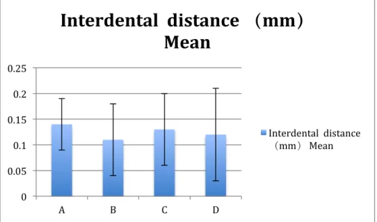
Figure 1. the intermolar distance changes in different groups. The 7 day’s,14 days 21 days and 42 days grourps result seems showed no statistical difference among the group.
0 0.05 0.1 0.15 0.2 0.25
A B C D
Interdental distance (mm)
Mean
Interdental distance
(mm) Mean
0 0.05 0.1 0.15 0.2 0.25 0.3 0.35 0.4
A B C D
Mean (21D)
Mean (21D)
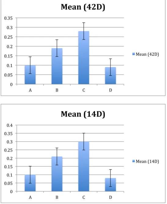
Figure 2. Micro-focus computed tomography data showing bone volume fraction (BVF) and tissue density.
Each value in different graphs represents the mean ± SD.
0 0.05 0.1 0.15 0.2 0.25 0.3 0.35
A B C D
Mean (42D)
Mean (42D)
0 0.05 0.1 0.15 0.2 0.25 0.3 0.35 0.4
A B C D
Mean (14D)
Mean (14D)
0 20 40 60 80 100 120
Mean (7D) Mean (14D) Mean (21D) Mean (42D)
A B C D
由本動物試驗結果可以發現,不同組別於不同時間下,其結果顯示有 LLLT 照射組之牙齒間回復量 較小,組別為藥物處理者,其牙持回復量異教小,此代表牙齒移動後,牙齒發生復位之機會較小,
經常時間觀察下結果亦同。
由 micro CT 針對骨頭密度分析下,不論是在哪個天數之觀察下,各組間骨頭密度變化無統計學上 差異,此結果不論是否使用藥物或是使用LLLT 下,均無太大變化。
四、結語
經由動物試驗下發現,移動之牙齒再有低劑量雷射處理下,其牙齒回復量較小,同時合併藥物使用或 是單獨藥物使用下,則無統計學上差異。但本研究建議,於病人許可下,矯正完成之牙齒位使其不易 再移動,使用LLLT 之處理,可以減少牙齒之復位,或增加矯正後牙齒之穩定性。
全文完
五;參考文獻:
Reference
1. A°rtun J, Garol JD, Little RM. Long-term stability of mandibularincisors following successful treatment of Class II, Division 1,malocclusions. Angle Orthod 1996;66:229-38.4.
2. Little RM. Stability and relapse of dental arch alignment. BrJ Orthod 1990;17:235-41
3. Rothe LE, Bollen AM, Little RM, Herring SW, Chaison JB,Chen CS, et al. Trabecular and cortical bone as risk factors for orthodontic relapse. Am J Orthod Dentofacial Orthop 2006;130:476-84.
4. Spillane LM, McNamara JA Jr. Maxillary adaptation to expansion in the mixed dentition. Semin Orthod 1995;1:176-87.
5. Haas AJ. Long-term posttreatment evaluation of rapid palatal expansion. Angle Orthod 1980;50:189- 217.
6. McNamara JA Jr, Baccetti T, Franchi L, Herberger TA. Rapid maxillary expansion followed by fixed appliances: a long-term evaluation of changes in arch dimensions. Angle Orthod 2003;73: 344-53.
7. Gurel HG, Memili B, Erkan M, Sukurica Y. Long-term effects of rapid maxillary expansion followed by fixed appliances. Angle Orthod 2010;80:5-9.
8. Saito S, Shimizu N. Stimulatory effects of low-power laser irradiationon bone regeneration in midpalatal suture during expansion in the rat. Am J Orthod Dentofacial Orthop 1997;111:525-32.
9. Uysal T, Amasyali M, Enhos S, Sonmez MF, Sagdic D. Effect of ED-71, a new active vitamin D analog, on bone formation in an orthopedically expanded suture in rats. A histomorphometric study.
Eur J Dent 2009;3:165-72.
10. Sawada M, Shimizu N. Stimulation of bone formation in the expanding mid-palatal suture by transforming growth factor-beta 1 in the rat. Eur J Orthod 1996;18:169-79.
11. Fırat Ozt, urk, Hasan Babacan, Sevinc¸ _Inan, Cesur G€um€us¸ dEffects of bisphosphonates on
sutural bone formation and relapse: A histologic and immunohistochemical study Am J Orthod Dentofacial Orthop 2011;140:e31-e41
12. Han G, Chen Y, Hou J, Liu C, Chen C, Zhuang J, et al. Effects of simvastatin on relapse and remodeling of periodontal tissues after tooth movement in rats. Am J Orthod Dentofacial Orthop 2010;
138:550.e1-7.
13. Hassan AH, Al-Hubail A, Al-Fraidi AA. Bone inductive proteins to enhance postorthodontic stability.
Angle Orthod 2010;80:1051-60.
14. Whitters CJ, Hall A, Creanor SL et al. A clinical study of pulsed Nd: YAG laser-induced pulpal analgesia. Journal of Dentistry 1995; 23, 145–50.
15. Renton-Harper P, Midda M NdYAG laser treatment of dentinal hypersensitivity. British Dental Journal 1992; 172, 13–6.
16. H. Miyata, T. Genma, M. Ohshima, Y. Yamaguchi, M. Hayashi, O. Takeichi, B. Ogiso & K. Otsuka.
Mitogen-activated protein kinase/extracellular signal-regulated protein kinase activation of cultured human dental pulp cells by low-power gallium-aluminium-arsenic laser irradiation. International Endodontic Journal, 2006; 39, 238–244.
17. Noriyoshi Shimizu, Kotoe Mayahara, Takeshi Kiyosaki, Akikuni Yamaguchi, Yasuhiro Ozawa, and Yoshimitsu Abiko. Low-Intensity Laser Irradiation Stimulates Bone Nodule Formation Via Insulin- Like Growth Factor-I Expression in Rat Calvarial Cells. Lasers in Surgery and Medicine, 2007;
39:551–559
18. Bolton P, Young S, Dyson M. The direct effect of 860 nm light on cell proliferation and on succinic dehydrogenase activity of human fibroblasts in vitro. Laser Therapy 1995;7: 55–60.
19. Yu HS, Chang KL, Yu CL, Chen JW, Chen GS. Low-energy helium-neon laser irradiation stimulates interleukin-1 alpha and interleukin-8 release from cultured human keratinocytes. J Invest Dermatol 1996;107(4):593–596.
20. Balboni, G. C., Zonefrati, R., Brandi, M. L. & Repice, F. Effects of He-Ne/I.R. laser irradiation on two lines of normal human fibroblasts in vitro. Archivo Italiano Di Anatomia E Di Embriologia 1986; 91, 179–188
21. Abergel, P., Lyons, R. F., Castel, J. C., Dwyer, R. M. & Uitto, J. Biostimulation of wound healing by lasers: experimental approaches in animal models and in fibroblast cultures. Journal of Dermatological Surgery and Oncology 1987;13, 127–133.
22. Bednarska, K., Rozga, B., Kolodziejczyk, K., Szosland, D., Leyko, W. & Bryszewska, M. Effect of low-power red light irradiation on the viability of human skin fibroblasts. Radiation and Environmental Biophysics1998;37, 215–217.
23. Neiburger EJ The effect of low-power lasers on intraoral wound healing. New York State Dental Journal 1995; 61, 40–3.
24. Shimizu N, Yamaguchi M, Goseki T et al. Inhibition of prostaglandin E2 and interleukin 1-b production by lowpower laser irradiation in stretched human periodontal ligament cells. Journal of Dental Research 1995; 74, 1382–8.
25. Whitters CJ, Hall A, Creanor SL et al. A clinical study of pulsed Nd: YAG laser-induced pulpal analgesia. Journal of Dentistry 1995; 23, 145–50.
26. Renton-Harper P, Midda M NdYAG laser treatment of dentinal hypersensitivity. British Dental Journal 1992; 172, 13–6.
27. Kawasaki K, Shimizu K. Effects of Low-Energy Laser Irradiation on Bone Remodeling During Experimental Tooth Movement in Rats Lasers Surg. Med 2000; 26:282–291
28. Kawasaki K, Shimizu K. Effects of Low-Energy Laser Irradiation on Bone Remodeling During Experimental Tooth Movement in Rats Lasers Surg. Med 2000; 26:282–291
29. Matthew D. Dunn a, Chan Ho Park b,c, Paul J. Kostenuik d, Sunil Kapila a,⁎, William V.
Giannobile Local delivery of osteoprotegerin inhibits mechanically mediated bone modeling in orthodontic tooth movement Bone 41 (2007) 446–455
30. Kobayashi Y, Hashimoto F, Miyamoto H, Kanaoka K, Miyazaki- Kawashita Y, Nakashima T, et al.
Force-induced osteoclast apoptosis in vivo is accompanied by elevation in transforming growth factor beta and osteoprotegerin expression. J Bone Miner Res 2000;15:1924-34.
三;科技部補助專題研究計畫成果報告自評表
請就研究內容與原計畫相符程度、達成預期目標情況、研究成果之學術或應用價值(簡要敘 述成果所代表之意義、價值、影響或進一步發展之可能性)、是否適合在學術期刊發表或申 請專利、主要發現(簡要敘述成果是否有嚴重損及公共利益之發現)或其他有關價值等,作 一綜合評估。
1.
請就研究內容與原計畫相符程度、達成預期目標情況作一綜合評估 v 達成目標□ 未達成目標(請說明,以 100 字為限)
□ 實驗失敗
□ 因故實驗中斷
□ 其他原因 說明:
2.
研究成果在學術期刊發表或申請專利等情形:論文:□已發表 □未發表之文稿 □撰寫中 □無 專利:□已獲得 □申請中 □無
技轉:□已技轉 □洽談中 □無 其他:(以 100 字為限)
3.
請依學術成就、技術創新、社會影響等方面,評估研究成果之學術或應用價值(簡要敘述成果所代表之意義、價值、影響或進一步發展之可能性),如已有嚴重損及公共利益之 發現,請簡述可能損及之相關程度(以 500 字為限)
本研究計畫對於矯正後牙齒移動之穩定性問題,經系列性研究,由基礎細胞學之細胞變 化,得知低能量雷側可ˋ以改變細胞之生物活性,進而使細胞進行骨生成反應,於動物 試驗下,觀察骨密度之變化,早期影響不大,但是長時間下,似乎已低劑量雷射處理組 群之骨頭其密度會較無處理的組別好。因此就使用低劑量雷射處理齒朝骨,ㄧ週一次 5J,可以作為使用之參數。成果意義上可以改善臨床矯正醫師煩惱患者矯正後牙齒復位 之問題,價值上,因需改良雷射過大之機款,讓操作者方便使用,才可以於臨床上方達 成效果。
科技部補助專題研究計畫出席國際學術會議心得報告
日期 105 年 06 月 27 日
一、參加會議經過
自朝鮮時代(1392-1910 年)至今約 600 年間,首爾一直是韓國的首都。朝鮮時代的首爾被稱爲“漢陽”,
後韓國歷經日 本佔領期於 1945 年建立大韓民國時,漢陽才改名爲首爾。首爾是韓國的經濟、政治、
文化中心。
世界牙醫學會 2016 年度大會於韓國首爾 COEX 會議中心舉辦,日期為六月 22-25 日。來自全世界許 多國家代表都齊聚於此,今年更特別是,東南亞 IADRSEA 大會同時於首爾這會議上一起舉辦。因此 來參加的人特別多,個人帶者博士班、碩士班學生一起出發,因會議第一天即有學生要報告,因此前 一天即抵達會場周圍,開始會議第一天,我們先辦理報到,因為電子化關係,許多工作已在網路上弄 好,因此只需依照大會給予號碼,即可以完成報到,非常有效率。報到後,我們研究室人員也發表一 篇口頭報告,因此也在那時段趕去聆聽,似乎研究方向上大家大同小異。下午時段最重要就是四點鐘 開幕式,台灣明年要主辦 IADRSEA 大會,因此於中華牙醫學會林理事長帶領下,來自臺灣七院校之 教授學生們浩浩蕩蕩的進入會場,並發文宣與禮物,鼓勵大家明年來台灣參加UADRSEA 年會,會場 上我們拿出國旗,大家一起合照,凸顯台灣的存在。
隔天有學生要報告,一早隨即去將要展示的論文海報貼好,學生們也競業的在複習一下內容,我們展 示時間是下午,因此也抽空去看一下相關自己研究領域之研究論文。 下午海報展示開始,我們研究 的主題系列也受到與會的同好一起來發問與討論,不管是自己或是學生,都感覺收獲許多,研究領域 浩瀚無窮,學生也見識到國際會議之模式,相信在科技部補助下讓學生有此機會出去參與,對台灣後 輩之之思想與觀念一定能起作用。
第三天、出席參加人數較多,實際上是COEX 場地太大,今年台灣也有學生參加論文競賽獲獎,也是 台灣之光。大陸部分就第一天見到許多團隊,北京上海武漢等校人院,但似乎之後就很少見到他們出 席於會場上,應該都是去參訪首爾!我們團隊則一樣,帶著學生去聆聽一些有趣的報告,晚上是韓國 主辦單位招待宴,人數超級多參加,我們也借機會幫牙醫學會推展明年活動,也介紹一些有名學者給 學生認識,韓國人辦事企圖心真不是吹的,台灣加油。
計畫編號 MOST 102-2314-B-040-007-MY3
計畫名稱 低能量雷射對於齒列矯正之牙齒穩定性-生物基礎與應用上系列研究 出國人員
姓名 高嘉澤 服務機構
及職稱
中山醫學大學牙醫學系 會議時間
105 年 06 月 22 日至105 年 06 月 26 日
會議地點
韓國首爾會議名稱 (
中文)94th 國際牙醫學研究學會年度大會(IADR) (英文)94th General Session and Exhibition of the IADR發表題目 ( 中文)張力作用於聚噬細胞經由活化 RANKL 誘導骨生成反應
( 英文)
Tensile force on human macrophage cells enhances osteoclastogenesis through RANKL induction17
第四天,今天又有學生要報告,我們同樣一早去貼海報,但是發現會場上,廠商已撤出,整個廣場顯 得空蕩,學生也嚇一跳,不過這似乎都是這樣情形,因有些人已經陸續離開,不過為了展示我們台灣 精神,老師學生仍然堅持到結束,當然今天來參與討論的就少許多,不過也是讓大家學的一經驗,最 好去做口頭論文報告,這樣會比較有回饋。
見識到韓國舉辦之實力與態度,場地上得到學術上的回饋與進步,這次參加大會的經驗與感想還是有 趣的與豐富的,台灣學子要更加加油,老師也要加油。感謝科技部對於師生研究學術之支持。
二、與會心得
每次韓國舉辦國際會議,都令人看到韓國舉辦之實力與態度,於會場上經由參加與討論,得到不 少學術上的回饋與進步,這次參加大會的經驗與感想還是有趣的與豐富的。謝謝科技部對於研究 學術方面出席國際會議之支持。
三、發表論文全文或摘要
ABSTRACT
Objectives: Little is known about the effects of tensile forces on osteoclastogenesis by human monocyte in the absence of mechanosensitive cells, including osteoblasts and fibroblasts. In this study we consider the effects of tensile force on osteoclastogenesis in human monocytes.
Methods: First, the marcophange cells were treated with RANKL to promote osteoclastgenesis. Then, cathepsin K expression and secretion were examined. The RANKL and the formation of osteoclasts during the osteoclast differentiation process under continual tensile stress were evaluated by western blot.
Results: It was found that ≤ -100 kPa induces RANKL-enhanced TRAP activity in a dose-dependent manner. Furthermore, an increased tensile force raises the expression and secretion of cathepsin K elevated by RANKL, and is concurrent with the increase of TRAF6 induction and NF-kB activation. Overall, the current report demonstrates that tensile force reinforces RANKL-induced osteoclastogenesis by retarding osteoclast differentiation. The tensile force is able to modify every cell through dose-dependent in vitro RANKL-mediated osteoclastogenesis, affecting the fusion of
preosteoclasts, and function of osteoclasts. However, tensile force increased TRAF6 expression.
Conclusions: These results are in vitro findings and were gained under a condition of tensile force, the current results help us to better understand the cellular roles of human macrophage cell populations in osteoclastogenesis as well as in alveolar bone remodeling when there is tensile stress.
四、建議
鼓勵師生與牙醫研究人員多參與此國際性會議,相關單位盡量給予補助。
五、攜回資料名稱及內容
一本program book 和一支大會附贈之USB
六、其他
無
科技部補助計畫衍生研發成果推廣資料表
日期:2016/09/12
科技部補助計畫
計畫名稱: 低能量雷射對於齒列矯正之牙齒穩定性-生物基礎與應用上系列研究 計畫主持人: 高嘉澤
計畫編號: 102-2314-B-040-007-MY3 學門領域: 牙醫學
無研發成果推廣資料
102年度專題研究計畫成果彙整表
計畫主持人:高嘉澤 計畫編號:102-2314-B-040-007-MY3 計畫名稱:低能量雷射對於齒列矯正之牙齒穩定性-生物基礎與應用上系列研究
成果項目 量化 單位
質化
(說明:各成果項目請附佐證資料或細 項說明,如期刊名稱、年份、卷期、起 訖頁數、證號...等)
國 內
學術性論文
期刊論文 0
研討會論文 0 篇
專書 0 本
專書論文 0 章
技術報告 0 篇
其他 0 篇
智慧財產權 及成果
專利權 發明專利 申請中 0
件
已獲得 0
新型/設計專利 0
商標權 0
營業秘密 0
積體電路電路布局權 0
著作權 0
品種權 0
其他 0
技術移轉 件數 0 件
收入 0 千元
國 外
學術性論文
期刊論文 8
篇
1. Laser Physics Letters
2014:11;075602, 2).. Journal of Bone and Mineral Metabolism 2014:32;671-682. 3).2014 Laser Phys. 24 085605. 4).Laser Physics 2014:24;115607 5). . Journal of Dental Science 2015;10:81-87.
6).Laser Physics Letter 2015;12:035601. 7).Materials Science: Materials in Medicine, accept, Sep 26 2015 8).Laser Physics Letter 2016;13:025604
研討會論文 0
專書 0 本
專書論文 0 章
技術報告 0 篇
其他 0 篇
智慧財產權 專利權 發明專利 申請中 0 件
及成果
已獲得 0
新型/設計專利 0
商標權 0
營業秘密 0
積體電路電路布局權 0
著作權 0
品種權 0
其他 0
技術移轉 件數 0 件
收入 0 千元
參 與 計 畫 人 力
本國籍
大專生 0
人次
碩士生 2
博士生 0
博士後研究員 0
專任助理 0
非本國籍
大專生 0
碩士生 0
博士生 0
博士後研究員 0
專任助理 0
其他成果
(無法以量化表達之成果如辦理學術活動
、獲得獎項、重要國際合作、研究成果國 際影響力及其他協助產業技術發展之具體 效益事項等,請以文字敘述填列。)