Mechanical Property Evaluation of IRM
®/Chitosan fiber Composite
Temporary Filling Material
Jia Horng Lin
1,2a, Po Ching Lu
1b, Chao Tsang Lu
3c, Tzu Hsuan Chao
1dand
Ching Wen Lou
4 e *1
Laboratory of Fiber Application and Manufacturing, Department of fiber and composite material, Feng Chia University, Taichung City 407, Taiwan, R.O.C.
2
School of chinese medicin, China Madical University, Taichung, Taiwan, R.O.C. 3
Institute of Life Sciences, Central Taiwan University of Science and Technology, Taichung 406, Taiwan, R.O.C. 4 Institute of Biomedical Engineering and Material Science, Central Taiwan
University of Science and Technology, Taichung 406, Taiwan, R.O.C. a
email: [email protected], bemail: [email protected], c email: [email protected], d
email: [email protected], eemail: [email protected]* *Corresponding author email: [email protected]
Keyword: temporary filling material, chitosan fiber, micro-leakage
Abstract
The temporary filling material seals up the tooth cavity so as to prevent bacterial leakage and the second infection in the root-end treatment. Chitosan is biodegradable, bio-resorbable, non-toxic, and antibiotic. In this study, chitosan fiber is used to enhance the temporary filling material compressive strength and the tooth bite force. IRM® is reinforced by the 2, 4 and 6 wt% chitosan fibers, respectively. And after solidified the IRM®/ Chitosan fiber composite temporary filling material was obtained. Then setting time, solution test, compressive strength and dye micro-leakage of IRM®/ Chitosan fiber composite temporary filling material were be evaluated.
In result, maximum compressive strength of IRM®/ Chitosan fiber composite temporary filling material was obviously increased 200 N than IRM® matrix. As to the micro-leakage test, no trace of leakage was found on the 7th day. Consequently, IRM®/ Chitosan fiber composite temporary filling material can be a good candidate in the short-term dental clinical surgery.
Introduction
Chitosan the partially deacetylated chitin, is a unique polysaccharide [1] The Chitosan molecule is a copolymer of N-acetyl-glucosamine; more than 90 % of it is glucosamine. The polysaccharide, having similar structures to glycosaminoglycans, seems to mimic their functional behaviors. Lysozyme plays an important role in Chitosan degradation in vivo; oligomers are further hydrolyzed into N-acetyl–glu cosamine, a common amino sugar in the body, which can be incorporated for the synthesis of glycoproteins or to be excreted as carbondioxide [2]. Many vivo studies have been done to examine various filling materials, techniques, and hand dexterity. The root-end micro-leakage is frequently used. It involves A. dye leakage method, B. fluid filtration method, C. electrochemical method, D. microorganism penetration method, and E. radioisotope labeling method. The dye leakage, the fluid filtration and microorganism penetration are popular. Having a long history, the dye leakage method is the easiest. None of those methods can replicate the complicated root-end infection mechanism, so it is hard to tell which one is of the best [3-10]. Advanced Materials Research Vols. 123-125 (2010) pp 487-490
© (2010) Trans Tech Publications, Switzerland doi:10.4028/www.scientific.net/AMR.123-125.487
All rights reserved. No part of contents of this paper may be reproduced or transmitted in any form or by any means without the written permission of the publisher: Trans Tech Publications Ltd, Switzerland, www.ttp.net. (ID: 140.134.68.89-25/07/10,09:12:44)
This study aims to examine mechanical properties of the Chitosan/IRM® composite material. The Chitosan/IRM® composite material was proved better than the original dental filling material (IRM®). The Chitosan/IRM® composite material had better mechanical property concerning to its compressive strength setting time, solubility, and micro-leakage.
Experimental
Zinc-Eugenol is composed of zinc-oxide powder and the Eugenol solvent. The powder and the solvent were blended in ratios of 1.5:1, 2:1, and 3:1. After the mixtures were hardened, their compressive strengths were examined. According to the result, the mixture prepared in ratio of 3:1 (zinc-oxide powder: Eugenol solvent) was sticky. This gluey substance hardened too fast to handling, so we used the ratio of 2:1 instead. The used Chitosan fiber was only 4mm±0.5mm long so as to prevent fiber exposures that were potential to micro-leakage. The fiber was later added into the zinc-Eugenol in different ratios. Initially, the zinc-oxide powder and the Eugenol solvent were stirred for one minute, and then the fiber was added within. The Chitosan fiber/zinc-oxide/Eugenol mixture was poured into a mold whose diameter is 4 mm long and thickness 6 mm. The hardened substance was later retrieved to undergo several tests.
Method
Micro-leakage Measurement
The tested material was selected according to the method recommended by the American Dental Association (ADA) #30. The harden specimen was put in the dye solution after being retrieved from the solution and cut into the desired shape.
Compressive strength measurement
The specimen, 6 mm thick with 4 mm diameter, was first put in a distilled water, whose temperate was set at 37 °C±1 °C, for 24 hours in the water base, and then in another distilled water set at 23 °C±1 °C for 15 minutes before the experiment. On the basic of American Dental Association (ADA) #30, this experiment used Instron5566 to measure.
Results and Discussion
In the setting time test, additional Chitosan fiber did successfully reduce the setting time; nevertheless, the improvement was too insufficient to influence the experiment.
Table 1: Setting time of the Chitosan/IRM composite material consisting of 0, 2, 4 and 6wt% Chitosan fibers
Chitosan (wt%) Setting Time (S)
0 370 ± 20 sec
2 365 ± 15 sec
4 350 ± 20 sec
6 345 ± 10sec
Compressive Strength
Fig. 1(a) demonstrates the compressive strengths of the zinc-oxide/Eugenol mixtures arranged in ratios of 1:1.5, 1:2, and 1:3. Chitosan fiber greatly shortened the material’s setting time, leaving it insufficient time for sample preparation. As a result, zinc-oxide powers and Eugenol solvent were mixed at 1:2. Each Chitosan fiber was cut into 4 mm long because the mold’s diameter was 4 mm and 6 mm tall. Longer fibers were inclined to produce bent or exposed fibers while shorter fibers fail to strengthen the composite material. The 4 mm long fiber was chosen accordingly.
Fig. 1(b) displays compressive strengths of the Chitosan/IRM composite material prepared in different ratio of components. We found a significant improvement at the composite material involving the 2 wt% Chitosan, from 150 N to 350 N.
Figure. 1 (a)The compressive strength resutls of each of the IRM®, consisting of powdered zinc oxide and Eugenol solvent mixed in following ratios: 1:1.5, 1:2 and 1:3.The best compressive strength result was reached when the Eugenol solvent and zinc oxide were blended in 3:1. (b) compressive strength results of the
chitosan/IRM® composite material consisting of chitosan fibers in the following ratios: 0, 2, 4 and 6 wt%.
Solubility
Solubility of the Chitosan/IRM® composite material is irregular as it shows in Fig. 2. Chitosan fibers did not affect chemical structure and properties of IRM® thus, solubility of the Chitosan/IRM® composite materal did not increase or decrease. Solubility results went higer on the 1st and the 7th day, but dropped on the 13th day. The solubility test was conducted by weighting the material before and after immersion. On the 13th day, the solubitlity was of the lowest becuse Chitosan fibers absorb water. More absorbed water lead to heavier weight and poorer solubility. As to the water absorption, we can find clear demonstrations on the the microleakage tests shown in figure 5 to 7.
Figure 2 Solubility results of Chitosan/IRM® composite materials observed on the 1st, 7st and
13th days.
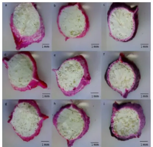
Figure 3 Image of the Chitosan/IRM® (2, 4, 6 wt % Chitosan fibers) composite material 10X in (a, d, g)
1st day (b, e, h) 7st day(c, f, i) 13st days.
Micro-leakage
The testing results of microleakage were shown in Fig. 3. When the 2 %, 4 % and 6 % Chitosan fibers were added in the Chitosan/IRM® composite materal, there were no microleakages
on the 1st and the 7th day show for figure 3 (a, b, d, e, g, h). On the 13th day show for figure 3 (c, f, i), microleakages found in all the composite materials mentioned above. When Chitosan fibers absorbed dye, its original yellow color was turned into dark red. IRM® failed to cover up all the Chitosan fiber; therefore, certain Chitosan fibers were observed on the surface of the Chitosan/IRM® material. Due to capillarity, dye leaked into the Chitosan /IRM® composite material accordingly.
Summary
Table 1 suggested that the Chitosan fiber won’t affect the material preparation procedure.The tested Chitosan/IRM® consisted of Chitosan fibers at ratios of 2 to 6 wt%. Mechanical property of each specimen was studied. Compressive strength results of the Chitosan/IRM® composite material were 363 N, 368 N and 376 N, respectively. Compressive strength of the original IRM® (without Chitosan fibers involved) was 165 N; Chitosan fibers better the composite compressive strength about 200 N. In the solubility test, there was no apparent variation, so additional Chitosan fiber did not affect the IRM’s property. As to the micro-leakage test, there was obvious micro-leakage on the 13th day. This proved that the Chitosan/IRM won’t sustain micro-leakage permanently. Compressive strength of IRM was improved even though micro-leakage was prevented only for a certain period of time.
Acknowledgements
The authors would like to thank the Industry-Academy Cooperation of the Republic of China, Taiwan, for financially supporting this research under Contract 99B-12-065.
Reference
1. Aiba S, Minoura N, Taguchi K, Fujiwara Y.Biomaterials.Vol. 8 (1987), p. 481-488.
2. Nakajima M, Atsumi K, Kifune K, Miura K, Kanamara H, , Japan. J. Surg, Vol.16 (1986), p. 418-424.
3. M.K. Wu, E. G. Kontakiotis and P. R. Wesselink. Journal of Endodontics. Vol. 24 (1998), p. 557–560.
4. F. B. Dagher and G. Yared. Journal of Endodontics. Vol. 21 (1995), p.335–336.
5. K. G. Antonopoulos, T. Attin and E .Hellwig, Journal of Endodontics. Vol. 24 (1998), p. 655–658.
6. G. R. Twell, Journal of Endodontics. Vol. 24 (1998), p.799–801.
7. B.M. Jacquot, M. M. Panighi, P. Steinmetz and C. G'sell, Journal of Endodontics. Vol. 22 (1996), p.586–589.
8. P. M. Michaïlesco, J. Valcarel, A. R. Grieve, B. Levallois and D. Lerner, Journal of Endodontics. Vol. 22 (1996), p.535–539.
9. Y. Haïkel, W. Wittenmeyer, G. Bateman, A. Bentaleb and C. Allemann, Journal of Endodontics. Vol. 25 (1999), p.172–177.
10. L. Pommel. Journal of Endodontics. Vol. 27 (2001), p.347–350.