Characterization of Fe/Mn
−
Superoxide Dismutase from Diatom
Thallassiosira weissflogii
: Cloning, Expression, and Property
C
HUIAN-F
UK
EN,
‡T
UNG-M
INGH
SIUNG,
†Z
ONG-X
IANH
UANG,
†R
ONG-H
UAYJ
UANG,
§ANDC
HI-T
SAIL
IN*
,†Institute of Bioscience and Biotechnology, National Taiwan Ocean University, 2 Pei-Ning Road, Keelung, Taiwan 202, Institute of Biotechnology, National Changhua University of Education,
Changhua, Taiwan 500, and Department of Biochemical Science and Technology, and Institute of Microbiology and Biochemistry, National Taiwan University, Taipei, Taiwan 106
A cDNA clone of 1114 bp encoding a putative Mn-superoxide dismutase (Mn-SOD) from diatom Thallassiosira weissflogii was cloned by the PCR technique. Nucleotide sequence analysis of this cDNA clone revealed that it was translated into 201 amino acid residues. When the sequence was compared with Mn-SODs fromVibrio mimicus and Escherichia coli, as well as two Fe-SODs from E. coli and Photobacterium leiognathi, this SOD showed higher homology to Mn-SOD. The amino acid residues required to coordinate the single manganese ion were conserved in all reported Mn -SOD sequences. This cDNA was introduced in an expression vector, pET-20b(+), and transformed intoE. coli BL21(DE3)pLysS. The expressed SOD protein was then purified by a His-tag column. The recombinant enzyme was heated at 55°C with a time-dependent assay; the time interval for 50% inactivation was 23 min, and its thermal inactivation rate constantKdwas 3.03×10⁻²min-1. The enzyme was inactivated either in acidic pH (below 4.0) or in the presence of imidazole (above 1.6 M) and had only a moderate effect under SDS (above 4%), whereas it was not affected under an alkaline pH (above 9.0). The atomic absorption spectrometric assay showed that 0.6 atom of iron/ manganese (3:1) was present in each subunit of SOD. Reconstitution study was suggested that diatom SOD was cambialistic (Fe/Mn)-SOD. The finding of this SOD cDNA could be used for a reference in comparing the differences among marine phytoplankton species and as a probe to detect the transcription level of this enzyme, which can be applied in cosmetics for skin protection or defending unesthetic effects caused by oxygen-containing free radicals.
KEYWORDS: Diatom; Thallassiosira weissflogii; expression; cambialistic-superoxide dismutase (Fe/Mn-SOD)
INTRODUCTION
Superoxide dismutases (SODs) form the first line of defense
system in various organisms against reactive superoxide radicals
and are vital to the survival of cells (1). SODs can be classified
into four types, Cu/Zn-, Mn-, Fe-, and Ni-SOD, depending
on the metal bound at the active site (2-5). Cu/Zn-SOD occurs
primarily in the cytosol of eukaryotic cells and the chloroplasts
of plants and is also found in the periplasm of gram-negative
bacteria and in the intermembrane space of mitochondria.
Mn-SOD is distributed among prokaryotic and eukaryotic organisms
associated with mitochondria and is insensitive to cyanide and
hydrogen peroxide. Fe-SOD is found in plants as well as in
prokaryotes and is not sensitive to cyanide but is inhibited by
hydrogen peroxide. Ni-SOD was purified from several aerobic
soil bacteria of the Streptomyces species that is distinct from
the Mn-, Fe-, or Cu/Zn-SODs on the basis of amino acid
sequence, immunological cross-reactivity, and spectroscopic
properties (6). Fe-SOD and Mn-SOD appear to be closely
related in structure but have no resemblance to Cu/Zn-SOD.
An anaerobic bacteria synthesized either Fe- or Mn-SOD with
identical protein moiety depending on the metal supplied (7).
Fe-containing Mn-SOD in E. coli grown in tryptic soy yeast
extracts medium suggested the possibility that Fe-substituted
Mn-SOD could have some physiological function (8). Even if
a Mn-SOD had been classified as having highly metal-specific
enzyme activity, it may exhibit at least some activity with iron
at acidic pH. The camphor SOD more resembled Mn-SODs
in primary sequence, but its bound metal (0.5-1 atom of Fe
per camphor SOD subunit) and the ultraviolet and visible spectra
were similar to those of iron-containing enzymes (9). These
represented a type of SODs that could accept either Fe or Mn
as metal cofactors and was named “cambialistic” SOD. In this
study, we report that a SOD cDNA was cloned from diatom
* Corresponding author. Telephone: 886-2-24622192 ext. 5513. Fax:886-2-24622320. E-mail: [email protected]. †National Taiwan Ocean University.
‡National Changhua University of Education. §National Taiwan University.
10.1021/jf048269f CCC: $30.25 © 2005 American Chemical Society Published on Web 02/09/2005
and the active form of SOD was purified from Escherichia coli.
Taking this primary sequence to compare with two Mn-SODs
from Vibrio mimicus and E. coli as well as that of two
Fe-SODs from E. coli and Photobacterium leiognathi (10-12), this
SOD is more similar to Mn-SOD in sequence, but SOD
purified from E. coli grown in LB medium containing iron
indicated that there was 0.6 atom per SOD subunit; the ratio of
Fe to Mn was 3-fold. Thus, this SOD could belong to be
cambialistic-SOD and was first reported from unicellular
eukaryotes.
Irradiation by visible light in the presence of a photosensitizer
leads to the production of reactive oxygen species (ROS), which
in plants and algae is linked to photosynthesis. Because of the
elevated oxygen concentration and intense electron flux within
chloroplasts, electrons inevitably react with oxygen, thereby
generating O
2‚
-, which dismutates to oxygen and hydrogen
peroxide, producing the highly reactive HO‚ through the
metal-ion-catalyzed Haber-Weiss reaction. Even under nonstress
conditions, this ROS-generating mechanism can harm and
inactivate the photosystem II reaction center, resulting in
photoinhibition. Thus, tolerance of photosynthetic organisms to
oxidative challenge is enhanced by defense responses that
prevent oxidative damage to chloroplasts. Because O
2-is a
precursor of several other reactive species, control over the
steady-state O
2-levels by SOD is critical.
In contrast to plants, the antioxidant response to oxidative
and environmental stress has not been investigated in diatoms
at the molecular level. These are a diverse group of unicellular
eukaryotes containing bioluminescent, photosynthetic,
hetero-tropic, and symbiotic members having important ecological roles
as primary producers and consumers in aquatic environments.
Diatoms are responsible for red tides, with those that are toxic,
having the potential for producing serious health and economic
problems. Diatoms have unique genomic features, including
large amounts of DNA packed in permanently condensed
chromosomes and an absence of classical histones, which make
their mechanisms of genetic regulation of great interest.
Although the regulation of Fe-SOD expression was studied
in the dinoflagellate (13), there is a paucity of data for marine
diatom Thallassiosira weissflogii. A diatom was chosen because
of its importance to the marine phytoplankton community.
Further reason was that the sequence and properties of SOD
was not reported in marine diatom.
The cDNA could be used as a reference for comparison of
differences among the marine phytoplankton species as well as
a probe to detect the transcription level of this enzyme. This
enzyme can be used for several beneficial applications in
cosmetics for the protection of the skin or unesthetic effects
caused by oxygen-containing free radicals (14-16).
MATERIALS AND METHODS
Culture Conditions. Cells of the diatom T. weissflogii were cultured
at 25°C on a 12:12-h light-dark cycle with cool white fluorescent light at an irradiance of 150µE‚m-2s-1. Typically, 115 mL medium (100 mL seawater containing 75µg NaNO3,50µg NaH2PO4‚H2O,300 µg Na2SiO3‚9 H2O, 980 ng CuSO4‚5 H2O, 220 ng ZnSO4‚7 H2O, 100 ng CoCl2‚6 H2O, 18µg MnCl2‚4 H2O, 630 ng Na2MoO4‚2 H2O, 315 µg FeCl3‚6 H2O, 436µg Na2EDTA, 200 ng biotin, 560 ng vitamin B12, 10µg thiamine‚HCl) was inoculated with 15 mL of dense culture and used after 1 week of growth.
Total RNA Preparation and Single-Strand cDNA Synthesis. Fresh
diatom (2.0 g) was put into liquid nitrogen and ground to powder in a ceramic mortar. The sample was dissolved in 20 mL of TRIzol reagent (GIBCO, Frederick, MD), incubated for 5 min at room temperature, and then 3 mL of chloroform was added. The mixture was shaken
vigorously for 15 s, incubated at room temperature for 2-3 min, then centrifuged at 12 000g for 15 min at 4°C. The aqueous phase was transferred to a new tube, 7.5 mL of isopropyl alcohol was added, and the tube was incubated at 4°C for 10 min and then centrifuged at 12 000g for 10 min at 4°C. The total RNA (264µg) was obtained. Then, we pipetted 3µg total RNA for synthesizing single-strand cDNA using a kit (GeneRacer) from Invitrogen (Grand Island, NY).
Subcloning and cDNA Sequence Analysis. In accordance with the
amino acid sequences of SOD from Nostoc sp. PCC 7120 (EMBL accession no. AF173990) and Nostoc commune (EMBL accession no. AF177945), two primers (5′TTC CAC CAC GAT AAG CAC CAC3′ and 5′GAG GTA GTA AGC GTG TTC CCA3′) were synthesized. Using 0.1 µg of the single-strand cDNA as a template, 10 pmol of each of the two primers was added. One 0.4-kb DNA fragment was amplified by polymerase chain reaction (PCR) techniques (25 cycles of 94°C for 30 s, 50°C for 30 s, 72°C for 30 s). The 0.4-kb DNA fragment was subcloned into pCR2.1 cloning vector (Invitrogen, Grand Island, NY) using TOPO10 as a host. The nucleotide sequence of the insert was determined in both directions according to autosequencing (ABI PRISM 377-96 DNA sequencer, Perkin-Elmer, CA). On the basis of this DNA sequence, a DiFe-14 primer (5′ATA ACC GCC ACC ATT ATT ACG3′) and a DiFe-18 primer (5′GAG GTT TGC TCA AGT GCG AAC CAG3′) were synthesized. To a 0.5-mL microtube containing 0.1µg of the single-strand cDNA as template was added 10 pmol GeneRacer 5′ primer and 10 pmol DiFe-14 primer. Ten picomoles GeneRacer 3′ primer and 10 pmol DiFe-18 primer were added to another 0.5 mL microtube containing 0.1µg of the single-strand cDNA. One 0.5-kb DNA (5′-RACE; 5′-DNA end) and one 0.5-kb DNA (3′-RACE; 3′-DNA end) were amplified by the PCR technique (25 cycles of 94°C for 30 s, 50°C for 30 s, 72°C for 30 s). Both 0.5-kb DNA fragments were subcloned into pCR2.1 cloning vector using TOPO10 as a host. The nucleotide sequences of these inserts were determined in both directions according to the dideoxy technique using autosequencing. Sequence analysis revealed that 5′-RACE and 3′-RACE covered an open reading frame of SOD cDNA (1.1 kb, EMBL accession no. AF478456).
Recombinant DNA Preparation for Transformation. According
to the 1.1- kb cDNA sequence, a 5′upstream primer (5′GGATCCG ATG GCT TTT GAA TTA CCA GAT C 3′) and a 3′downstream primer (5′CTCGAG TTT TCC AGC GGC ATA TCT TCT TG 3′) were synthesized. Using 0.1µg of the single-strand cDNA as a template, 10 pmol of each 5′upstream and 3′ downstream primer was added and a 0.6-kb fragment was amplified by PCR (25 cycles of 94°C for 30 s, 50°C for 30 s, 72°C for 30 s), ligated with pCR2.1, and then transformed into an E. coli TOPO10 host. A positive clone was selected by hybridization with32P-labeled SOD cDNA as the probe, and the plasmid DNA was prepared. A suitable amount of the plasmid DNA was digested with Bam HI and Xho I and then run on a 0.8% agarose gel. A 0.6- kb insert DNA containing Bam HI and Xho I sites was recovered and subcloned into pET-20(+) expression vector (pretreated with Bam HI and Xho I) from Novagen (Madison, WI). The recom-binant DNA was then transformed into E. coli BL21(DE)pLysS. The recombinant DNA was expressed in E. coli BL21(DE)pLysS and its protein was identified by activity staining.
Culture and Enzyme Purification. The transformed E. coli was
grown at 32°C in 250 mL of Luria Bertani medium containing 50 µg/mL ampicillin and 12.5 µg/mL chloramphenicol until A600reached 0.9. Isopropyl β-D-Thiogalactopyranoside (IPTG) was added to a concentration of 1 mM, and 25µL of 0.1 M Fe2+was added. The culture was incubated at 32°C for 7 h at 150 rpm, and then the bacterial cells were harvested by centrifugation at 6000g for 5 min. Cells were suspended in 2 mL of PBS buffer containing 1% glycerol and 1 g glass beads, and then they were vortexed for 5 min and centrifuged at 10 000g for 5 min. The extraction procedure was repeated two times, and the supernatants were pooled together. The final crude enzyme (6 mL) was loaded on a Ni-nitrilotriacetic acid Sepharose superflow (Qiagen) column (with a bed volume of 2 mL), and then the column was washed with 12 mL PBS buffer containing 5 mM imidazole. The enzyme was eluted with 6 mL PBS buffer containing 100 mM imidazole (at a flow rate of 0.4 mL/min and 1.5 mL/fraction at room temperate). The purified enzyme (3 mL) that was dialyzed against 200 mL PBS buffer containing
1% glycerol at 4°C for 4 h twice was used for analysis or stored at -20°C for further analysis.
Protein of the Purified Enzyme Concentration Measurement.
Protein concentration was determined by a Bio-Rad Protein Assay Kit (Richmond, CA) using bovine serum albumin as a reference standard.
Enzyme Assay in Solution. The SOD activity was measured by
using a RANSOD kit (RANDOX, Ardmore, U.K.). One milliliter of the assay solution contained 40 mM CAPS at pH 10.2, 0.94 mM EDTA, 0.05 mM xanthine, 0.025 mM INT [2-(4-iodophenyl)-3-(4-nitrophenol)-5- phenyltetrazolium chloride], and 0.01 unit of xanthine oxidase. The amount of SOD added was adjusted to obtain a rate of INT reduction at 25 °C over the first 3-min time interval, was measured as the absorbance at 505 nm, and fell within the percentage of inhibition that could be transformed into units of SOD by referring to a standard curve according to the instruction manual.
Enzyme Assay by Activity Staining on a Native Gel. Samples of
the enzyme were electrophoresed on a 15% native gel for 2.5 h at 100 V. The slab acrylamide gel was then cut into parts: one part was assayed, as previously described (17), for SOD activity staining. The gel was soaked in 0.6 mg/mL nitro blue tetrazolium (NBT) solution for 15 min in the dark with gentle shaking, followed by an immersion with illumination in a solution containing 0.45% tetramethylenediamine and 10µg/mL riboflavin. During illumination, the gel became uniformly blue except at positions containing SOD, which showed acromatic zones revealing insolubility of the blue reduction product of NBT by superoxide anion. The other part was stained with Coomassie blue. The area and intensity of activity and protein bands were measured by a computing densitometer (Molecular Dynamics, CA).
Enzyme Characterization. Each enzyme sample underwent the
following tests. After treatments as shown below, each sample was divided into two parts, and then each part of the sample was electrophoresed onto a 15% native gel to determine the changes of activity and protein levels.
(1) Thermal Stability. Enzyme samples were heated to 55°C for 2, 4, 8, or 16 min.
(2) pH Stability. Enzyme sample was amended with half a volume of buffer in different pH values: 0.2 M citrate buffer (pH 2.2, 3.0, 4.0, or 5.0), 0.2 M Tris-HCl buffer (pH 7.0, 8.0, or 9.0), or 0.2 M glycine-NaOH buffer (pH 10.0, 11.0, or 12.0). Each sample was incubated at 37°C for 1 h.
(3) SDS Effect. SDS was added to the enzyme sample to the levels of 1, 2, or 4% and incubated at 37°C for 1 h. Both SDS and imidazole are protein denaturing reagents.
(4) Imidazole Effect. Imidazole was added to the enzyme sample to the levels of 0.2, 0.4, 0.8, or 1.6 M and incubated at 37°C for 1 h.
(5) Proteolytic Susceptibility. The enzyme was incubated with1/20 its weight of trypsin or chymotrypsin at pH 8.0 and 37°C for up to 1, 2, or 3 h. In the chymotrypsin digestion, CaCl2 was added until it reached 20 mM. Aliquots were removed from time to time and analyzed.
Determination of Fe and Mn in Diatom SOD. Composition of Fe
and Mn in the purified SOD was measured by a Hitachi Z-8200 graphite furnace atomic absorption spectrometer (GFAAS). Calibration standards were prepared by diluting 1000 mg L-1stock solution (J. T. Baker) with PBS buffer.
Metal Replacement and Reconsituted Activity. The diatom SOD
was dialyzed for 24 h with 50 mM acetate buffer (pH 5.5) containing 1 mM o-phenanthroline and 10 mM sodium ascorbate and subsequently was reconstituted by dialysis with 50 mM acetate buffer containing 1 mM ferrous ammonium sulfate, 0.5 mM ferrous ammonium sulfate/ 0.5 mM manganese chloride, or 1 mM manganese chloride followed by extensive dialysis with 50 mM potassium phosphate buffer (pH 7.8) (7, 18).
RESULTS AND DISCUSSION
Cloning and Characterization of a cDNA Coding for
Mn-SOD. Figure 1 shows the nucleotide sequence, and the deduced
amino acid sequence, of the diatom Mn-SOD clone. Sequence
analysis found that the DNA comprised a complete open reading
frame coding for 201 amino acid residues.
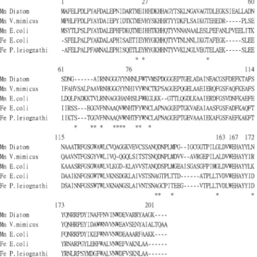
Figure 2 shows that four residues (H-27, H-76, D-163, and
H-167) which are required to coordinate the metal are conserved
in all reported Fe/Mn-SODs (V. mimicus, E. coli, P. leiognathi).
Residues H-27 through Y-35 which contained four histidines,
as well as the alpha helix (residues P-158 through Y-173 which
contain H-167, a patch of four aromatic residues), are conserved
and presumably form an ion binding region. Stallings et al. (19)
Figure 1. Nucleotide sequence of a Mn−SOD cDNA and the deduced amino acid sequence. Numbers to the left refer to nucleotide and amino acid residues. The asterisk denotes the stop signal.
Figure 2. Optimal alignment of diatom Mn−SOD cDNA and other organisms. Mn Diatom (this study); Mn V. mimcus,V. mimicus Mn−SOD DNA (accession no. AAL26843); Mn E. coli, E. coli Mn−SOD DNA (accession no. P00448); Fe E. coli,E. coli Fe−SOD DNA (accession no. P00448); Fe P. leiognathi,P. leiognathi Fe−SOD DNA. A dash denotes deletion. Residues designated with a star pinpoint as potential discrimina-tors between the iron and manganese SOD.
suggested that the observed helical conformation is required to
juxtapose the three residues (H-27, H-31, and Y-35), and the
arrangement is crucial for catalysis on the basis of the structure
of Mn-SOD from Thermus thermophilus at 2.4 Å resolution.
The primary structure of this SOD in comparison with two
known Mn-SODs and two Fe-SODs showed the major
differences of residues 71(Gly), 72(Gly), 79(Phe), 144(Gln), and
145(Asp). It can be concluded that this diatom SOD belongs to
Mn-SOD in sequence.
Transformation and Expression of Diatom Mn-SOD. One
goal of this study was to clone and express the diatom T.
weissflogii Mn-SOD coding sequence in E. coli. Using cDNA
as the template and two specific primers corresponding to the
translation initiation and termination sequences, the 0.6-kb DNA
fragment coding for the diatom T. weissflogii Mn-SOD was
amplified by PCR and successfully subcloned into the expression
vector, pET-20b(+). Positive clones were verified by DNA
sequence analysis. The transformants were incubated in LB
containing 10
µM iron and induced with IPTG, and their total
cellular proteins were analyzed by a 15% native PAGE with
activity staining or protein staining.
Purification of Diatom SOD. The diatom SOD was fused
in the pET-20b(+)-6His-tag vector and expressed in E. coli
BL21(DE3)pLysS. The enzyme containing His-tag in the
C-terminus was purified by affinity chromatography with nickel
chelating Sepharose (Qiagen) according to the instruction
manual. The yield was 0.25 mg from 0.25 L of culture. The
specific activity was 2780 units/mg. The purified enzyme
showed active enzymatic form (Figure 3, lane 4) on a 15%
native PAGE.
Metal Composition of Diatom SOD and Reconsituted
Activity. GFAAS assay indicates that 0.6 atom of iron/
manganese (3:1) is present per subunit of SOD. Dialysis of the
SOD with o-phenanthroline and sodium ascorbate for 24 h and
subsequent dialysis with ferrous ammonium sulfate, ferrous
ammonium sulfate/manganese chloride (1:1), or manganese
chloride reconstituted activity (recoveries of 37, 43, or 64%,
respectively), suggesting that diatom SOD was cambialistic (Fe/
Mn)-SOD.
Characterization of the Purified Diatom SOD. The enzyme
inactivation kinetics at 55
°
C fit the first-order inactivation rate
equation ln(Et/Eo) ) -k
dt, where Eo and Et represent the
original activity and the residual activity that remained after
heating for time t, respectively. The thermal inactivation rate
constant (k
d) values calculated for the enzyme at 55
°
C was
3.03
× 10
-2min
-1, and the half-life for inactivation was 23
min (Figure 4A-C).
As shown in Figure 5 (lanes 4-10), SOD was stable in a
broad pH range from pH 5 to 12.
The enzyme activity showed decrease in either SDS or
imidazole (data not shown). The enzyme was resistant to
digestion by trypsin (data not shown) and chymotrypsin even
at a high enzyme/substrate (w/w) ratio of 1/20 (Figure 6 A-B).
CONCLUSION
In summary, neither the Mn-SOD cDNA sequence of diatom
nor the properties of recombinant Fe/Mn(3:1)-SOD were
reported; the results from the experiments illustrate that the
cloned cDNA from this microbe not only overexpressed Fe/
Mn-SOD in prokaryotes but also remained stable under a broad
range of pH, higher temperature, and proteases. These properties
are beneficial for applications described in the Introduction.
Figure 3. Purification of diatom Fe/Mn−SOD. Six milliliters of crude extract was obtained from 250 mL culture.Τen microliters of each sample was performed on a 15% native PAGE followed by (A) activity stain, (B) Coomassie blue stain. Lanes 1∼4: 1, crude extract; 2, pass-through; 3, wash; 4, fraction containing Fe/Mn−SOD. An arrow denotes purified diatom SOD.
Figure 4. Effect of temperature on the purified Fe/Mn−SOD. The enzyme samples heated at 55°C for various times were analyzed by 15% native PAGE. (A) Staining for activity (4 µg/lane), (B) staining for protein (4 µg/lane). Lanes 1∼5 (control, 2, 4, 8, 16 min). (C) Plot of thermal inactivation kinetics. The effect of temperature was determined by activity stain. The PAGE data were quantitated by a densitometer for calculation. E0andEtare original activity and residual activity after being heated for different times. The areas of activity measured by a densitometer were 324± 23 (control), 315±28 (2 min), 285± 39 (4 min), 238±26 (8 min), and 206±11 (16 min). An arrow denotes both activity and protein.
Diatom Fe/Mn
−
Superoxide Dismutase
J. Agric. Food Chem., Vol. 53, No. 5, 2005
1473
NOTE ADDED AFTER ASAP PUBLICATION
The original posting of February 9, 2005, contained an
incorrect version of Figure 4. The correct version is shown in
the posting as of February 14, 2005.
LITERATURE CITED
(1) Halliwell, B.; Gutteridge, J. M. Role of free radicals and catalytic metal ions in human disease: an overview. Methods Enzymol.
1990, 186, 1-85.
(2) Brock, C. J.; Walker, J. E. Superoxide dismutase from Bacillus stearothermophilus. Complete amino acid sequence of a man-ganese enzyme. Biochemistry 1980, 19, 2873-2882.
(3) Fridovich, I. Superoxide anion radical (O2-.), superoxide dis-mutases, and related matters. J. Biol. Chem. 1997, 272, 18515-18517.
(4) Harris, J. I.; Auffret, A. D.; Northrop, F. D.; Walker, J. E. Structural comparisons of superoxide dismutases. Eur. J. Bio-chem. 1980, 106, 297-303.
(5) Youn, H. D.; Kim, E. J.; Roe, J. H.; Hah, Y. C.; Kang, S. O. A novel nickel-containing superoxide dismutase from Streptomyces spp. Biochem. J. 1996, 318, 889-896.
(6) Youn, H. D.; Youn, H.; Lee, J. W.; Yim, Y. I.; Lee, J. K.; Hah, Y. C.; Kang, S. O. Unique isozymes of superoxide dismutase in Streptomyces griseus. Arch. Biochem. Biophys. 1996, 334, 341-348.
(7) Meier, B.; Barra, D.; Bossa, F.; Calabrese, L.; Rotilio, G. Synthesis of either Fe- or Mn-superoxide dismutase with an apparently identical protein moiety by an anaerobic bacterium dependent on the metal supplied. J. Biol. Chem. 1982, 257, 13977-13980.
(8) Beyer, W. F., Jr.; Fridovich, I. In vivo competition between iron and manganese for occupancy of the active site region of the manganese-superoxide dismutase of E. coli. J. Biol. Chem. 1991, 266, 303-308.
(9) Chen, H. Y.; Hu, R. G.; Wang, B. Z.; Chen, W. F.; Liu, W. Y.; Schroder, W.; Frank, P.; Ulbrich, N. Structural studies of an eukaryotic combialistic superoxide dismutase purified from the mature seeds of camphor tree. Arch. Biochem. Biophs. 2002, 404, 218-226.
(10) Isobe, T.; Fang, Y. H.; Muno, D.; Okuyama, T.; Ohmori, D.; Yamakura, F. Difference between amino acid residues in the metal-ligand environments of iron- and manganese-superoxide dismutases. Biochem. Int. 1988, 16, 495-501.
(11) Parker, M. W.; Blake, C. C. F. Iron- and manganese-containing superoxide dismutase can be distinguished by analysis of their primary structures. FEBS Lett. 1988, 229, 495-501.
(12) Yamakura, F.; Kobayashi, K.; Ue, H.; Konno, M. The pH-dependent changes of the enzymatic activity and spectroscopic properties of iron-substituted manganese superoxide dismutase. Eur. J. Biochem. 1995, 227, 700-706.
(13) Okamoto, O. K.; Robertson, D. L.; Fagan, T. F.; Hastings, J. W.; Colepiclo, P. Different regulation mechanisms modulate the expression of a dinoflagellate iron-superoxide dismutase. J. Biol. Chem. 2001, 276, 19989-19993.
(14) Wilder, M. S.; Mass, A. Composition for preventing or alleviating skin irritation by formulations containing superoxide dismutase. U.S. Patent 4,957,740, 1990; pp 1-12.
(15) N’Guyen, Q. L.; Colin, C. Cosmetic composition containing a superoxide dismutase and a porphyrin. U.S. Patent 5,650,137, 1997; pp 1-6.
(16) Colin, C.; N’Guyen, Q. L. Cosmetic composition containing, in combination, a superoxide-dismutase and a melanin pigment. U.S. Patent 5,925,363, 1999; pp 1-9.
(17) Beauchamp, C.; Fridovich, I. Improved assays and an assay applicable to acrylamide gel. Anal. Biochem. 1971, 44, 276-287.
(18) Martin, M. E.; Byers, B. R.; Olson, M. O.; Salin, M. L.; Arceneaux, J. E.; Tolbert, C. A Streptococcus mutans superoxide dismutase that is active with either manganese or iron as a cofactor. J. Biol. Chem. 1986, 261, 9361-9367.
(19) Stallings, W. C.; Pattridge, K. A.; Strong, R. K.; Ludwig, M. L. The structure of manganese superoxide dismutase from Thermus thermophilus HB8 at 2.4-Å resolution. J. Biol. Chem. 1985, 260, 16424-16432.
Received for review October 19, 2004. Revised manuscript received December 12, 2004. Accepted January 6, 2005. This work was partially supported by the National Science Council of the Republic of China under grant NSC 92-2313-B-019-037 to C-T.L. and supported by the Council of Agriculture, Executive Yuan under grant 92AS-4.2.3-FD-Z4 to C-T.L.
JF048269F
Figure 5. Effect of pH on enzyme stability. The enzyme samples were incubated in buffers with different pH values at 37°C for 1 h and then analyzed by 15% native PAGE followed by activity staining (A panel, 4µg/lane) and Coomassie blue staining (B panel, 4µg/lane). Lanes 1∼10 (pH 2.3, 3.0, 4.0, 5.0, 7.0, 8.0, 9.0, 10.0, 11.0, or 12.0). The total areas of activity measured by a densitometer were 16±3 (pH 2.3), 32±14 (pH 3.0), 40±7 (pH 4.0), 194±25 (pH 5), 236±25 (pH 7), 253±33 (pH 8), 245±31 (pH 9), 250±28 (pH 10), 251±17 (pH 11), and 238±32 (pH 12). An arrow denotes both activity and protein.
Figure 6. Effect of chymotrypsin (A and B). The enzyme samples were incubated with chymotrypsin (1/20SOD) at 37°C for different times and then subjected to 15% native PAGE. (A) Staining for activity (4µg/lane). (B) Staining for protein (4µg/lane). Lanes 1∼4 (control, 1, 2, or 3 h). The enzyme activities after treatment with chymotrypsin measured by a densitometer were 312±16 (control), 257±21 (1 h), 250±21 (2 h), and 160±29 (3 h). An arrow denotes both activity and protein.