行政院國家科學委員會專題研究計畫成果報告
假影免除面回訊影像在功能性影像的應用及 3T 磁場下的扭曲修正 Ghost-free echo planar imaging in fMRI and dynamic distortion
correction at 3T
計畫編號:NSC 96-2314-B-011-001 執行期限:96 年 1 月 1 日至 96 年 7 月 31 日
主持人:林益如 台灣科技大學 電子工程系 一、中文摘要
在這個研究中我們發展一種修正面回訊影像的假影的方式。我們利用雙倍取樣面 回訊影像,k 空間中的每一行都會連續取樣兩次,但兩次的取樣方向是相反的,
因此兩次取樣可分別重組出沒有N/2 假影的影像,而且兩張影像的讀取時間有數 毫秒的間隔,因此可以利用這兩張影像的相角差,來推得區域磁場的不均勻程 度,並藉由此資訊,來修正影像的幾何扭曲。使用這種方法,可以在不增加掃描 時間的狀況下,得到沒有N/2 假影且減低幾何扭曲的影像,並且訊雜比不會比一 般面回訊影像差。在功能性磁振造影的實驗中,我們證實運用這項技術可提昇功 能性磁振造影的敏感性,也有助於其他利用面回訊影像的應用如擴散性影像或微 灌流影像等。
關鍵詞:磁振造影 面回訊影像 假影免除 功能性磁振影像 扭曲修正
Abstract
In this project we proposed a method called double sample EPI. In this sequence, positive and negative readout gradient is applied with the same phase encoding which forms the same line in the k-space. Therefore even and odd echoes can form the image independently which is automatic ghost removal. Moreover these two images separated by several milli-seconds can be perfectly used to calculate field map which can be use to correct geometry distortion. Using this method, we can acquire N/2 ghost free and improved geometry distortion images without increasing scan time or sacrificing SNR. The specificity and sensitivity of functional activation is improved for fMRI using this technique. The proposed method is also expected to help many applications involving EPI technique such as diffusion imaging and perfusion imaging.
Keywords: MRI, fMRI, EPI, artifact, Nyguist ghost, distortion correction
二、緣由與目的
Nyquist ghosts in EPI are known to interfere with image and lower the SNR, which in turn decrease fMRI sensitivity(1,2). A double-sampled EPI (DSEPI) method has been proposed to remove ghost artifact (3), trading off stronger geometry distortion due to longer readout time. In this work, DSEPI is used to improve the SNR in visual fMRI experiment, with field map information inherent in DSEPI used to correct for geometry distortion.
As opposed to conventional EPI, DSEPI sequence uses phase encoding blip on
RF G(slice)
G(phase) G(freq)
t t t t
Signal t
RF G(slice)
G(phase)
G(freq)
t t t t
Signal t
(a)
(c)
(b)
(d)
Fig 1. Sequence diagram and corresponding K-space sampling trajectory of conventional EPI and double-sampled EPI. (a) Cartesian sampling waveform where phase encoding is turned on as blips at each readout gradient switch. (b) The corresponding K-space trajectory. (c)
Cartesian sampling waveform where phase encoding is turned on as blips at every two readout gradient switch. (d) K-space trajectory of DS-EPI
Kx Ky
every other echo as shown in Fig 1(c, d). The even and odd echoes were divided into two sets of k-space data and reconstructed separately. For field map calculation, the two central k-space lines each traversing in opposite direction were used as reference lines to correct misalignment between odd and even echoes (4), following which phase images were polynomial-fitted to remove noise influences. With TE differing by one echo spacing, the field map can be calculated from two echoes. The short TE separation also helps minimizing phase wrap (phase aliasing) problem in field map estimation. The calculated field maps were then used to remove geometric distortion (5-8).
Five subjects participated in this project. Visual fMRI experiments were performed with conventional EPI and DSEPI sequence on 1.5T system (Siemens Vision+). Scan parameters of fMRI included TR 2s, TE 48m, echo spacing 0.6ms, matrix size 64x64, FOV 23x23cm, slice thickness 3mm. A block design of 5 blocks (2 on, 3 off, 10 frames in each block) was applied. Flashing checkerboard visual stimulus was given in second and fourth blocks. In DSEPI experiment the two echoes were averaged before functional analysis. Functional activation maps were generated by independent component analysis (ICA) supplied in fMRlab software package . 三、結果與討論
The conventional EPI, DSEPI, and corresponding T1-weighted images were shown in Figure 2. Nyquist ghosts were totally removed using DSEPI acquisition. The contour from T1WI showed that geometric distortion was stronger in DSEPI images comparing to conventional EPI images, but the distortion was successfully corrected by field map correction using the inherent information in DSEPI.
Fig 2. (a) T1-weighted, (b) conventional EPI (c) DSEPI, (d) corrected DSEPI images of one subject. Nyquist ghost can be easily seen in conventional EPI images (arrow head).
Geometrical distortion existed in both conventional EPI and DSEPI images (arrow), which was corrected using field map correction (d).
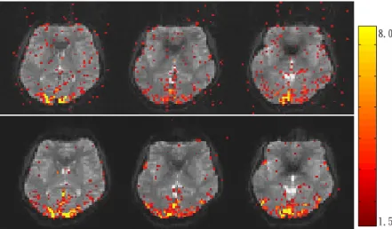
The activation maps and percentage signal changes for the fMRI experiments are shown in Figs.3 and 4, respectively. DSEPI showed stronger percentage signal changes and larger activation areas than conventional EPI. The SNR was 78 for conventional EPI and 112 for DSEPI, showing SNR improvement slightly larger than square root of two. The additional SNR gain is anticipated to be from the suppression of the Nyquist ghosts.
In this study, DSEPI is demonstrated to provide better fMRI quality than
conventional EPI by at least three factors: SNR improvement, ghost suppression, and reduced geometric distortion. The better SNR comes from the retention of signal Fig 4. Time course for the percentage signal changes from the primary visual cortex of the same subject in Fig.3: conventional EPI (red) and DSEPI (blue). Better functional sensitivity is seen with DSEPI
Conventional EPI DSEPI
____
____
Fig 3. Activation maps of 3 selected slices of one subject: (a) conventional EPI and (b)
DSEPI. Larger activation areas are seen with DSEPI. Scale represents Z value from 1.5 to 8.0
energy from Nyquist ghosts, as well as simultaneous acquisition of two images at similar TE. Since a high field strength desirable for fMRI study also enhances Nyquist ghosts due to stronger susceptibility, we expect the advantage of DSEPI to be more prominent in higher field systems. Stronger geometric distortion in DSEPI can be reduced using field map correction, the information inherently stored in the two acquisitions in DSEPI. This property is especially important in fMRI where simultaneously monitoring of the field map in dynamic scan is advantageous to correct for distortion resulting from dynamic field fluctuations. Furthermore, this method is compatible with parallel imaging as long as the central line of k-space is acquired.
四、結論
We conclude that DSEPI provides a ghost-free image with comparable SNR with conventional EPI, and the field map estimated from 2 echo images were capable to reduce the geometric distortion. Furthermore, DSEPI with inherent field map correction capability is an effective approach for fMRI studies.
The result of this study has been published in the meeting of international society of magnetic resonance in medicine, Seattle, U.S.A.
五、參考文獻
1. Kwong KK, Belliveau JW, Chesler DA, Goldberg IE, Weisskoff RM, Poncelet BP, Kennedy DN, Hoppel BE, Cohen MS, Turner R, et al. Dynamic magnetic resonance imaging of human brain activity during primary sensory stimulation.
Proc Natl Acad Sci U S A 1992;89(12):5675-5679.
2. Schmitt F, Stehling MK, Turner R. Echo-Planar imaging. Verlag Berlin Heidelberg: Springer; 1998.
3. Yang QX, Wang J, Smith MB, Meadowcroft M, Sun X, Eslinger PJ, Golay X.
Reduction of magnetic field inhomogeneity artifacts in echo planar imaging with SENSE and GESEPI at high field. Magn Reson Med
2004;52(6):1418-1423.
4. Roopchansingh V, Cox RW, Jesmanowicz A, Ward BD, Hyde JS. Single-shot magnetic field mapping embedded in echo-planar time-course imaging. Magn Reson Med 2003;50(4):839-843.
5. Chen NK, Wyrwicz AM. Optimized distortion correction technique for echo planar imaging. Magn Reson Med 2001;45(3):525-528.
6. Reber PJ, Wong EC, Buxton RB, Frank LR. Correction of off
resonance-related distortion in echo-planar imaging using EPI-based field maps. Magn Reson Med 1998;39(2):328-330.
7. Chen NK, Wyrwicz AM. Correction for EPI distortions using multi-echo gradient-echo imaging. Magn Reson Med 1999;41(6):1206-1213.
8. Jezzard P, Balaban RS. Correction for geometric distortion in echo planar images from B0 field variations. Magn Reson Med 1995;34(1):65-73.