行政院國家科學委員會專題研究計畫 成果報告
以原子力顯微鏡分析材料表面、生物分子與骨細胞間之交 互作用力 II
研究成果報告(精簡版)
計 畫 類 別 : 個別型
計 畫 編 號 : NSC 99-2221-E-011-120-
執 行 期 間 : 99 年 08 月 01 日至 100 年 07 月 31 日 執 行 單 位 : 國立臺灣科技大學化學工程系
計 畫 主 持 人 : 何明樺
計畫參與人員: 碩士班研究生-兼任助理人員:黃靜薇 碩士班研究生-兼任助理人員:張君凌 博士班研究生-兼任助理人員:蕭盛文 博士班研究生-兼任助理人員:Thien DV
報 告 附 件 : 出席國際會議研究心得報告及發表論文
處 理 方 式 : 本計畫涉及專利或其他智慧財產權,2 年後可公開查詢
中 華 民 國 100 年 10 月 26 日
以原子力顯微鏡分析材料表面 以原子力顯微鏡分析材料表面 以原子力顯微鏡分析材料表面
以原子力顯微鏡分析材料表面、 、 、 、生物分子與骨細胞間之交互作用力 生物分子與骨細胞間之交互作用力 生物分子與骨細胞間之交互作用力 生物分子與骨細胞間之交互作用力 II 專
專 專
專題 題 題 題研 研 研究 研 究 究計畫 究 計畫 計畫成果報告 計畫 成果報告 成果報告 成果報告 表面起伏結構對骨母細胞的影響
目錄
Introduction p. 1
Materials and Methods p. 2
Results and Discussion p. 3
Conclusion p. 9
References p. 10
1. Introduction
Cells would be inherently sensitive to their surroundings. In the body, the cell was proved to respond to environmental cues from macro-scale down to molecular-scale [1].
The interaction between cell and extra-cellular matrix (ECM), happened on nano-micrometer scale (10-300nm), supporting cells and presenting an instructive background to guide cellular behaviors [1]. However, for the behaviors of osteolastic cells on micro- or nano-patters, the results from previous researches were discrepant and the osteoblastic differentiation was rarely investigated. As to the late mineralization maker, calcium nodules, has not been discussed yet. To investigate the phenomena systematically, we cultured human osteoblasts and dental pulp stem cells, on surfaces with micro patterns continuously varying in their width and depth. The observations of cell behaviors, including the attachment, proliferation, alignment, and mineralization were performed in this research. Moreover, the interaction between osteoblasts and surfaces with different groove width was also measured by using AFM in the liquid phase. The statistical analysis was further carried out to compare the influences from groove width and depth.
2. Materials and Methods
The micro-grooves on silicone wafers were fabricated by photolithographic techniques [2]. Then, silicon wafers were cleaned ultrasonically in acetone solution, then continued by immersing wafers in piranha solution (7/3 (v/v) of 98% H2SO4 / 30% H2O2).
After that, the wafers were copiously rinsed with DI water, PBS, and sterilized under UV light. osteoblasts were cultured on the treated silicone wafer supplied with MEM, incubated at 37oC, and 5% CO2. With various culture times, the cells cultured on silicone wafers were fixed, dehydrated, and observed under SEM or light microscope. For the idenfication of cellular mineralization, the von Kossa staining [3] and ESCA were applied on osteoblasts on silicone wafers after the culture with mineralization -induced medium [3].
AFM experiments were performed with a Nanoscope III and all the data was measured by a J scanner (maximum XY scan range of 125 * 125 µm with vertical Z range of 5 µm).
The experiments were performed in air at a constant temperature of 20°C, and relative humidity below 40%. The tip was made to approach the cell layer at a constant speed of 1 µm/s.
This cantilever was modified by osteoblast with manipulator after gelatin was deposited on the end of cantilever. Thus, the cultured osteoblast was attached to the designed place on AFM cantilever. The modified cantilever was then inserted in the AFM to measure the force-distance curve, where the substrate was the surfaces with different topography prepared by lithography process. By monitoring the deflection of the cantilever, a force-distance curve was obtained and the adhesion force evaluated by Hook’s law, F =k×∆Z, where k is the spring constant and ΔZ is the deflection of the cantilever. The deflection, ΔZ, was defined as the lowest point on the force-distance curve. This definition was consistent with methods in previous research [4]. In Fig. 1, Δ Z is marked by a dashed line.
Fig.1 The schematic figure for the force-distance curve. The cantilever deflection, ∆Z, applied in Hook’s law is marked as the dotted line between points 5 and 6. [5]
3. Results and Discussion
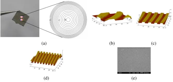
Using photolithographic techniques, micro -grooved patterns were made on silicon wafers. The wafers were patterned with varied groove depths ranged from 0.8 to 1.1 µm, and relative groove widths were ranged from 1 to 15 µm. The groove patterns were described with AFM pictures as Fig. 2.
The influences of groove pattern on cell attachment were quantitatively investigated through the percentage of attached cell on different topographies in this study. After the desired incubation time, samples were gently washed by PBS to remove unattached cells.
The density of attached cells was then determined and was divided by the initial seeding density to achieve the percentage of attached cells. According to the results in Fig. 3, the percentage of attached cell gradually increases with the culture time. In addition, there is a tendency that the cell attachment decreases with the decrease in groove width. The cells prefer to attach onto the smooth surface than the groove pattern, and hence the highest cell attachment was found on smooth surface. These results propose that microgroove pattern would retard the attachment of DPSCs. The inhibition of cell attachment resulting from groove patterns was also observed on human gingival fibroblast (hGF) cultured on micro grooved PDMS [6], and on rat dermal fibroblasts (RDFs) cultured on microgrooved polystyrene [7]. In contrast, Fransiska proposed that there were more osteoblast-like cell on grooved surface than on smooth surface [8]. Microgroove patterns exhibit different effects on these cell types.
The attached cells were all in spherical appearance in the beginning, and then started to spread on the silicone wafer. Throughout the culture for 1 week, all the cells were well proliferated on material surface, and the “contact guidance” phenomena was found, which was shown as Fig. 4. That is, the cells elongated according to the surface pattern, except on the smooth surface. The tendency of cell elongation would be significantly induced by decreasing the groove width, but only slightly decreased by increasing the groove depth.
From the SEM pictures of attached osteoblast, such as Fig.4, the cell elongation, alignment and spreading area on grooved surfaces were analyzed by using ImageJ software. The results are described in Figs. 5-7. The cell elongation was defined to be the ratio of cell major to minor axis, and the cell alignment was defined as the angle between the major axis of the ellipse fitted to the cellular trace and the given direction which was groove direction in this research. On microgrooved surface of PDMS, osteoblasts significantly elongate, revealed in Fig. 5. The ratios of major to minor axis of cells cultured on microgrooved surfaces were larger than 1 during the whole culture time. It indicates the elongation of osteoblasts can be induced by microgroove pattern. On the
contrary, the ratios for cells cultured on smooth PDMS were all close to 1 in 24-hour culture, demonstrating there was almost no elongation of osteoblasts on smooth surfaces.
It is clear that the cell elongation significantly increases when the groove width decreases.
The cells exposed the highest elongation on narrowest groove pattern (1-2 µm). On smooth surface, the cells had the spindle-like shape, just like their native shape, resulting in the cell elongation was just a little bit higher than 1. The above observations generally indicates that microgroove pattern on PDMS can induce the elongation of osteoblasts. In addition, the higher degree of elongation was found on the cells cultured on narrower grooves.
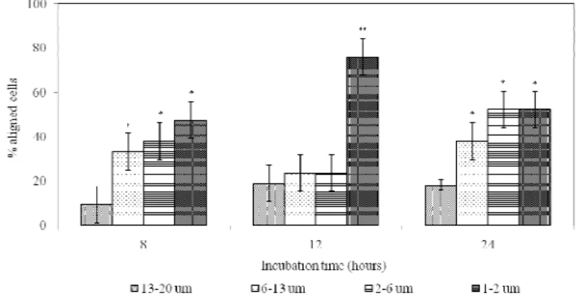
The alignment of osteoblasts was evaluated by calculating the percentage of aligned cells. The cell is considered as an “aligned” cell when the angle between cell major axis relative to the groove direction is smaller than 100. Fig.6 interprets the degree of cell orientation on surfaces with different groove widths. The smooth surface is not mentioned in this analysis because the cells locating on smooth surface expressed random orientation only. From Fig.6, groove patterns of PDMS substrate cause the alignment of osteoblasts and are able to direct the cell orientation. The decrease of groove width results in the enhancement of cell alignment. Thus, when the groove width ranges from 12 µm, the cells achieve the highest alignment. However, the guidance effect of groove pattern of PDMS varied with the culture time and was with the clearest appearance at 8th hour of incubation.
At 12th hour culture, the cell alignment on 2-6 µm grooved surface was similar to those on 6-13 µm and 13-20 µm. That probably resulted from the influence of cell aggregation on those grooved surfaces. With a lot of cell aggregations, the cells would bridge over grooves easily and would not be restricted by topography anymore.
The spreading of osteoblasts on grooved PDMS was measured and presented in Fig.
7. Compared with cells on flat surfaces, the cells on groove patterns appeared in smaller spreading area. Moreover, the cell spreading would obviously decrease with the decrease of groove size. The smallest cells always located on the narrowest groove (1-2 µm). These results proved that the groove pattern of PDMS significantly obstructed the cell spreading, which supports the results on pristine PDMS. It reveals that for osteoblasts, although the increase in PDMS hydrophilicity successfully promote the cell spreading, the hindrance on cell spreading caused by grooved topography would still exist.
To analyze the phenotype of cultured osteoblasts, the mineralization marker, calcium deposition caused by cultured osteoblastic cells, was evaluated by using ESCA. From the values of Ca/C shown in Fig. 8, the decrease in groove width clearly increased the number of calcium nodules on the silicone wafer; that is, the phenotypes of osteoblastic cells would be promoted by the micro-grooves. Besides, the promotion would be stronger with the groove with smaller width.
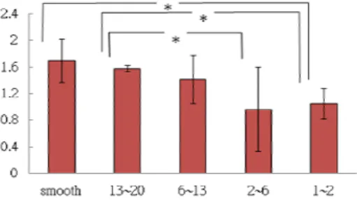
In this research, the cell probe was prepared by using the micro-manipulator, where living osteoblast was immobilized on the end on AFM cantilever. By using the Hook’s law, the interforces between osteoblast and surfaces with different groove width was thus evaluated from the force-distance curve. The results were described in Fig. 9. The results in Fig. 9 proved that the interforce between osteoblast and surface decreased with the decreasing in groove width. This outcome is consistent with the measurements of the spreading area in Fig. 7. The higher interforce between the cell and materials would result in faster cell attachment at the beginning of culture, which was previously hypothesized.
The difference in the attachment rate of cells onto chitosan surfaces could be applied to cell separation; this was proposed in the previous section.
The culture of DPSCs (dental pulp stem cells) was also performed in this research;
however, the detail results were not shown here due to the limitation of length. Briefly speaking, there is clear contact guidance effect for cultured DPSCs on grooved surfaces.
The spreading, attachment and proliferation were hindered by small grooves, which were corresponded to the results from osteoblasts. To further estimate the effect of groove topography on the differentiation of DPSCs, surface marker staining was performed. The samples used in this experiment consisted of cells cultured on plasma-treated PDMS.
After 1 day of culture, the cells were examined for for stem cell markers, CD34 and CD44.
These two markers are commonly used for testing the stem cell property. CD34 is a convenient marker for human hematopoietic stem/progenitor cells/endothelium cells [9].
On the other hand, CD44 is a cell surface marker associated with cancer stem cell populations and treatment resistance in other tumors [10].
Experimental result showed the staining result for CD34 of DPSCs on smooth, wide grooved and narrow grooved surfaces. Obviously, on either smooth or grooved surface, the staining images exhibit only black color and without positive signal of CD34, indicated by green color. That is to say there is no expression of CD34 on these samples.
However, staining images for differentiation marker CD44 in Figure IV.48 show an opposite result. The stained cells express green color of CD44 marker with different intensity on the entire substrate surface. Moreover, the intensity of CD44 significantly decreases with the decrease of groove width. The strongest expression of CD44 could be found on the cell cultured on smooth surface. It reveals that microgroove patterns are able to promote the differentiation of DPSCs and thus result in the decrease of CD44 intensity.
This result also corresponds to ALPase staining results; that is, narrow grooved surface could enhance the early bone differentiation of DPSCs.
These findings are in agreement with the outcomes in previous researches about the properties of DPSCs. In 2000, based on an immunohistochemical analysis of human DPSCs and bone marrow stromal cells (BMSCs) in vitro, Gronthos and colleagues indicated that DPSCs and BMSCs did not react with hematopoietic stem/progenitor cells
marker CD34 [11]. In contrast, two kinds of stem cell expressed strong positive signal of CD44. In a research of Shi and co-workers, it was revealed that not only DPSCs and BMSCs but also stem cells from human exfoliated deciduous teeth (SHED) and periodontal ligament stem cells (PDLSC) exhibited negative signal for CD34 and positive signal for CD44 [12].
(a) (b) (c)
(d) (e)
Fig. 2 The groove patterns on PDMS. (a) The patterns were distributed in co-center circles. The aspect ratio (width/depth) was largest in the center of circle, while it was smallest in the edge of circle.; (b) the AFM analysis for the grooved pattern at point b; (c) the AFM analysis for the grooved pattern at point c; (d) the AFM analysis for the grooved pattern at point d; (e) the SEM picture for the smooth part.
Fig. 3 The percentage of attached osteoblasts on PDMS surfaces with different groove width after the seeding for 10, 16 and 24 hours. n = 9 and t-test indicated : * p <
0.05, ** p < 0.01 compared with smooth surface at the same incubation period.
(a) (b)
Fig. 4 The contact guidance effects on (a) grooved surface and on (b) smooth surface
Fig. 5 The elongation of osteoblasts cultured on PDMS surfaces with different groove width. n = 9 and t-test indicated : * p < 0.05, ** p < 0.01 compared with smooth surface at the same incubation period.
Fig. 6 The alignment of osteoblasts cultured on PDMS surfaces with different groove width. n = 9 and t-test indicated : * p < 0.05, ** p < 0.01 compared with smooth surface at the same incubation period.
Fig. 7 Spreading area of attached osteoblasts on surfaces with different groove width. n = 9 and t-test indicated : * p < 0.05, ** p < 0.01 compared with smooth surface at the same incubation period.
Fig. 8 The values of Ca/C of silicone wafers with cultured and mineralization -induced osteoblasts. n = 9 and t-test indicated : * p < 0.05, ** p < 0.01 compared with smooth surface at the same incubation period.
Fig. 9 Interforces between osteoblast and PDMS surfaces with different groove width. n
= 4 and t-test indicated : * p < 0.05, ** p < 0.01.
4. Conclusion
This study establishes the technique to prepare cell probe for the AFM measurement, so the interforce between living cells and different surfaces can be analyzed quantitatively.
With the cell probe, we measure the interforce between osteoblast and materials with different topography at first. The AFM measurement was carried out in a liquid chamber filled with culture medium at a constant temperature, so the cell viability was maintained through the measurement. The result indicated that the small groove width resulted in a low interforce to osteoblasts. The osteoblasts were also cultured for the analysis of cell attachment, spreading and proliferation. The results showed that the cell growth and adhesion would be both retarded with the decrease in groove width. Comparing the in vitro results with the interforce measured by AFM, we found that the small groove width would hinder the cell attachment and proliferation, which is caused by the decrease in the cell-material interforce.
Except for osteoblasts, DPSCs (dental pulp stem cells) were also applied in this research. In the early stage of cell culture, microgroove patterns result in the lower cell attachment. Despite grooved surface can provide larger specific area, groove patterns seems to be ineffective to support cell attachment. On both hydrophobic and hydrophilic surface, the cell attachment decrease with the decrease of groove width and smooth surface exposes the highest cell attachment.
The effect of groove topography on DPSCs’ differentiation was further investigated through surface marker staining. The cells cultured on plasma-treated PDMS exposed the negative signal for stem cell marker CD34. It reveals that the stem cell property of DPSCs is not significant. However, there was the expression of differentiation marker CD44 for cells on substrate. The intensity of CD44 varied with surface topography.
On smooth surface, the cells exhibited strongest CD44 expression and the marker intensity decreased with the reduction of groove width. The result implies that microgroove pattern may enhance the differentiation of DPSCs, which was also observed in ALPase staining.
References
[1] Molly MS, Julian HG, “Exploring and Engineering the Cell Surface Interface”, Science, 2005; 310: 1135
[2] Su JC, “White light LED with dielectric omni-directional reflectors”,US Patent, US6, 2004; 833: 565 B2
[3] Chang PC, Hou LT, Liu BY, Jehng SM, Ho MH, Wang DM, “Biocompatibility of
newly-made porous poly-(L-lactic acid) scaffold”, Journal of Dental Research, 2002;
81, A210-A210
[4] Behrend O P, Oulevey F, Gourdon D, Dupas E, Kulik A J, Gremaud G and Burnham N A Intermittent contact: tapping or hammering? Appl Phys A 1998; 66: 219-221.
[5] Digital Instruments Veeco Metrology Group 1999 AFM/LFM Instruction Manual p 10-4 chapter 1
[6] Millati N, The Effects of Micro-Grooves on Gingival Fibroblasts and Osteoblast-Like Cells (Master Thesis, National Taiwan University of Science and Technology, 2009) [7] Walboomers XF, Monaghan W, Curtis ASG, Jansen JA, Attachment of fibroblasts on
smooth and microgrooved polystyrene, J Biomed Mater Res, 1999; 46: 212-220 [8] Fransiska S, The Effect of Surface Micro-Patterns on Osteoblastic Cells’ Behaviors
(Master Thesis, National Taiwan University of Science and Technology, 2007)
[9] Lee MW, Jang IK, Yoo KH, Sung KW, Koo HH, Stem and progenitor cells in human umbilical cord blood, Int J Hematol, 2010; 92:45-51
[10] Vaillant BD, Bhat K, Sulman EP, Balasubramaniyan V, Wang S, Aldape KD, Colman H, CD44 as a prognostic and predictive marker for GBM. J Clin Oncol, 2011 (2049):
29
[11] Gronthos S, Brahim J, Li W, Fisher LW, Cherman N, Boyde A, DenBesten P, Robey PG, Shi S, Stem cell properties of human dental pulp stem cells, J Dent Res, 2002;
81(8): 531-535
[12] Shi S, Bartold PM, Miura M, Seo BM, Robey PG, Gronthos S, The efficacy of mesenchymal stem cells to regenerate and repair dental structures, Orthod Craniofacial Res, 2005; 8: 191-199
出席國際學術會議報告 出席國際學術會議報告 出席國際學術會議報告 出席國際學術會議報告
填寫日期:100 年 7 月 24 日
報告人姓名 何明樺 系(所) 化工系 職稱 副教授 電話 (02)27301255
會 議 時 間 自 100 年 6 月 26 日至 100 年 7 月 01 日 地點 新加坡 會 議 名 稱 2011 國際材料先進技術會議
International Conference on Materials for Advanced Technologies 2011 (ICMAT 2011)
發表論文題目:
1. Chien-Hao Huang and Ho MH, “The Preparation of Pt-Catalyst Layer for Fuel Cells by Using the Electrospinning Process”, International Conference on Materials for Advanced Technologies 2011 (ICMAT 2011), Singapore, Jun. 26 – Jul 01, 2011
2. Chang JL, Ho MH, “Quantification of Interactions between Osteoblasts and Surfaces with Different Topography”, International Conference on Materials for Advanced Technologies 2011 (ICMAT 2011), Singapore, Jun. 26 – Jul 01, 2011
3. Tan NH, Ho MH, “Osteoblast-like cell’s behaviors on micro-grooved PDMS”, International Conference on Materials for Advanced Technologies 2011 (ICMAT 2011), Singapore, Jun.
26 – Jul 01, 2011
4. Wang JW, Ho MH, “Preparation and Characterization of Chitosan Substrate with Immobilized Naringin”, International Conference on Materials for Advanced Technologies 2011 (ICMAT 2011), Singapore, Jun. 26 – Jul 01, 2011
一、 參加會議經過
2011 國際材料尖端科技會議 (International Conference on Materials for Advanced
Technologies 2011, ICMAT 2011)於 2011 年六月二十六日至七月一日在新加坡新達城(Suntec city)的國際會議中心舉行。此次會議由新加坡材料研究學會 (Singapore Material Research Society)主辦,新加坡大學(National University of Singapore)、新加坡材料工程研究中心
(Singapore Institute of Material Research and Engineering)與南洋理工大學協辦(Nanyang Technology University),為材料相關研究領域中相當重要的會議。
申請人搭乘中華航空公司班機前往新加坡,於六月二十五日上午約八點四十五分搭乘華 航 CI0661 班機由桃園國際機場起飛,飛抵時間約為下午一點三十,再搭乘巴士前往大會會 場-新達城國際會議中心,註冊並參加傍晚所舉辦的歡迎酒會,總計旅程大約費時六小時。
2007 國際材料尖端科技會議為本年度亞洲地區相當大規模的研討會,固定由新加坡材料 研究學會與新加坡大學所主辦,參加者來自世界各地多達三十餘國,總人數達數千人,參與 國家除亞澳地區外,也有許多來自歐洲與美洲的材料相關領域人士。
此次會議安排了九場的專題演講(Plenary Lecture),分別為
(1) “Manipulating Atoms with Light” C. C. Tannoudji (College de France and Laboratoire Kastler Brossel, France)
(2) “Nanotechnology and Materials Science at the Intersections of Engineering, Biology and Medicine” S. Suresh (Massachusetts Institute of Technology, USA)
(3) “Orthogonal Reactivity” B. Sharpless (The Scripps Research Institute, USA)
(4) “Organic Electronics: Interface, Heterojuctions and Semiconductor Device Engineering” R. H.
Friend (University of Cambridge, UK)
(5) “Cell Suicide: Programmed Cell Death in Development and Disease” H. R. Horvitz (Massachusetts Institute of Technology, USA)
(6) “Drug Discovery in the p53 Pathway” D. Lane (Institute of Molecular and Cell Biology, Singapore)
(7) “Computing at the Nanoscale will Employ Physics and Logic Operations” R. S. Williams (Hewlett-Packard Lab., USA)
(8) “Nano-Carbon Materials: Their Fundamentals and Various Applications Including Nano-Biotechnology” S. IiJima (Maijo University and AIST/Research Center for Advanced Carbon Material, Japan)
(9) “Hybrid Inorganic-Organic Materials and Their Applications” A. K. Cheetham (International Center for Materials Research, USA)
這幾場專題演講涵蓋了生物材料與電子材料等多種不同的領域,演講者皆為各領域之翹 楚,其中第一、三、五場的主講人更為諾貝爾物理、生理與化學獎的得主,由此可一窺主辦 單位強烈的企圖心。
於第一篇專題演講中,C. C. Tannoudji 教授首先介紹利用雷射進行分子級結構的建立。S.
Suresh教授於第二篇專題演講中,介紹了奈米結構材料於生物醫學領域中的目前運用與將來
的可能發展。在第五篇與第六篇演講中,H. R. Horvitz 與 D. Lane 教授則介紹了在細胞分子訊 號路徑上的最新發現,若能與材料相互結合,對於以奈米顆粒進行癌症治療可望有相當的突 破。
論文發表在六月二十六日的開幕式後正式開始,在本次的會議中,包含了 Oral、Student poster paper contest、Student oral paper contest 以及 Poster session 等,由於 Oral 有許多不同的
主題,大會將所有論文發表區分成十八個討論會(symposium),以平行方式進行不同領域論
文的發表,涵蓋的領域有尖端生物材料、奈米生物介面、功能性生物材料的設計、奈米半導 體材料、奈米元件的設計與製備、奈米光學材料的開發、SPM 技術的應用、MEMS 技術與元 件、感測器的發展、電化學材料、觸媒材料等等,Oral session 總計近千篇論文,Poster session 近兩千篇。申請人此次所發表的論文屬於 symposium A: 尖端生物材料,皆獲得不錯的迴響。
除了論文發表之外,本次大會亦安排有分析儀器的展示會,共十餘個攤位展示各式材料相關 的分析儀器,提供研究學者最新的儀器資訊。
與會期間,申請人除了論文的發表,亦與本領域著名的學者晤談,此外,也撥空參觀了 展示會場及海報展示,除了吸收新知外,並與展示者與會場主持人交換了意見。
在會議結束後,申請人受邀前往新加坡大學牙醫學學系進行參訪,除就研究發展交換意 見外,並訂立了研究合作協定,回國後即已著手對材料的動物實驗進行準備,目前已得到初 步的體內生物相容性分析結果。
二、 與會心得
在本次會議中,明顯可見材料在未來應用與發展的兩大主軸:生醫與奈米。以主辦單位 新加坡大學而言,其化工系更已更名為化工與生物科技學系,指出此一趨勢已銳不可擋。就 申請人主要參加的討論會 A(Advanced Functional Biomaterials)與 C (Bio Functional Materials: From Understanding to Design)而言,多數研究集中在藥物釋放、基因治療與組織 工程方面。在藥物釋放與基因工程方面,較受廣泛注意與討論的研究為奈米粒子的應用與靶 向投藥技術的發展,會議中提出了多種奈米粒子的製備方法,並可有效將高分子奈米粒子的 大小降低至數十奈米左右,證實可將所攜帶之藥物成功穿過血腦屏障,用於基因轉殖或基因 治療,其基因表現能力明顯提高,而在載體表免施以特殊抗體的固定化,可賦予釋藥粒子相 當之專一性,增加了藥物的治療效果也降低了可能的副作用。在組織工程方面,較受注意的 研究包括以下兩者,一是利用中空纖維管狀膜製備生物反應器,可大幅縮短組織培養所需要 的時間,另一則是利用薄膜製備對細胞代謝廢物與分泌物的感測器,可以更精準地控制細胞 發育的情形。
三、 建議
申請人十分感謝能獲得國科會支持出席國際會議,但申請人畢竟分身乏術,無法兼顧所 有講題,若能以團隊形式參加此一會議,可以集中研究實力,並可提昇國家的知名度與研究 聲譽,相信將可由此一會議獲得更豐碩的成果。
四、 攜回資料名稱及內容
本次會議後攜回 ICMAT 2011 會議論文集一本和光碟片一片,亦由參展的廠商中取得相關 分析儀器的資訊,這些皆將提供給國內從事材料研究人員或業者做為參考。
國科會補助計畫衍生研發成果推廣資料表
日期:2011/10/26
國科會補助計畫
計畫名稱: 以原子力顯微鏡分析材料表面、生物分子與骨細胞間之交互作用力 II 計畫主持人: 何明樺
計畫編號: 99-2221-E-011-120- 學門領域: 生化及生醫工程
無研發成果推廣資料
99 年度專題研究計畫研究成果彙整表
計畫主持人:何明樺 計畫編號:99-2221-E-011-120-
計畫名稱:以原子力顯微鏡分析材料表面、生物分子與骨細胞間之交互作用力 II 量化
成果項目 實際已達成
數(被接受 或已發表)
預期總達成 數(含實際已
達成數)
本計畫實 際貢獻百
分比
單位
備 註 ( 質 化 說 明:如 數 個 計 畫 共 同 成 果、成 果 列 為 該 期 刊 之 封 面 故 事 ...
等)
期刊論文 0 0 100%
研究報告/技術報告 0 0 100%
研討會論文 1 1 100%
篇 發 表 於
APCChE2010( 台 灣)
論文著作
專書 0 0 100%
申請中件數 0 0 100%
專利 已獲得件數 0 0 100% 件
件數 0 0 100% 件
技術移轉
權利金 0 0 100% 千元
碩士生 2 2 100%
博士生 2 2 100%
博士後研究員 0 0 100%
國內
參與計畫人力
(本國籍)
專任助理 0 0 100%
人次
期刊論文 1 1 100% 目前已投稿
研究報告/技術報告 0 0 100%
研討會論文 2 2 100%
篇 發 表 於
ICMAT2011( 新 加 坡)與 AMS6(澳洲) 兩 場 國 際 研 討 會 中
論文著作
專書 0 0 100% 章/本
申請中件數 0 0 100%
專利 已獲得件數 0 0 100% 件
件數 0 0 100% 件
技術移轉
權利金 0 0 100% 千元
碩士生 0 0 100%
博士生 0 0 100%
博士後研究員 0 0 100%
國外
參與計畫人力
(外國籍)
專任助理 0 0 100%
人次
其他成果
(
無法以量化表達之成果如辦理學術活動、獲 得獎項、重要國際合 作、研究成果國際影響 力及其他協助產業技 術發展之具體效益事 項等,請以文字敘述填 列。)
無
成果項目 量化 名稱或內容性質簡述
測驗工具(含質性與量性) 0
課程/模組 0
電腦及網路系統或工具 0
教材 0
舉辦之活動/競賽 0
研討會/工作坊 0
電子報、網站 0
科 教 處 計 畫 加 填 項
目 計畫成果推廣之參與(閱聽)人數 0
國科會補助專題研究計畫成果報告自評表
請就研究內容與原計畫相符程度、達成預期目標情況、研究成果之學術或應用價 值(簡要敘述成果所代表之意義、價值、影響或進一步發展之可能性) 、是否適 合在學術期刊發表或申請專利、主要發現或其他有關價值等,作一綜合評估。
1. 請就研究內容與原計畫相符程度、達成預期目標情況作一綜合評估
■達成目標
□未達成目標(請說明,以 100 字為限)
□實驗失敗
□因故實驗中斷
□其他原因 說明:
2. 研究成果在學術期刊發表或申請專利等情形:
論文:□已發表 □未發表之文稿 ■撰寫中 □無 專利:□已獲得 □申請中 ■無
技轉:□已技轉 □洽談中 ■無 其他:(以 100 字為限)
3. 請依學術成就、技術創新、社會影響等方面,評估研究成果之學術或應用價 值(簡要敘述成果所代表之意義、價值、影響或進一步發展之可能性)(以 500 字為限)
本研究已依據先前之規劃發展了 AFM 的液相量測系統, 並成功建立製備活細胞探針之技 術, 目前, 我們已完成對於骨母細胞/骨癌細胞與不同起伏結構表面間之作用力測量, 並 已與細胞培養結果進行比對分析, 在牙髓幹細胞方面, 我們目前與國防醫學院合作, 已 成功建立對幹細胞分化之分析技術, 目前正在不同起伏結構的表面進行牙髓幹細胞的培 養, 而其作用力的分析將在新一年度的計畫中完成
此一技術預計將能藉由材料的使用與材料性質的控制,調節幹細胞的增生與分化,配合我 們先前所開發的生物因子釋放系統與材料製備改質技術,以提高組織工程的效率