探討電漿處理對細胞間質蛋白質, 血清蛋白在材料界面的 表面以及其與生物分子之交互作用
研究成果報告(精簡版)
計 畫 類 別 : 個別型
計 畫 編 號 : NSC 95-2218-E-011-019-
執 行 期 間 : 95 年 10 月 01 日至 96 年 07 月 31 日 執 行 單 位 : 國立臺灣科技大學化學工程系
計 畫 主 持 人 : 王孟菊
計畫參與人員: 碩士班研究生-兼任助理:張慶全、廖証億
處 理 方 式 : 本計畫涉及專利或其他智慧財產權,2 年後可公開查詢
中 華 民 國 96 年 10 月 26 日
行政院國家科學委員會專題研究計畫成果報告
探討電漿處理對細胞間質蛋白質,血清蛋白在材料界面的表面以 及其與生物分子之交互作用
Investigations of the effects of plasma treatment to the incorporation of extracellular-matrix proteins and serum proteins and the interactions between
biomolecules and materials 計畫編號: NSC 95-2218-E-011-019
執行期限: 2006 年]10 月 01 日至 2007 年 07 月 31 日
主持人: 王孟菊 國立台灣科技大學化學工程系
計畫參與人員:張慶全(研究助理)、廖証億(研究助理) 國立台灣科技大學化學工程系
1. 中文摘要
本計畫目的在探討電漿對材料表面處 理,以及血清蛋白在高分子材料界面的表 現。為了驗證由蛋白質所引發細胞和高分 子材料的交互作用,我們選用可應用於臨 床醫學的矽膠材料,研究血清蛋白於材料 界面的吸附。由接觸角測量可以計算材料 的表面能,由此可以判斷材料親疏水性的 改變。掃描式電子顯微鏡和原子能顯微鏡 用以紀錄材料和蛋白質產生作用後,表面 形態之變化。蛋白質的附著性質,用以驗 證材料的生物相容性,以及電漿和蛋白質 對表面的改質的效果。
關鍵詞:界面現象、電漿處理、血清蛋白、
表面分析、表面形態、掃描式電 子顯微鏡、原子力顯微鏡、粗糙 度。
Abstract
Polymer is extensively used in medical research and for clinical devices due to its natural clarity, its chemical resistance and its good mechanical properties. However the
applications are limited for the surface hydrophobicity. Plasma treatment has been recognized as the most effective method to functionalize surface of materials without altering the bulk property and chemical residues. This proposed project focus on investigating the effects of plasma treatments, the interactions between proteins and materials on the interfaces.
In order to demonstrate the specificity of protein mediated interaction between cells and silicone membranes, commercially available silicone membranes (PDMS) were selected. To verify hydrophobicity, contact angles were measured on uncoated and protein coated silicone surface. The topography and roughness were obtained by SEM and AFM analyses. Finally, the protein adhesion on the different membranes was analyzed. This project was investigating and comparing the effects of surface modifications by physico-chemical and biological methods.
Keywords: Interface phenomenon, plasma treatment, surface analyses, surface topography, roughness, SEM, AFM.
research and in broad number of medical devices. Thin PDMS membranes have been used to support monolayer cell adhesion and to transmit mechanical force in the form of bioaxial stain. This ability of PDMS membrane enhances the materials clinical usefulness and allows one to study cell-surface interactions in detail. However, a disadvantage of these procedures is that the material often has hydrophobic surface properties which results in poor cell adhesion.
Therefore, it is necessary to investigate the possibility of modifying a hydrophilic film on silicon for further study of cell-biomaterials interactions.
Previous reported surface modification methods for PDMS include (1) to promote crosslinking by gelatin-glutaralhehyde (2) to integrate functional groups by plasma treatments, and (3) to increase cellular adhesion to polymers. The increment of crosslinking produces about 100 microns thin film which is too thick to preserve any micro-nanostructures that have been designed into the surface. On the other hand, plasma treatment resulted in a high quality thin film..
Cellular adhesion to polymers has been enhanced by modifying the surface of substrates with attached extracellular-matrix proteins which provide cell attachment sites, and act as reservoirs for soluble signaling molecules.
In this research paper, the plasma treatments were employed to enhance the adhesion of biomolecules on PDMS membranes. In order to examine the effects of activation and functionalization of plasmas, PDMS membranes were characterized by XPS, water contact angle, SEM and AFM for the membranes treated by O2, and CF4
plasmas respectively. FITC-BSA was immobilized membranes either untreated or treated by plasmas in order to examine the interactions between membranes and biomolecules.
The PDMS samples for plasma treatments were prepared as following: the Polydimethylsiloxane (Sylgard 184 ® elastomer , Dow Corning –Midland,MI,USA) solution and its curing agent were mixed as 10:1(v/v) ratio. The mixed solution was degassed in vacuum chamber to ensure well mixing and to remove air bubbles. The mixed solution was then spin coated on 11 cm2 silicon wafer disc and the samples were then heated at 150 oC for 15min for curing.
FTIC-BSA (Fluorescein isothiocyanate conjugate - Bovine Serum Albumin) (Sigma) was used directly without further purification.
All solution were made with deionized water (18Mcm)y. The purity of gases used for plasma treatments (O2 and CF4) are higher than 99.9%.
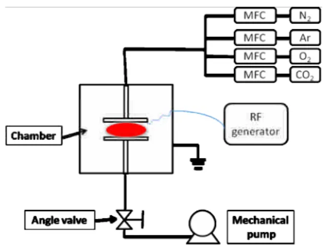
3.2 Plasma equipment
Figure 1. shows schematically the plasma configurations used in the present study. The plasma is composed by three main parts: (1) reaction chamber; (2) radio-frequency generator; and (3) vacuum system. The samples were directly mounted on a round stainless steel electrode powered by a 13.56MHz rardio-frequency power supply. Process gases (O2, CF4) were introduced into the reaction chamber by a mass flow system. The gas flow was adjusted between 10-50sccm depending on the conditions required for the plasma treatments.
The total pressure was controlled from 50~100mTorr.
Figure 1. Plasma equipment
3.3 AFM
The morphology of untreated and plasma treated PDMS was analyzed by a Digital Instruments Nanoscope III Atomic Force Microscopy (Veeco Ltd.). The contact mode was employed for the scanning.
3.4 XPS
X-ray photoelectron spectroscopy (XPS) spectra were measured ex situ on a Thermo VG Scientific Theta Probe Instrument, using Al (1486.6eV) and Mg (1253.6eV) as excitation sources. Data processing including background subtraction, integration, and deconvolution was carried out using Peak-fitsoftware.
4. Experiment results and discussion 4.1 Surface Hydrophilicity
The oxygen plasma alters the surface of PDMS to hydrophilic very effectively. For 2 minutes treatment by 10W or higher power (150W), the contact angle of PDMS varies from 83.5to 18.75(Table 1). The BSA immobilization did not alter the contact angle for the oxygen plasma treated PDMS.
On the other hand, the surface of PDMS membrane shows increment of hydrophobicity by lower power applied of CF4 plasma treatment. According to the reaction mechanisms proposed for the CF4 plasmas in the previous studies [ref], the degradation effect might play more important role at higher applied power, and this could lead to the decrease of contact angle.
The total surface energy can be calculated according to the concept of acid-base interactions developed by RJ Good
(1) which defined the total surface energy can be contributed by LW and AB (2, 3). LW represents the Lifshitz-van der Waals interaction, i.e. the London dispersion force and A represents the acid-base interactions due to the hydrogen binding and other types of forces from chemical bindings,
respectively. By applying three different liquids, deionized water, diiodomethane and glycerol, we can obtain the total surface energy according to the following equations:
Fig.2 Total surface energy vs. plasma treatment time.
Table.1 Water contact angle measurement for plasma treated PDMS
O2plasma
Water
Contact angle CF4plasma
Water Contact angle PDMS
Control
83.52.1 PDMS
Control
83.52.1
10W 18.83.3 10W 102.07.1
10W+BSA 17.02.5 10W+BSA 66.83.3
150W 30.85.3 150W 66.83.3
150W+BSA 455.0 150W+BSA 50.32.5
The untreated PDMS shows the total surface energy as 17.6(mJ/m2). The CF4
plasmas can increase the surface energy in 10 seconds to 24.6 (mJ/m2) and 21.3(mJ/m2) by applying the power of 10W and 150W, respectively. The total surface energy reaches the plateau value after 30 seconds of CF4 plasma treatment and achieves about 20.0 (mJ/m2) for the power of 10W and 17.0 (mJ/m2) for the power of 150W. This indicates the CF4plasma modification can be accomplished within 30 seconds and the applied power did not influence the total surface.
0 50 100 150 200 250 300
0 5 10 15 20 25 30 35 40 45 50
TotalSurfaceenergy(mJ/m2)
Time(s)
CF410W CF4150W O210W O2150w
S S LW
S AB S LW S tot S
L S L
S LW
L LW S tot
L
γ γ γ
γ γ γ
γ γ γ
γ γ
γ θ
γ
2
2 2
2 cos 1
for 10 seconds of treatment by the power of 10W. The total surface can increase up to 50 (mJ/m2) for 300 seconds of treatment. For higher voltage, 150W, of oxygen plasma, the total surface energy increases up to 45.4 (mJ/m2) within 10 seconds of treatment and reaches a plateau of 42.0 (mJ/m2) after 30 seconds. These results show that the surfaces of PDMS stay hydrophobic by CF4 plasma for different applied power. The oxygen plasma, on the other hand, brought the hydrophilicity onto the surfaces therefore the total surface energy increase markedly.
4.2 Surface composition
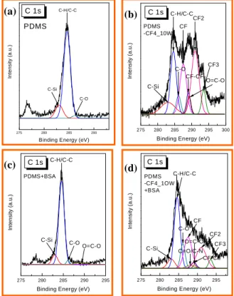
The surface chemical composition analyses of plasma treated and untreated PDMS membranes were carried out using XPS. The wide scan spectrum demonstrated the total compositions on the surfaces; on the other hand, the decomposition of the spectra of C1s peak provides the information of the chemical bindings.
For the PDMS treated by CF4plasma, Fig.4 shows the wide scan and the integration of fluorine can be clearly seen to increase from 0 to 47.5% and 47.6% by 5 minutes of 10W and 150W CF4treatments, respectively.
This indicates that the different applied power (10W and 150W) have same effect on fluorination. Both oxygen content and Si content both increase by 10W and 150W applied power. This could be due to that the higher energy can promote the dissociation of oxygen and Si atoms from the substrates.
Fig.3 XPS wide scan spectra of PDMS membranes by CF4plasma.
The FITC-BSA was immobilized onto
the amino acid compositions of BSA proteins and this can be increased from 0 to 4.6% for both 10W and 150W power of CF4plasma.
To investigate the incorporation of FITC-BSA, from wide scan analyses, the untreated PDMS shows no content of N elements. However, for the 10W and 150W CF4 plasma treated membranes, the increase of nitrogen elements can be observed from 0% to 4.61% and 4.66% respectively. This can indicate the adhesion of FITC-BSA on the membranes.
Fig. 4. Peak fitted of XPS C 1s narrow scan spectra of PDMS membranes. (a) PDMS control, (b) PDMS + CF
410W, (C) PDMS + BSA, (d) PDMS + CF
410W+BSA。
4.3 Surface morphology
The surface morphology and roughness can be analyzed by atomic force microscopy (AFM). The roughness for the untreated PDMS is 0.29nm and this could be increased by 10W CF
4plasma treatment (0.532 nm) and more drastically (2.24 nm) by higher power (150W) CF
4plasma treatment (Table3).
Si 2p N 1s
F 1s
CF4_150W+BSA O 1s
CF4_150W
CF4_10W+BSA
CF4_10W
PDMS+BSA
PDMS Wide
Intensiry(a.u.)
Binding Energy (eV) C 1s
Si 2s
275 280 285 290
Intensity(a.u.)
Binding Energy (eV) C-Si
C-H/C-C
C-O
C 1s PDMS
275 280 285 290 295 300 C-O
C-H/C-C C 1s
Intensity(a.u.)
Binding Energy (eV) PDMS
-CF4_10W
C-Si CF
CF-CF O=C-O CF2
CF3
275 280 285 290 295
PDMS+BSA C 1s
Intensity(a.u.)
Binding Energy (eV) C-Si
C-H/C-C
C-OO=C-O
275 280 285 290 295
O=C-O CF3 C-H/C-C
C 1s
Intensity(a.u.)
Binding Energy (eV) C-Si
C-O CF
C=O/C-N CF-CF
CF2 PDMS
-CF4_1OW +BSA
(a) (b)
(c) (d)
Fig.5. AFM analyses for plasma treated surface morphology.
(a) PDMS control; (b) PDMS + O
210W;
(c) PDMS + O
2150W; (d) PDMS + CF
410W; (e) PDMS + CF
4150W。
Table.2 Roughness for plasma treated PDMS sample Ra(nm) PDMS-CONTROL 0.29 PDMS-10W-O
20.179 PDMS-150W-O
20.371 PDMS-10W-CF
40.532 PDMS-150W-CF
42.24
4.4 Protein AdhesionThe results obtained by XPS, contact angle analyses indicated that oxygen plasma treatment could increase hydrophilic property of PDMS membrane surface. However, CF4
plasma treatment plays a different role. The amount of attached protein BSA on PDMS
membrane also increased on the CF4 plasma treated membrane, Figure 6.
(a) (b)
Fig.8 Fluorescent Microscope images of (a)
untreated and (b) plasma treated PDMS membranes that FITC-BSA immobilized.
Conclusion
In summary, the main results of this study are that we use CF4 and O2 plasma to enhance the adhesion of protein BSA on PDMS membranes. The experimental results showed that the amount of BSA immobilized on native PDMS surface is limited, and BSA preferred to adhere on hydrophobic surfaces.
From AFM images, PDMS surface morphology (roughness) just slightly changed after plasma treatments. Contact angle measurement had showed that CF4 plasma treatment resulted in hydrophobic surface of PDMS, and O2 plasma resulted in hydrophilic surface. After BSA immobilized on PDMS membranes, the surfaces become more hydrophilic. XPS analyze revealed that the amount of BSA immobilized on surface increased by CF4 plasma treatment. We think plasma modification will receive wide application in the future.
五、參考文獻
1. Hsiue, G.-H., Lee, S.-D., Chang, P.C.-T. and Kao, C.-Y., Journal of Biomedical Materials Research, 42, 134-147 (1998).
2. Blau A, Weinl C, Mack J, Kienle S, Jung G, Ziegler C., J Neurosci Methods. 112(1), 65-73.
(2001)
3. Sherratt, M., Bax, D., Chaudhry, S.S., Hodson, N., Lu, J.R., Saravanapavan, P. and Kielty, C.M., Biomaterials, 26, 7192-7206 (2005).
4. H. Hillborg, J. F. Ankner, U. W. Gedde, G. D.
Smith, H. K. Yasuda and K. Wikström Polymer, Volume 41, Issue 18, August 2000, Pages 6851-6863
5. H. Hillborg, M. Sandelin and U. W. Gedde;
Polymer, Volume 42, Issue 17, August 2001, Pages 7349-7362
(a)
(b)
(c)
(d)
(e)
7. H.B. Lim, Donghoon Kim, Taeyoon Jung, Limb, M. C. Kwan and K. K. Gleason; Thin Solid Films, Volume 395, Issues 1-2, 3 September 2001, Pages 288-291
8. K. K. S. Lau, H. G. Pryce Lewis, S. J. Klee, D., Ademovic, Z., Bosserhoff, A., Hoecker, H., Maziolis, G. and Erli, H.-J., Biomaterials (2003).
9. Miyamoto, S., Katz, B.-Z., Lafrenie, R.M. and Yamada, K.M., Fibronectin and Integrins in Cell Adhesion, Signaling, and Morphogenesis. In:
K.M. Yamada (ed.), Fibronectin and Integrins, pp. 119-129, Annals New York Academy of Sciences, New York (1999).
10. Mosher, D.F., Fibronectin. Progress in Hemostatis and Thrombosis, 5, 111-51 (1980).
11. Ricci, J.L., Alexander, H. and Howard, C., Materials Research Society Symposium Proceedings, 252, 221-227 (1992).
12. Altankov, G., Grinnell, F. and Groth, T., Journal of Biomedical Materials Research, 30, 385-391 (1996).
13. Banes, A.J., Apparatus for Applying Stress to Cell Cultures, US Patent Office, 4,839,280, US (1989).
14. Banes, A.J., Biocompatible polyorganosiloxane composition for cell culture apparatus, US Patent Office, 4,822,741, US (1989).
15. Barnes, D., Wolfe, R., Serrero, G., McClure, D.
and Sato, G., Journal of Supramolecular Structure, 14, 47-63 (1980).
16. Behnisch, J.; Hollander, A.; Zimmermann, H. J.
Appl. Polym. Sci., 49, 117, (1993).
17. Blau A, Weinl C, Mack J, Kienle S, Jung G, Ziegler C., J Neurosci Methods. 112(1), 65-73.
(2001)
18. Braut-Boucher, F., Pichon, J., Rat, P., Adolphe, M., Aubery, M. and Font, J., Journal of Immunological Methods, 178, 41-51 (1995).
19. Burrill, P.H., Bernardini, I., Kleinman, H.K.
and Kretchmer, N., Journal of Supramolecular Structure, 16 (1981).
20. Carter, W.G., Rauvala, H. and Hakomori, S.-i., The Journal of Biological Chemistry, 88, 138-148 (1981).
21. d'Agostino, R. Plasma deposition, treatment;
and etching of polymers; Academic Press: San Diego, CA, (1990).
22. D'Agostino, R.; Cicala, G.; Creatore, M.; Favia, P.; Lammendola, R. Proc. 12th ISPC, 355 (1999).
23. Degasne, I., Basle, M.F., Demais, V., Hure, G., Lesourd, M., Grolleau, B., Mercier, L. and Chappard, D., Calcified Tissue International, 64, 499-507 (1999).
24. den Braber, E.T., de Ruijter, J.E., Ginsel, L.A., von Recum, A.F. and Jansen, J.A., Journal of Biomedical Materials Research, 40, 291-298 (1998).
26. Dupont-Gillian, Ch; Adriaensen, Y.; Derclaye, P.; Rouxhet, G.; Langmuir, 16, 8194.
27. Esty, A., Receptor-specific serum-free cell attachment using a highly stable engineered protein polymer., 44 (1991).
28. Favia, P.; Stendardo, M. V.; d'Agostino, R.
Plasmas Polym. 1996, 1, 91.
29. Ferreira, N. G.; Corat, E. J.; Trava-Airoldi, V.
J.; Leite, N. F. Diamond Relat. Mater, 9, 368 (2000).
30. Fodil-Bourahla, I., Drubaix, I. and Robert, L., Mechanisms of Ageing and Development, 106, 241-260 (1999).
31. France, R. M.; Short, R. D. Langmuir, 14, 4827 (1998).
32. Franz, D.; Hollenstein, M.; Hollenstein, C. Thin Solid Films, 379, 37(2000).
33. Garscadden, A.; Nagpal, R. Plasma Sources Sci.
Technol., 4, 268 (1995).
34. Gatmaitan, Z., Jefferson, D.M., Ruiz-Opazo, N., Biempica, L., Arias, I., M., Dudas, G., Leinwand, L.A. and Reid, L.M., The Journal of Cell Biology, 97, 1179-1190 (1983).
35. Gerenser, L. J. J. Adhesion Sci. Techol., 7, 1019 (1993).
36. Hayward, I.P., Bridle, K.R., Campbell, G.R., Underwood, P.A. and Campbell, J.H., Cell Biology International, 19, 839-846 (1995).
37. Hersel U, Dahmen C, Kessler H. Biomaterials.
24(24):4385-415. (2003)
38. Hirohata, Y.; Tsuchiya, N.; Hino, T. Appl. Surf.
Sci., 612, 169-170 (2001).
39. Horbett, T.A. and Schway, M.B., Journal of Biomedical Materials Research, 22, 763-793 (1988).
40. Hsiue, G.-H., Lee, S.-D., Chang, P.C.-T. and Kao, C.-Y., Journal of Biomedical Materials Research, 42, 134-147 (1998).
41. Idage, S. B.; Badrinarayanan, S. Langmuir, 14, 2780 (1998).
42. Isnard, N., Fodil, I., Robert, L. and Renard, G., Experimental Gerontology (2002).
43. Jokinen, J., Dadu, E., Nykvist, P., Kapyla, J., White, D.J., Ivaska, J., Vehvilaninen, P., Reunanen, H., Larjava, H., Hakkinen, L. and Heino, J., Journal of Biological Chemistry, 279, 31956-31963 (2004).
44. Juliano, D.J., Saavedan, S.S. and Truskev, G.A., Journal of Biomedical Materials Research, 27, 1103-1113 (1993).
45. Klee, D., Ademovic, Z., Bosserhoff, A., Hoecker, H., Maziolis, G. and Erli, H.-J., Biomaterials (2003).
46. Kumar, S.; Baldwin, M. J.; Fewell, M. P.;
Haydon, S. C.; Short, K. T.; Colins, G. A.;
Tendys, J. Surf. Coat. Technol., 123, 29 (2000).
47. Lam, K., Zhang, L., Yamada, K.M. and Lafrenie, R.M., Journal of Cellular Physiology, 189, 79-90 (2001).
六、計畫成果自評:
本研究內容依據核准之新進人員專題 研究計畫進行,執行內容符合原計畫之第 一年進度,完成電漿參數之研究,使用不 同氣體之電漿對於生物材料進行表面改 質,分別獲得具親水性以及疏水表面性質 之矽膠材料。多種物理化學方法被應用於 定性以及定量此經由電漿處理對於表面所 產生之影響:在定性分析方面,藉由測量接 觸角以計算表面能,材料表面的親水性可 由氧氣電漿獲得;相反的,利用四氟化碳 氣體電漿處理的矽膠表面則極具疏水性。
此 定 性 分 析 由 X-ray Photoelectron spectroscopy 分析獲得的化學鍵結資訊進 一步得到映證。在定量方面,血清蛋白用 以檢驗電漿表面處理對於血清蛋白的附著 性以及其生物功能性。利用螢光顯微鏡進 行附著性實驗結果證實,利用四氟化碳氣 體電漿處理的矽膠表面有助於血清蛋白之 附著,相反的氧氣電漿處理之矽膠表面則 顯示與未經處理的材料表面有同樣效果。
因此可獲得結論,血清蛋白較容易吸附於 具有碳氟化學鍵結的表面,同時能夠維持 其生物功能性。
本研究利用界面科學、物理化學、材 料科學以及生物醫學等技術與知識,整合 電漿技術與生物材料領域,除了具有學術 研究價值之外,同時具有可能貢獻於組織 工程學,改善生物材料以增進人類醫學進 步之潛能。參與本研究之研究助理於 96 年 8 月參加亞洲薄膜會議,利用海報發表初步 研究成果,即已獲得大會肯定,得到最優 海報之ㄧ獎項,證實本研究方向獲得大會 以及與會諸多專家以及教授之肯定,因此 本研究除了適合發表於學術期刊,更有繼 續進行深入研究之價值。
8
※※※※※※※※※※※※※※※※※※※※※※※※※※
※ ※
※ 探討電漿處理對細胞間質蛋白質, 血清蛋白 ※
※ 在材料界面的表面以及其與生物分子之交互作用 ※
※ ※
※※※※※※※※※※※※※※※※※※※※※※※※※※
計畫類別:個別型計畫 □整合型計畫 計畫編號:NSC 95-2218-E-011-019
執行期間:206 年 10 月 01 日至 2007 年 07 月 31 日
計畫主持人:王孟菊
計畫參與人員: 張慶全 (研究助理)、廖証億(研究助理)
本成果報告包括以下應繳交之附件:
□赴國外出差或研習心得報告一份
□赴大陸地區出差或研習心得報告一份
□出席國際學術會議心得報告及發表之論文各一份
□國際合作研究計畫國外研究報告書一份
執行單位:國立台灣科技大學化學工程系
中 華 民 國 96 年 10 月 22 日