行政院國家科學委員會專題研究計畫 期中進度報告
發展動態顯影及具標定功能之生醫分子磁振影像: 評估肺
癌之腫瘤生成、轉移、血管新生及治療反應--針對肺癌診
斷與治療評估之核磁共振分子影像技術平台:具標定功能
氧化鐵奈米粒子之發展(總計畫及子計畫二)(2/3)
期中進度報告(精簡版)
計 畫 類 別 : 整合型 計 畫 編 號 : NSC 96-2627-B-002-006- 執 行 期 間 : 96 年 08 月 01 日至 97 年 07 月 31 日 執 行 單 位 : 國立臺灣大學電機工程學系暨研究所 計 畫 主 持 人 : 陳志宏 共 同 主 持 人 : 袁昂、余忠仁、葉晨聖、吳育德 報 告 附 件 : 出席國際會議研究心得報告及發表論文 處 理 方 式 : 本計畫可公開查詢中 華 民 國 97 年 06 月 04 日
行政院國家科學委員會補助專題研究計畫
□ 成 果 報 告
■期中進度報告
(計畫名稱) 發展動態顯影及具標定功能之生醫分子磁振影像:評估肺癌之腫瘤生成、轉移、血管新生及治療反應 (子計畫二)-針對肺癌診斷與治療評估之核磁共振分子影像技術平台:具標定功能氧化鐵奈米粒子之發 展Evaluation of tumorigenesis, metastasis, angiogenesis and therapeutic response of lung cancer in mice model with dynamic contrast perfusion MR Imaging and targeted MR molecular imaging (subproject 2)-The development and preparation of targeted Fe3O4 nanoparticles as MR contrast agent; its application for MR
molecular imaging in lung cancer diagnosis and treatment
計畫類別:□ 個別型計畫 ■ 整合型計畫 計畫編號:NSC 95-2627-B-002-012 執行期間:96 年 8 月 1 日至 97 年 7 月 30 日 計畫主持人:陳志宏 共同主持人:袁昂、余中仁、葉晨聖 計畫參與人員:林韋廷、呂宜倩、蘇家豪 成果報告類型(依經費核定清單規定繳交):■精簡報告 □完整報告 本成果報告包括以下應繳交之附件: □赴國外出差或研習心得報告一份 □赴大陸地區出差或研習心得報告一份 ■出席國際學術會議心得報告及發表之論文各一份 □國際合作研究計畫國外研究報告書一份 處理方式:除產學合作研究計畫、提升產業技術及人才培育研究計畫、列管計畫及下列情形者外,得 立即公開查詢 □涉及專利或其他智慧財產權,□一年 ■二年後可公開查詢 執行單位:國立台灣大學 中 華 民 國 97 年 5 月 30 日
I 中、英文摘要及關鍵詞(keywords) 中文摘要 分子及活體細胞追蹤造影技術在臨床診斷與基礎醫學研究上扮演著日益重要的角色。本研究目的 為發展生醫分子及活體細胞追蹤之磁振造影技術,並研究其在早期偵測並評估肺癌細胞小鼠模式之腫 瘤血管新生型態功能及其轉移。主要內容為發展動態顯影磁振造影之血管新生評估技術、開發對細胞 分子表現特異性鑑別之奈米磁振顯影劑、並且利用高溫超導射頻線圈及平行影像重建法來達到高訊雜 比及高效率之磁振造影技術,用來研究肺癌細胞與肺臟轉移小鼠模式。結合上述技術之開發則可更進 一步提供病灶分子病理資訊並設計多功能甚或智慧型藥劑,使醫師得以即時知道用藥之效能與投遞分 布情形以最佳化治療策略。 此計畫針對肺癌之疾病模式作為主要之疾病研究標的,其主因為肺癌之臨床特徵為不易早期診 斷,並常早期發生轉移,尤其是肺腺癌更常在疾病發現初期,即有遠處轉移之現象,並且常有手術後 復發之情況。就目前的治療成效,非小細胞肺癌對放射線治療及化學治療之反應也較差。因此研究發 展早期診斷肺癌、早期偵測肺癌轉移、評估腫瘤血管新生能力以及監測肺癌治療反應之新方法,即成 為刻不容緩之課題。於此,本團隊已建立VEGF(vascular permeability factors)與 EGFR 超表現或低表 現之鼠肺癌活體模型之標準化誘導程序,可快速以正位手術移植法重現,將作為本研究標的。
於子計畫二針對細胞分子表現特異性鑑別之奈米磁振顯影劑的發展平台部份,可藉由測試修改奈 米粒徑及表面化學以改良並強化其顯影效果並合成可控制均勻大小之正(胺基)或負電荷(梭基)表面之 氧化鐵奈米粒作為負顯影劑之核心部分;根據過去的文獻報導,EFGR(Epidermal growth factor receptor) 為一腫瘤生長激素表面受器,其功能可被抗 EGFR 抗體抑制(本團隊已可自融合瘤量產並純化此抗 體),因此並進一步連結抗腫瘤特異性表面抗原之單株抗體,如 EGFR 或其重組抗體,以作為融合標 的投遞之導向器及攻擊武器於一體之雙功製劑。
為求有較高效率之對比試劑,於此計畫中亦同時發展新型態之釓奈米材料作為正向對比顯影,如 此可增加多功能製劑的奈米載體,並作為發展新型顯影藥物之技術平台。
II 英文摘要
Molecular and cell specific imaging play increasingly important roles in both clinical diagnosis and fundamental biomedical researches. In this study, we aim to develop a platform of nanocontrast agents and molecular MR imaging system that specifically recognizes cancer cells and exerts its therapeutic efficacy while enables real-time tracking of the drug distribution. This kind of system will provide not only valuable molecular pathological information but also real-time therapeutic efficacy evaluation for personalized healthcare treatment strategy.
Among various disease models, lung cancer is one of the most common causes of cancer-related death in Taiwan (top 2nd) as well as other industrialized nations. The prognosis of lung cancer is poor as compared with other malignancies. Therefore, the research for early diagnosis, early detection of metastasis, and tumor-associated angiogenesis process, and assessment of therapeutic response become more and more important for lung cancer diagnosis and treatment. In this study, pulmonary tumor implantation animal models in mice with high and low VEGF (vascular permeability factors) and EGFR expression lung cancer cell carcinoma model were established by our team and will be used through the whole three years.
In subproject 2, there are evidences of non-invasive measurement of vascular permeability characteristics and apoptosis of tumor before the change of tumor size by using MRI with the information from apparent diffusion coefficient (ADC) and superparamagnetic iron oxide (SPIO) particles. For specifically recognized cancer cells, we have previously developed synthesis of iron oxide nanoparticles with excellent stability, biocompatibility, and interface for additional biochemical modifications. In this project, we will improve the synthesis and modifications of iron oxide nanoparticle to achieve better contrast and targeting effect. Furthermore, for combined molecular expression specific cancer targeting and therapy, bioconjugation of nanoparticles with anti-EGFR(Epidermal growth factor receptor) monoclonal antibody or specific recombinant ligand peptides will be performed, and the materials will be evaluated whether or not an increased signal contrast and hence high detection rate could be achieved for early detection of metastatic lesions.
Furthermore, we also developed Gd-based nanocontrast agents for MR positive contrast agent using in this year, and this Gd-based nanocontrast agent would be as platform for nanomedicine and photothermal therapy applications.
Keywords
Molecular imaging, Non-invasive, Lung cancer, Iron oxide nanoparticle, Anti-EGFR antibody, Gd-based nanocontrast agents, Nanomedicine.
1
前言、研究目的、文獻探討
Nanoparticle systems are promising new paradigms in pharmacotherapy and are being used in gene therapy, drug delivery,1,2 imaging,3,4 and novel drug discovery techniques.5,6 The aim of nanodiagnostics is to identify disease at its earliest stage, particularly at the molecular level. Nanoparticle-based molecular imaging has set a unique platform for cellular tracking, targeted diagnostic studies, and image-monitored therapy.7-10 Magnetic resonance imaging (MRI) has been recognized as the most important development in medical diagnosis since the discovery of the X-ray.
Despite significant progress in the understanding of molecular genetics of cancers, this malignant disease is still the top killer of human in many countries in the world. Early detection of cancer and its metastasis is important for the ultimate clinical outcome. Within a wide spectrum of medical imaging modalities, MRI offers a high spatial resolution, excellent depth of penetration with tomography capability, outstanding soft tissue contrast, and provides good anatomical detail and orientation. However, conventional MRI also suffered from the relatively low signal contrast as a result of partial-volume dilution effects.11 Different image contrast agents have been developed for MRI through modulation of proton T1 or T2
relaxations using contrast agents such as Manganese (Mn2+),12 Gadolinium ethoxybenzyl diethylenetriamine pentaacetic acid (Gd-EOB-DTPA)13 or iron oxide nanoparticles.14 Iron oxide nanoparticles are in general biocompatible and provide excellent negative image contrast effect under T2 or T2* sequences. Iron oxides
of various composition, size, shape and surface modifications have been developed and evaluated in the past decades for their MRI applications from laboratory to clinical usage.15-18 However, in vivo molecular targeting imaging is still in the proof of concept stage.19 Some pioneer works were performed in early nineties. Currently, MR contrast agents are categorized into T1-positive agents of paramagnetic species, the
Gd-based nanorods (Gd(BDC)1.5(H2O)2 nanorods), where BDC denotes 1,4-benzenedicarboxylate embedded
with Eu3+ or Tb3+ ions, were successfully synthesized using a reverse microemulsion method.20 Those hybrid nanorods demonstrated the performance of MR and luminescent images, and the literatures pertaining to the inorganic Gd-based particles preparation and their further surface modification were seriously limited. Very recently, the bifunctional probes consisting of Gd2O3 nanoparticles conjugated with different
fluorescent dyes (FITC, RBITC, or Cy5 NHS ester) were directly demonstrated in vivo studies.21 On the other hand, the T2-negative agents of superparamagnetic particles, the superparamagnetic particles
(T2-negative agents) are nano-sized or sub-micrometer-sized and are classified as superparamagnetic iron
oxide (SPIO), ultrasmall SPIO (USPIO), or monocrystalline iron oxide nanoparticles (MION).22,23 Superparamagnetic particles consist of many magnetic ions with significant, large, unpaired spins; they are superparamagnetic when the magnetic ions are mutually aligned.
Lung cancer is the most common cause of cancer-related death in Taiwan (top 2) and other industrialized nations. Therefore, the research for early diagnosis, early detection of metastasis, and
2
tumor-associated angiogenesis process, and assessment of therapeutic response become more and more important for lung cancer diagnosis and treatment. On the other hand, the epidermal growth factor receptor (EGFR) is a tyrosine kinase (TK) receptor of the ErbB family that is commonly altered in epithelial tumors. EGFR was shown to be an oncogene, capable of inducing cancer when aberrant and the EGFR was shown to be expressed in multiple cancer types at elevated levels relative to normal tissues.24,25 Investigators demonstrated that use of specific monoclonal antibodies against the EGFR could inhibit its activity.26 Since EGFR appeared to play a central role in tumorigenesis, this observation implied that targeting the receptor itself might be a useful way to detect and treat EGFR-expressing cancers. The epidermal growth factor receptor (EGFR) is detected by immunohistochemistry on up to 80% of non–small cell lung cancers (NSCLC)27, 28. Over-expression of EGFR in NSCLC is correlated with a high metastatic rate, poor tumor differentiation, and a high tumor growth rate29, 30. In this subproject, we developed magnetic nanoparticles MR probe conjugated to anti-EGFR (epidermal growth factor receptor) antibody, and we used these magnetic nanoparticles MR probe targeting the surface marker of non-small cell lung cancer cell to evaluate the usefulness of this molecular imaging technique in early detection of lung cancer and its metastasis, predict response to specific chemotherapy base on genomic changes, and assessment the response to multiple modalities of cancer therapy.
Therefore, the aims of this subproject in the second year are:
1. To use magnetic nanoparticle MR probes targeting wild type EGFR on non-small cell lung cancer cell and MR molecular imaging to assess their ability to detect tumor, or distant metastasis at early stage in SCID mice xenograft model.
2. To development Gd-based nanocontrast agents for MR molecular imaging.
3. To compare the molecular MR image signal changes indicating apoptosis with the degree of apoptosis revealed by TUNEL staining of the tumor specimens.
Reference
(1) Shaffer, C. Drug Discov. Today 2005, 10, 1581-1582. (2) Vinogradov, S. Expert. Opin. Drug Deliv. 2004, 1, 181-184.
(3) Muldoon, L. L.; Tratnyek, P. G.; Jacobs, P. M.; Doolittle, N. D.; Christoforidis, G. A.; Frank, J. A.; Lindau, M.; Lockman, P. R.; Manninger, S. P.; Qiang, Y.; Spence, A. M.; Stupp, S. I.; Zhang, M.; Neuwelt, E. A. AJNR Am. J. Neuroradiol. 2006, 27, 715-721.
(4) Li, K. C.; Pandit, S. D.; Guccione, S.; Bednarski, M. D. Biomed. Microdevices 2004, 6, 113-116. (5) Wilkinson, J. M. Med. Device Technol. 2003, 14, 29-31.
(6) Roco, M. C. Curr. Opin. Biotechnol. 2003, 14, 337-346.
(7) Winter, P. M.; Morawski, A. M.; Caruthers, S. D.; Fuhrhop, R. W.; Zhang, H.; Williams, T. A.; Allen, J. S.; Lacy, E. K.; Robertson, J. D.; Lanza, G. M.; Wickline, S. A. Circulation 2003, 108, 2270-2274. (8) Krause, M. H.; Kwong, K. K.; Gragoudas, E. S.; Young, L. H. Magn. Reson. Imaging 2004, 22,
3 779-787.
(9) Bulte, J. W.; Zhang, S.; van Gelderen, P.; Herynek, V.; Jordan, E. K.; Duncan, I. D.; Frank, J. A. Proc.
Natl. Acad. Sci. U S A 1999, 96, 15256-15261.
(10) Anderson, S. A.; Glod, J.; Arbab, A. S.; Noel, M.; Ashari, P.; Fine, H. A.; Frank, J. A. Blood 2005, 105, 420-425.
(11) Shapiro, E. M.; Sharer, K.; Skrtic, S.; Koretsky, A. P. Magn. Reson. Med. 2006, 55, (2), 242-9. (12) Lee, J. H.; Koretsky, A. P. Curr. Pharm. Biotechnol. 2004, 5, (6), 529-37.
(13) Bluemke, D. A.; Sahani, D.; Amendola, M.; Balzer, T.; Breuer, J.; Brown, J. J.; Casalino, D. D.; Davis, P. L.; Francis, I. R.; Krinsky, G.; Lee, F. T., Jr.; Lu, D.; Paulson, E. K.; Schwartz, L. H.; Siegelman, E. S.; Small, W. C.; Weber, T. M.; Welber, A.; Shamsi, K. Radiology 2005, 237, (1), 89-98.
(14) Nishimura, H.; Tanigawa, N.; Hiramatsu, M.; Tatsumi, Y.; Matsuki, M.; Narabayashi, I. J. Am. Coll.
Surg. 2006, 202, (4), 604-11.
(15) Cheng, F. Y.; Su, C. H.; Yang, Y. S.; Yeh, C. S.; Tsai, C. Y.; Wu, C. L.; Wu, M. T.; Shieh, D. B.
Biomaterials 2005, 26, (7), 729-38.
(16) Lee, H.; Lee, E.; Kim do, K.; Jang, N. K.; Jeong, Y. Y.; Jon, S. J. Am. Chem. Soc. 2006, 128, (22), 7383-9.
(17) Muller, K.; Skepper, J. N.; Posfai, M.; Trivedi, R.; Howarth, S.; Corot, C.; Lancelot, E.; Thompson, P. W.; Brown, A. P.; Gillard, J. H. Biomaterials 2007, 28, (9), 1629-42.
(18) Lencioni, R.; Bartolozzi, C. Eur. Radiol. 2004, 14 Suppl 1, C10.
(19) Corot, C.; Robert, P.; Idee, J. M.; Port, M. Adv. Drug Deliv. Rev. 2006, 58, (14), 1471-504. (20) Rieter, W. J.; Taylor, K. M. L.; An, H.; Lin, W. J. Am. Chem. Soc. 2006, 128, 9024.
(21) Bridot, J. L.; Faure, A. C.; Laurent, S.; Rivière, C.; Billotey, C.; Hiba, B.; Janier, M.; Josserand, V.; Coll, J. L.; Vander Elst., L.; Muller, R.; Roux, S.; Perriat, P.; Tillement, O. J. Am. Chem. Soc. 2007, 129, 5076.
(22) Harisinghani, M. G.; Barentsz, J.; Hahn, P. F.; Deserno, W. M.; Tabatabaei, S.; van de Kaa, C. H.; de la Rosette, J.; Weissleder, R. N. Engl. J. Med. 2003, 348, 2491-2499.
(23) Shieh, D. B.; Cheng, F. Y.; Su, C. H.; Yeh, C. S.; Wu, M. T.; Wu, Y. N.; Tsai, C. Y.; Wu, C. L.; Chen, D. H.; Chou, C. H. Biomaterials 2005, 26, 7183-7191.
(24) Anderson, S. M.; Hayward, W. S.; Neel, B.G. et al. J. Virol. 1980, 36, 676-683. (25) Nicholson, R. I.; Gee, J.M.; Harper, M. E. Eur. J. Cancer 2001, 37, S9-15. (25) Gill, G. N.; Kawamoto, T.; Cochet, C. et al. J. Biol. Chem. 1984, 259, 7755-7760. (27) Lynch, T. J.; Bell, D.W.; Sordella, R. et al. N. Engl. J. Med. 2004, 350, 2129–39. (29) Amann, J.; Kalyankrishna, S.; Massion, P. P. et al. Cancer Res. 2005, 65, 226–35.
4 Bunn, Jr. Cancer Res. 2003, 9, 5813-5824.
5 研究方法
Second Year:
In this year, we assessed the ability of the EGFR-targeting MR molecular imaging to detect the subcutaneous transplanted tumor of different lung cancer cell lines in vivo in SCID xenograft model to evaluate the sensitivity of the EGFR-targeting MR molecular imaging to detect lung metastasis nodules in
vivo. Furthermore, in the same way, we used magnetic nanoparticle MR probe targeting the mutated EGFR
to assess the ability of MR molecular imaging to differentiate lung cancer with mutated EGFR or wild type EGFR. Furthermore, for the nanomedicine properties, we also developed the Gd-based nanocontrasr agents, and changed the composition by biocompatible polymer modification, then using for photothermal therapy.
The preparation of iron oxide nanoparticles
For preparing water-soluble and dispersed Fe3O4 nanoparticles, two-stage additions of protective agent
and chemical co-precipitation were employed in the process. Briefly, two solutions containing FeII and FeIII at pre-determined concentration ratio were mixed followed by addition of organic acid as adherent. Subsequently, the pH of the solution was carefully adjusted and proper amount of adherent was added to achieve complete coating of the particle surface that is required for Fe3O4 nanoparticle dispersion
functionalized with –NH3+ group.
Electron micrographs of the magnetite dispersions were carried out using a drop of the sample onto a copper mesh coated with an amorphous carbon film, and then dried in a vacuum desiccator. The prepared samples were studied in a transmission electron microscopy (JEOL-JEM-1200-EX). The selected area electron diffraction patterns were recorded using a high resolution TEM (JEOL 3010 Analytical Scanning Transmission Electron Microscope) at an accelerating voltage of 200 kV.
Fe3O4 surface properties were characterized from both XPS and zeta potential measurements. The
XPS spectra were recorded on an Omicron ultra high vacuum system, ESCA 2000-125. All XPS spectra were attained using a Mg Kα source (12 kV and 10 mA). The binding energy scale was calibrated to 284.6 eV for the main C 1s peak. The surface potential was measured by Zeta potential (Zetasizer 3000HS-Advanced).
The magnetization property of the nanoparticle was carried out at room temperature using a Quantum Design MPMS-7 SQUID magnetometer. The particle concentration was analyzed using an atomic absorption spectrometer (UNICAM Solaar M6 series), where the iron oxides were treated with nitric or hydrochloride acid until complete dissolution. Once the cationic iron concentrations were determined, the Fe3O4 nanoparticles concentrations were calculated on the basis of the average diameter of the iron oxide
particles.
Hybridoma for monoclonal antibody production
Protein G-Sepharose, horseradish peroxidase (HRP)-conjugated goat anti-mouse IgG and HRP-conjugated donkey anti-rabbit IgG were purchased from Pierce Chemical (Rockford, IL); CNBr-activated Sepharose 4B was from Amersham Biotech Company (Uppsala, Sweden); FO cells were from the American Type Culture Collection (Rockville, MD); BALB/c mice, aged between 6 and 8 weeks, were from Nanjing University. All other reagents and chemicals were analytical grade.
Magnetic resonance nanoparticles coated with anti-EGFR or other antibodies
The modification of Fe3O4-NH3+ nanoparticles was used the traditional chemical cross-linking method.
The iron oxide nanoparticle were reacted with antibody to form a covalent bond by catalyzing with 1-ethyl-3-(3-dimerthylaminopropyl)-car-bodiimide (EDC), and the molar ratio of Fe3O4-NH3+ nanoparticles
and anti-EGFR antibody (or antibody against mutated EGFR, apoptotic marker, or endothelial cell marker), was 1 : 10.
6
Tumor cell lines
We chose three non-small cell lung cancer cell lines CL1-0, CL1-5, A549, lung cancer cell with mutated EGFR and one monocyte THP-1. All of them were cultured with ATCC complete growth medium RPMI 1640 medium within 2 mM L-glutamine, 1.5g/L sodium bicarbonate, 4.5 g/L glucose,100U/ml penicillin G sodium, 100μg/ml streptomycin sulfate and 10% fetal bovine serum in a humidified atmosphere consisting of 5% CO2 in air at 37°C.
Tumor xenograft of NSCLC cell line CL1-5/A549 in SCID mice model
To assess tumorigenicity, 5 x 10⁶ CL1-5, A549 lung cancer cells with EGFR overexpression confirmed by flow-cytomery were injected orthotopically into the subdermal region in the bilateral flanks of 4-to-6-week-old NOD SCID mice (Charles River, St. Constant, Quebec,Canada). Tumor volume (mm3) was estimated using the standard formula (length x width2 x 0.5) per week after injection. We will use EGFR-targeting MR molecular imaging to image these tumor a different time points to determine the smallest size the MRI can detect these tumors.
Tumor will be resected after EGFR-targeting MR imaging, and the specimen will be stained for EGFR by IHC staining for comparison with the MR signal from EGFR-targeting MR imaging.
MRI for evaluation of primary tumor using EGFR-targeting magnetic nanoparticle MR probe
For evaluating the MR enhancement efficiency of Fe3O4 nanopartivles conjugated with anti-EGFR
antibody, in vivo MR images were performed in the SCID mice model. The mice were anesthetized using isoflurane and then given Fe3O4 nanopartivles conjugated with anti-EGFR antibody (10 mg/kg), dispersed in
normal saline, and injected via the jugular vein with a 30G-needle connected to the syringe with 100 cm polyethylene tubing. The MR experiments were acquired using a 3T Medspec/Biospec MRI system (Bruker, Ettlingen, Germany) with an inserted mini-gradient system, i.d. was 12 cm and a maximal gradient strength was 200 mT m−1. A actively decoupled volume RF transmit coil with an inner diameter of 7.2 cm (Bruker BioSpin) was used for RF transmission, and a receive-only surface coil including low noise preamps (RAPID Biomedical) was used for RF reception. For the MR imaging, the coronal view had a field of view of 40 × 40 mm, a slice thickness of 1.5 mm, and a matrix size of 256 × 256, corresponding to the voxel size of 0.15 × 0.15 × 1.5 mm3. T
2-weighted MR images were acquired using a fast spin echo imaging sequence
with repetition time (TR) = 3500 ms, echo time (TE) = 62 ms, and the number of NEX was 8. T2*-weighted
MR images were acquired using a gradient echo imaging sequence with repetition time (TR) = 500 ms, echo time (TE) = 5.2 ms, flip angle (α) = 30o and the number of NEX was 8.
Immunohistochemical staining of EGFR and evaluate of EGFR expression in tumor in SCID mice
Immunohistochemical staining was done on formalin-fixed, paraffin-embedded sections. Four- to 5-Am-thick sections were cut, deparaffinized in xylene, and rehydrated in graded alcohol. Antigen retrieval was done by boiling for 20 minutes in a microwave oven (Micro MED T/T Mega, 800 W) in a preheated 0.01 mol/L concentration of sodium citrate buffer (pH 6.0). Endogenous peroxidase activity was blocked by 3% hydrogen peroxide in ethanol for 5 minutes.
Slides were incubated for 1 hour with the mAb: 13 (1:10 dilution, Novocastra Laboratories, Newcastle upon Tyne, United Kingdom), or rabbit polyclonal antibody against survivin (1:100 dilution, NOVUS Biologicals, Littleton, CO). After the incubation, the slides were washed thrice with PBS and incubated for 30 minutes with MAX-PO (MULTI) secondary antibody mixture (Nichirei, Tokyo, Japan). After washing thrice with PBS, staining was done by incubation for 1 to 2 minutes with 3,3Vdiaminobenzidine used as the chromogen, and counterstaining was done with Myer’s hematoxylin.
7
For the synthesis of the Gd2O(CO3)2·H2O spherical and rhombus-like particles, the typical preparation
was performed by the addition of 1 mmol of urea (99%, Alfa Aesar) and 0.25 mmol of GdCl3·6H2O (99.5%,
Aldrich) in 10 mL of distilled water with constant stirring for 10 min. Then the transparent solutions were maintained at 91 °C for a period of 4 (spheres) or 10 h (rhombus), leading to the formation of white precipitates. The white precipitates were collected by centrifugation and then washed three times with distilled water. This was followed by evaporation of the solvent in a vacuum desiccator for 4 h. The experimental parameter was also adjusted by the change of the concentration ratio of [urea]/[GdCl3·6H2O]
for the studies of the morphology affected. Table 1 lists the different experimental conditions and the resulting morphology. A calcination process (800 °C for 3 h with a rate of 4 °C/min) was performed to transform Gd2O(CO3)2·H2O into white Gd2O3 crystalline. The morphologies were characterized by TEM
(HITACHI transmission electron microscope), HRTEM (JEOL 2100 electron microscope) and SEM (JEOL JSM-6460 microscope) images. Infrared spectra were taken on a Nicolet Magana 550 FT-IR spectrophotometer. The crystalline of products was characterized by XRD (Rigaku D-Max IIIV diffractometer using Cu Kα radiation (λ = 1.5418 Å) at 30 kV and 30 mA).
Synthesis of Gd2O3/C nanoshells
We prepared a stock solvent by mixing isopropanol and toluene (v/v = 1/2 hereafter called "IT solvent") for the Gd2O3/C nanoshell experiments. The gelatins in ethanol were centrifuged and dispersed in IT solvent,
called "IT gelatin solution". For Gd2O3/C preparation, 1 mL of IT gelatin solution was diluted by 9 mL of IT
solvent. To prepare P123 solution, 1.62 g of P123 was added to 82.5 mL of ethanol containing 5.4 mL of 4.5 M HCl. The 0.075 mL of gadolinium(III) tris(isopropoxide) solution was then added to the diluted IT gelatin solutions (10 mL) and P123 solution (0.075 mL), and stirred for 3 h. The mixing solution (0.075 mL of gadolinium(III) tris(isopropoxide), 10 mL of diluted IT gelatin solutions, and 0.075 mL of P123 solution) was treated with hydrothermal process (hydrothermally heated in a sealed vessel at 88 oC for 14-16 h) in a temperature controlled oven. In the course of the hydrothermally heating, the hand-shaking action for the mixing solution was performed every 45 min for the initial 6-8 h, followed by aging for 7-8 h to form gelatin/inorganic composites. The composites were washed using IT solvent for five times. After drying in air, the composites were calcinated at 440 °C in air for 8 h leading to a black product. To obtain crystallized Gd2O3 with graphite carbon coating, an additional annealing process was operated for the black products at
600 oC under a N2 atmosphere for 2h.
Synthesis of Gd2O3/C@PSMA nanoshells
A suspension solution of the Gd2O3/C@PSMA nanoshells was prepared by dispersing 2.5 mg of
Gd2O3/C nanoshells into a 4 mL of PSMA aqueous solution (250 μg/mL PSMA and 0.25 M NaOH) via
sonicating process in a sonication bath for an hour. The black Gd2O3/C@PSMA nanoshells were washed
with deionized water and collected by centrifugation performance at 12000 rpm for 10 min.
Preparation of anti-EGFR conjugated Gd2O3/C@PSMA nanoshells
The Gd2O3/C@PSMA nanoshells (500 μg/mL) were incubated with anti-EGFR antibodies (25 μg/mL)
in N-(2-hydrxyethyl)piperazine-N’-2-ethanesulfonic acid (HEPES) solution (pH 7.4). After 1 h incubation, the anti-EGFR conjugated Gd2O3/C@PSMA nanoshells were washed with phosphate-buffered saline (PBS,
pH 7.4) solution and collected by a centrifugation/resuspension process at 4 oC, then stocked in the PBS buffer at 4 oC.
The biocompatibility of Gd-based nanocontrast agents
We treated a A549 (human epithelial lung cancer) cell line with various Gd-based nanomaterials and then used MTT (3-(4,5-dimethylthiazol-2-yl)-2,5-diphenyltetrazolium bromide) assay to determine the
8
biocompatibility of the nanoshells. The A549 cells were cultured in a 96-well microplate with Dulbecco Modification of Eagle medium (DMEM), plus 5% fetal bovine serum (FBS, Gem Cell). The cell density was 5 × 10³ cells/well. The cells were maintained at 37 °C in a humid atmosphere of 95% air and 5% CO
2. After
24 h, serial diluents of Gd-based nanomaterials at concentrations of 500, 200, 100, 10, 1, 0.1, 0.01, 0.001, and 0 μg/mL−1 were added to the culture wells to replace the original culture medium with a final volume of
100 μL. Subsequently, the cells were incubated with the particles for 24 h. The culture medium was then removed and replaced by 100 μL of fresh culture medium (DMEM) containing 10% MTT reagent for the MTT assay. The resulting cells were incubated at 37 °C for 4 h for the MTT assay to allow the formation of formazan dye. The cultural medium in each well was centrifuged (to prevent the nanoparticles from interfering with the spectrophotometric measurement) and transferred to an ELISA plate. The quantification determining cell viability was done using an ELISA plate reader at an optical absorbance of 540/650 nm.
Photothermal therapy of A549 cancer cells
A549 cancer cells were cultured in 96-well plates with Dulbecco Modification of Eagle medium (DMEM) plus 5% fetal bovine serum (FBS, Gem Cell) at 37 oC under 5% CO2. Each well contained 5×10⁴
cells for a culture time of 24h. The anti-EGFR conjugated Gd2O3/C@PSMA nanoshells was added to the
cells and incubated for 1 h at 37 oC. To wash away the unbound anti-EGFR conjugated Gd2O3/C@PSMA,
the experiment wells were rinsed with PBS buffer three times, then the fresh DMEM was added to each wells. The photothermal killing cancer cell was performed by using a CW diode laser with a wavelength of 808 nm with the beam spot of 1 mm2. The cell viability was performed by staining the cells with calcein AM and ethidium homodimer-1 (EthD-1), giving green fluorescence for living cells and red fluorescence for dead cells.
9
成果與討論 (含結論與建議) The preparation of iron oxide nanoparticles
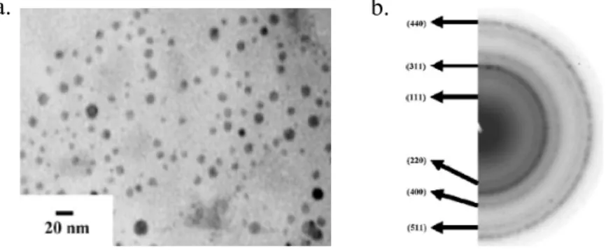
In this subproject, we synthesized the Fe3O4 nanoparticles that modified with –NH3+ exhibit
well-dispersed morphology last year, as shown in Fig. 1.
Figure 1. a.) TEM imaging showing Fe3O4 nanoparticles with average size of 6.2 ± 2.1 nm, and b.) Selected
area electron diffraction pattern of the Fe3O4 nanoparticles.
Furthermore, for the targeting properties, we used anti-EGFR antibodies as a targeting model system to evaluate the strategy for covalent crosslinking of functional biomolecules and the targeting efficiency of various lung cancer cell lines and in vivo tumor targeting.
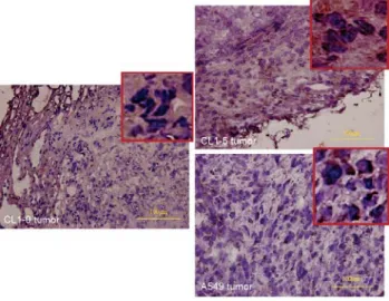
Immunohistochemical staining of EGFR and evaluate of EGFR expression in tumor in SCID mice
EGFR immunoreactivity was evaluated semiquantitatively according to the percentage of cells showing distinct cell membrane and/or diffuses cytoplasmic immunohistochemical reaction (Fig. 2). Cell menbrane and/or cytoplasmic immunoreactivities were assessed in at least five high-power fields at X400 magnification and assigned to one of the following categories: 0, <5%; 1, 5% to 20%; 2, >20%. Because tumors showed heterogeneous staining, the dominant pattern was used for scoring. A cutoff value of >20% was defined as a positive staining, and tumors with a score of 0 or 1 were considerednegative. The EGFR immunoreactivity also will be compared with the MR signal from EGFR-targeting MR imaging of the tumors.
10
Figure 2. The immunohistochemical staining of EGFR in various tumor cell line in SCID mice. a.) CL 1-0
tumor, b.) CL 1-5 tumor;,and c.) A549 tumor.
In the Fig.2, the imaging of immunohistochemical staining showed that the expression of EGFR had different level in various tumor cell line. Herein, we found that the CL 1-5 and A549 have more EGFR expression on cell membranes of tumor section. Furthermore, we could apply the Fe3O4-anti EGFR antiboby
nanocontrast agents for tumor region targeting in MR in vivo assay.
MRI for evaluation of primary tumor using EGFR-targeting magnetic nanoparticle MR probe
For MR in vivo assay, we used the 3 tesla superconductive scanner (ParaVison, Bruker, Germany) with a mini system including a resonator with active decoupling capability, animal bed for mice, and a 4-element array coil with integrated preamplifiers will be used to perform all MR studies.
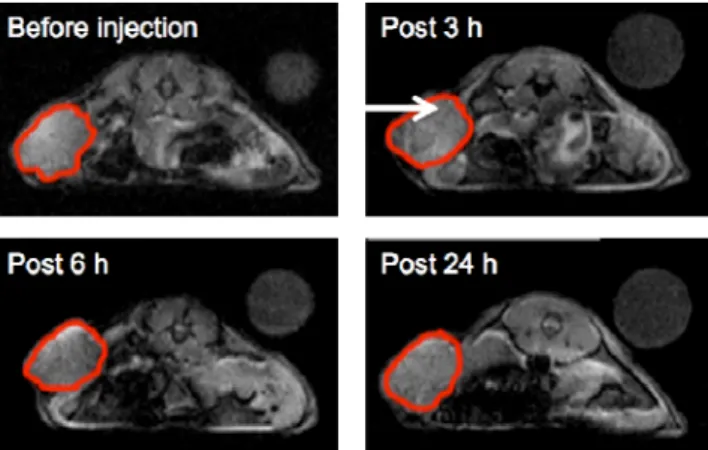
Considering the MR contrast enhancement in T2 and T2* effect for iron oxide nanoparticles, in Fig. 3,
the mice were administrated with Fe3O4 nanoparticles conjugated with anti-EGFR antibody (10 mg/Kg) via
the jugular vein. We monitored the T1-weighted imaging in a series imaging time at 3T MR system.
Comparing with the tumor region, at post 3 h, the imaging of tumor (white arrow in Fig. 3) became slightly darker. After 6 h of circulation, we found that the tumor became darker that was enhanced by iron oxide nanoparticle targeting (shown in Fig. 3) appeared darker than pre-contrast images.
11
Figure 3. In vivo progressive MRI events. T2*-weighted images of male SCID mice administrated with
Fe3O4 nanoparticles conjugated with anti-EGFR antibody at the indicated temporal points (pre-injection, 3 h,
6 h and 24 h) (The white arrows indicate the tumor region).
Furthermore, we detected the signal intensity of tumor region from T2*-weighted imaging (The red
circled areas in Fig. 3 indicate the orthotopically induced tumor lesion), and showed in Fig. 4.
Figure 4. Modification of the magnetite nanoparticles with anti-EGFR antibody significantly decrease their
absorption in the tumor region as revealed by quantitative measurement and compared to the time point before nanoparticle administration. (The red-circled areas in Fig. 3 indicate the tumor lesion)
As the result, we found that the signal intensity of tumor region could be enhancement by iron oxide nanoparticles that conjugated with anti-EGFR antibody in ca. 3~6 h, and the signal intensity of tumor region was decreased 17% after the mice were administrated negative nanocontrast agents. The signal contrast persisted for at least 12h after intravenous injection of the Fe3O4-anti-EGFR antibody nanoparticles.
To explore the tissue distribution of the Fe3O4-anti-EGFR antibody nanoparticles in the tumor and the
organs, histological examine of formalin fixed paraffin embedded tumor and normal tissues was performed in the surgically removed specimens 24 hr after delivery of the nanoparticles. The tissues were sectioned at 5 μm thickness, processed for H&E or Pearl’s iron stain then observed under a light microscopy (OLYMPUS BH-2, Japan) to reveal the tissue distribution of the aggregated iron oxide nanoparticles. As shown in Fig. 5, the tumor region (Fig. 5, H&E stain) presented a light bluish purple staining in iron stain
12
(Fig. 5, arrow head). On the other hand, we found that the liver and spleen would uptake the iron oxide nanoparticles, and showed the blue staining in histological examine (Fig. 5)
Figure 5. Lung cancer tissue and the organs of the same animal obtained 24h after i.v. injection of the
Fe3O4-anti-EGFR antibody nanoparticles was processed for histological and histochemical analysis. Pearl’s
iron stain of the adjacent section showed a diffuse bluish purpose color (arrow head) in the tumor.
Preparations and Applications of Gd2O(CO3)2·H2O and Gd2O3 particles for MR nanocontrast agents The solution approach was employed to yield multifunctional amorphous Gd2O(CO3)2·H2O colloidal
spheres by a reflux of the aqueous solution containing GdCl3·6H2O and urea. By elongating the reaction
time, crystalline rhombus-shaped Gd2O(CO3)2·H2O with at least 87% yield could be formed and
accompanied by some rectangular particles as well. The high resolution synchrotron powder X-ray diffraction have provided crystal structural information such as cell dimensions and indexed the exact crystal packing with hexagonal symmetry, which is absent from the JCPDS file, for crystalline rhombus sample. The particles formation was studied based on the reaction time and the concentration ratio of [urea]/[GdCl3·6H2O]. After a calcination process, the amorphous spheres and crystalline rhombus
Gd2O(CO3)2·H2O particles converted into crystalline Gd2O3 at the temperature higher than 600 oC. For in
vitro MR imaging, both Gd2O(CO3)2·H2O and Gd2O3 species showed the promising T1- and T2-weighted
effects and could potentially serve as bimodal T1-positive and T2-negative contrast agents. The amorphous
Gd2O(CO3)2·H2O contrast agent further demonstrated the enhanced contrast of the liver and kidney using a
dynamic contrast-enhanced MR imaging (DCE-MRI) technique for in vivo investigation. The multifunctional capability of the amorphous Gd2O(CO3)2·H2O spheres was also shown by the evidence of
the formation of the nanoshell using amorphous spheres as the template particles. The surface engineering of the amorphous Gd2O(CO3)2·H2O spheres could be performed by covalent bonding to form hollow silica
13
Scheme 1. The Gd2O(CO3)2·H2O colloidal particles have been prepared by a reflux of the mixture
containing GdCl3·6H2O and urea solution. The Gd2O(CO3)2·H2O spherical particles exhibited
multifunctional capability by the observation of showing MR contrast effect and serving as a template particle to fabricate silica hollow structures and to form composites with various nanoshell compositions.
The Gd2O(CO3)2·H2O particles were synthesized by refluxing the aqueous solutions containing
gadolinium salt and urea. In the preparation of the particles, the experimental parameters including the reaction time and the concentration ratio of [urea]/[GdCl3·6H2O] were adjusted to study their effects on the
particles formation. The formation of the sphere- and rhombus-shaped particles could be achieved by the change of the reaction time. Meanwhile, the increase of the [urea]/[GdCl3·6H2O] ratio favors the
polymorphs formation. Fig. 6a shows the spherical particles obtained exclusively from a ratio of [urea]/[GdCl3·6H2O] = 4 (urea = 1 mmol ; GdCl3·6H2O = 0.25 mmol) for a reaction of 4 h at 91 oC.
Figure 6. SEM images showing (a) the spherical particles obtained from a ratio of [urea]/[GdCl3·6H2O] = 4
for a reaction of 4 h at 91 oC, (b) the rhombus-shaped particles obtained from a ratio of [urea]/[GdCl3·6H2O]
= 4 for a reaction for 10 h at 91 oC, and (c) the rice-shaped mixtures obtained from a ratio of [urea]/[GdCl3·6H2O] = 8 for a reaction for 10 h at 91 oC.
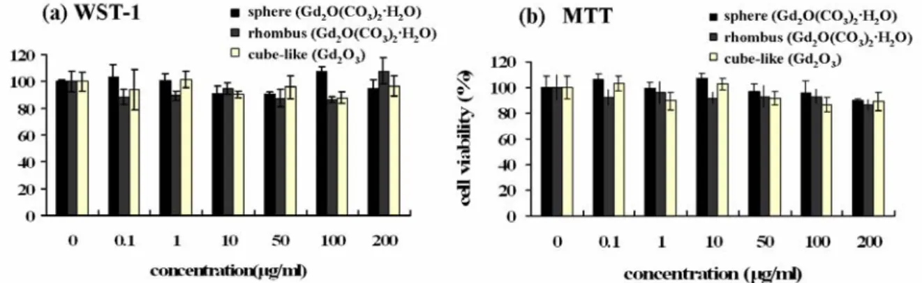
Due to the potentially showing MR signal enhancement as contrast agents, these Gd-containing particles were evaluated for deleterious biological properties. The sphere and rhombus Gd2O(CO3)2·H2O and
cube-like Gd2O3 samples were chosen to verify their cytotoxicity. Cell viability experiments were conducted
on a Vero cell line (monkey kidney cell line) using two well-established WST-1 and MTT assays. These particles were delivered over a range of dosages (0∼200 μg/mL). Both assays showed satisfactory results for in vitro biocompatibility in all dosages for the particles, as seen in Fig. 7. Additionally, the leaching of
14
Gd3+ from the spherical Gd2O(CO3)2·H2O was examined in aqueous solution using xylenol orange as
indicator for the detection of free Gd3+. A very low free Gd3+ of ∼23 μM leaching out from particles (with 2 mM of Gd3+) was observed after 4 days incubation.
Figure 7. The biocompatibility of the sphere and rhombus Gd2O(CO3)2·H2O and cube-like Gd2O3 samples
were analyzed using (a) WST-1 and (b) MTT assays. Vero cells were incubated with particles for 24 h.
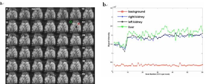
Considering that the rhombus sample includes the rectangle shape, we have chosen the amorphous Gd2O(CO3)2·H2O sphere particles as an example to monitor the T1 contrast enhancement in mice. The
dynamic contrast-enhanced MR imaging (DCE-MRI) technique was introduced to trace the imaging effect of the Gd2O(CO3)2·H2O spheres. The T1-weighted images were recorded every 12 sec for a total of 12 min
and 48 sec period. We injected the Gd2O(CO3)2·H2O spheres (0.3 mg/kg) at 6th repetition (at 61 sec) in the
course of DCE-MRI imaging acquisition. In Fig. 8a, the DCE-MR imaging showed that the liver and kidney were apparently enhanced by Gd2O(CO3)2·H2O spheres starting from 10th (post 48 sec) repetition acquisition.
Fig. 8b displays that the signal intensity of liver and kidney were measured as compared with the signal of the background noise marked as green circle throughout the acquisition. It is apparent that the signal contrast of liver and kidney were enhanced at 10th scan and remained brightness image up to 60th (post 10 min 48 sec) repetition acquisition. In Fig. 8b, it shows that the increase of signal intensity of liver was consistent larger than that of kidneys and made the change of MR signal brighter in liver. Fig. 8c shows that the liver and the cortex of kidneys could be enhanced within 2 min 24 sec in T1-weighted imaging. The evidence of the
enhanced contrast in liver and kidney suggests that the amorphous Gd2O(CO3)2·H2O spheres could be
15
Figure 8. The dynamic contrast-enhanced MR imaging (DCE-MRI) of the Gd2O(CO3)2·H2O (sphere-shaped
particles). (a) The DCE-MR images from 4th to 45th repetition acquisitions that used gradient echo imaging sequence after injected particles (0.3 mg/kg) via retro-orbital plexus at 3T MR imaging system (red arrow: liver; black: kidney; green circle: region of interest of background). (b) The signal profile in liver area (red arrow) and kidney area (black arrow) as compared with the background noise (green circle).
The Gd2O(CO3)2·H2O colloidal particles have been prepared by a reflux of the mixture containing
GdCl3·6H2O and urea solution. In particular, the biocompatible amorphous Gd2O(CO3)2·H2O spheres have
been demonstrated showing T1-enhancing and T2-lowering effects, which provide an alternative choice for
an MRI contrast agent. Furthermore, the ready surface modification has led to the Gd2O(CO3)2·H2O spheres
as an ideal template to form hollow nanoshells and hybrid composites. Based on the versatile combination strategy of the advantages of the surface engineering, MRI imaging and hollow structure shown by the Gd2O(CO3)2·H2O spheres hold the great potential in developing innovative composite materials and
multifunctional biomaterials with imaging, targeting, delivery, and therapeutic capabilities in one.
Preparations and Applications of Gd2O3/C nanoshells for MR nanocontrast agents
The dual function of Gd2O3/C nanoshells for the applications in the MR contrast images and
NIR-triggered killing cancer cells. The surface of Gd2O3/C nanoshells could be readily modified by PSMA
polymer to improve a water-dispersible property and promote their biocompatibility. The MR assays showed that Gd2O3/C and Gd2O3/C@PSMA nanoshells enhanced the image contrast in T1-, T2-, and T2*-weighted
images. Considering the different MR contrast enhancement for both nanoshells, the Gd2O3/C nanoshells
were injected into BALB/C mice to monitor T1 contrast and observed brightening images of kidneys cortex
and liver. On the other hand, Gd2O3/C@PSMA nanoshells showed the live signal darkened in T2 and T2*
16
near-infrared region. A large extinction coefficient of ε ~ 1563 M-1cm-1 at 808 nm was determined, indicating as a candidate for the potential photothermal agent. The Gd2O3/C@PSMA nanoshells conjugated
with anti-EGFR antibodies were used for targeting and destroying A549 lung cancer cells. Both laser power density- and material dose-dependence were carried out to evaluate the photothermolysis in cancer cells.
SEM image (Fig. 9a) shows the products after 600 oC under N2 as spherical particles. It can be seen that
some of particles are broken (marked by black arrows), indicating the particle constructed of nanoshells structure with an interior cavity. The spherical nanoshells exhibit the average particle sizes of ~ 138 nm and shell thickness of ~ 19.2 nm (by counting 150 particles). Figure 9b shows X-ray diffraction (XRD) analysis of the nanoshell crystal structures before and after the addition of annealing process at 600 oC under N2.
Figure 9. (a) SEM image of Gd2O3/C nanoshells prepared by a two-step thermal treatment: calcination at
440 oC in air, followed by annealing at 600 oC under N2. (b) XRD patterns for the nanoshells prepared at 440 oC in air and at 600 oC in N
2.
For MR contrast agent applications, we evaluated the longtitudinal relaxation rate (1/T1) and transverse
relaxation rate (1/T2) with various Gd ion concentrations of Gd2O3/C and Gd2O3/C@PSMA nanoshells (0 to
3.50 mM) in 5% agarose gel at 3T MR imaging system. The MR assays of T1-, T2-, and T2*-weighted
imaging show the imaging enhancement at various Gd ion concentrations of Gd2O3/C and Gd2O3/C@PSMA
17
Figure 10. MR in vitro assays of Gd2O3/C and Gd2O3/C@PSMA (a) T1-weighted images
(repetition-time/echo-time (TR/TE) = 472 ms/9.4 ms), (b) T2-weighted (TR/TE = 4500 ms/65 ms), and (c)
T2*-weighted images (TR/TE = 500 ms/5.2 ms; flip angle ( ) = 30 o of nanoshells in water containing 0.5% agarose gel. The images were taken using the designed sequences with a matrix size of 256 × 192, a field of view of 60 × 60 mm, and a slice thickness of 6 mm operated at 125.3 MHz (3T MR system).
One malignant humanized lung cancer cell line, A549, which overexpresses the epithermal growth factor receptor (EGFR) on cell surface, was used to study the photothermal killing effect of Gd2O3/C@PSMA nanoshells. To highly specifically targeting to the A549 cancer cells, the anti-EGFR
antibodies (humanized anti-EGF receptor (EGFR) antibody) were used to conjugate with Gd2O3/C@PSMA
nanoshells. The laser irradiation power densities were varied from 5 to 35 W/cm2 with a 7 min of irradiation to evaluate the cancer cells killing efficiency. The concentration of Gd2O3/C@PSMA nanoshells was fixed at
500 μg/mL. After NIR laser treatment, the cells were incubated with fluorescent labels of calcein AM and EthD-1 dyes, providing green emission for viable cells and red color for the dead cells, respectively. Fluorescence microscopy was employed to monitor images to observe cell viability. It was found that a significant loss of viability, with the area lacking green fluorescence (left) and exhibiting red fluorescence (right), began at 15 W/cm2, as seem in Fig. 11a (middle row). On the other hand, cells remaining alive (a full green color image (left) and colorless image (right)) were observed as power density down to 10 W/cm2 (Fig. 11a (bottom row)). This clearly demonstrated that the anti-EGFR conjugated Gd2O3/C@PSMA nanoshells
could specifically target to EGFR overexpressed A549 cells and provided an effective locally heating to destroy the cancer cells upon exposing to 808 nm NIR light. Interestingly, we found that nanoshell dosage also affected photothermal killing effect. The nanoshells dose-dependent experiments were conducted and carried out at a fixed 20 W/cm2 (Fig. 11b). The nanoshell dosage to cause cell death started at 400 μg/mL.
18
By dropping dosage to 300 μg/mL, there is not enough to cause cell death (high laser power required to induce cell demage).
Figure 11. (a) Anti-EGFR conjugated with Gd2O3/C@PSMA nanoshells (500 μg/mL) treated with A549
cancer cells were irradiated by the laser dosages of 20 W/cm2 (top row), 15 W/cm2 (middle row), and 10 W/cm2 (bottom row) for 7 min. (b) Anti-EGFR conjugated with Gd2O3/C@PSMA nanoshells were
irradiated by the laser dosages of 20 W/cm2 as a function of nanoshell dosages: 500 μg/mL (top row), 400 μg/mL (middle row), and 300 μg/mL (bottom row). The column shows staining by green fluorescence dye, calcein AM, for living cells. The column displays stainning by EthD-1, where the red fluorescence color indicates cell death.
We have presented the first report of Gd2O3/C@PSMA nanoshells showing bifunctionalities with
efficient in MR anatomic imaging enhancement and photothermolysis of killing cancer cells. Interestingly, the MR assays show that we could switch the imaging contrast effect from positive Gd2O3/C contrast agents
to negative Gd2O3/C@PSMA contrast agents. Cytotoxicity assay shows that the Gd2O3/C and
Gd2O3/C@PSMA nanoshells have no significant toxicity and biodistribution indicates that they can be
eliminated after 24 h. The graphite carbon coating on Gd2O3 nanoshells display large extinction coefficient
of ε ~ 1563 M-1cm-1 at 808 nm with good absorbance in the near-infrared (NIR) region. Therefore, Gd2O3/C@PSMA nanoshells could act as promising photothermal therapeutic agent. By the oppositely
charged interaction between Gd2O3/C@PSMA nanoshells and anti-EGFR antibodies, the antibody
conjugated nanoshells could effectively target to malignant A549 lung cancer cells and exhibited therapeutic effect of killing A549 cells by photothermolysis.
19
計畫成果自評部份
In this year, we plan to build the platform of iron oxide nanoparticles and biocojugation of specific antibodies in NSCLC cell targeting and applied these nanocontrast agents in tumor targeting for in vivo MR molecular imaging. Furthermore, we plan to have Gd-based nanocontrast agents for positive contrast enhancement and kill the lung cancer cell (A549) by photothermal methodology. Up to date, the progress of this grant is under control, and we scale new heights in nanocontrast agents’ development. Herein, we have completed the bioconjucation and modification of iron oxide nanoparticles, in vivo tumor targeting assay, and in vivo MR molecular imaging of tumor xenograft of NSCLC cell line A549 in SCID mice model.
As the results, we completed all of the goals in developments and applications of iron oxide nanocontrast agents for lung cancer targeting. Although the signal intensity was contrasted down by iron oxide nanoparticle in 17 %, we plan to change the modification strategy to increase the targeting efficiency. Furthermore, we also developed the Gd-based nanomedicine for MR contrast application and phototheemal therapy. In conclution, we achieved 100% of scheduled progress in subproject 2 in the second year.
Herein, these results of Gd-based nanomedicien were published on Adv. Funct. Materials. Furthermore, we also applied of iron oxide nanoparticle for oral cancer targeting and MR molecular imaging eveluation, and these results were published on Bioconjugated Chem. In brief, 2 abstracts have been represented in 6th Annual Meeting of Society of Molecular Imaging and 4 articles have been published. (shown in Table 1)
Table 1. The statistical table of publications in this integrated research project (2007/8 to 2008/5)
With the support of this grant, one master student will be graduated this year, another Ph.D. student and master student work on further parallel imaging study and might graduate next year.
Publication
1. Chi-Chia Huang, Tzu-Yu Liu, Chia-Hao Su, Yi-Wei Lo, Jyh-Horng Chen, Chen-Sheng Yeh, "Superparamagnetic Hollow and Paramagnetic Porous Gd2O3 Particles", Chem. Mater. 2008, Accepted.
(IF: 5.104)
2. Fong-Yu Cheng, Saprina Ping-Hsien Wang, Chia-Hao Su, Tsung-Liu Tsai, Ping-Ching Wu, Dar-Bin Shieh, Jyh-Horng Chen, Patrick Ching-Ho Hsieh, and Chen-Sheng Yeh, "Stabilizer-free poly(lactide-co-glycolide) nanoparticles for multimodal biomedical probes", Biomaterials, 2008, 29, 2104-2112. (IF: 5.196; 1/14)
2007 2008
Journal paper - 4 Conference paper 2 -
20
3. I-Fang Li, Chia-Hao Su, Hwo-Shuenn Sheu, Hui-Chi Chiu, Yi-Wei Lo, Wei-Ting Lin, Jyh-Horng Chen, and Chen-Sheng Yeh, "The Gd2O(CO3)2·H2O Particles and their Corresponding Gd2O3: Preparation and
Applications of MR Contrast Agents and Template Particles for Hollow Spheres and Hybrid Composites", Adv. Funct. Mater. 2008, 18, 766-776. (IF: 6.779; 7/176)
4. Chien Yuan Lin, Chen Chang, Jyh Horng Chen, Tien Nan Lin " Dynamic Changes in Vascular Permeability, Cerebral Blood Volume, Vascular Density and Size after Transient Focal Cerebral Ischemia in Rats: Evaluation with Contrast-Enhanced Magnetic Resonance Imaging" J. Cereb. Blood Flow Metab.,
2008. Accepted. (IF: 4.843; 34/200)
5. Chia-Hao Su, Jun-Cheng Weng, Dar-Bin Shieh, Jyh-Horng Chen, Chen-Sheng Yeh, " The Novel Platform of Nanomedicine: For Drug Carrier and MR Contrast Agents Applications " The 6th
Annual Meeting of The Society for Molecular Imaging, Rhode Island, American, 2007.
6. Chia-Hao Su, Hwo-Shuenn Sheu, Chia-Yun Lin, Chih-Chia Huang, Yi-Wei Lo, Ying-Chih Pu, Jun-Cheng Weng, Dar-Bin Shieh, Jyh-Horng Chen, and Chen-Sheng Yeh, "Nanoshell Magnetic Resonance Imaging Contrast Agents" 2007 International Microprocesses and Nanotechnology Conference, No. 139, Kyoto, Japan,
出席國際學術會議心得報告
計畫編號 NSC 95-2627-B-002-012 計畫名稱 發展動態顯影及具標定功能之生醫分子磁振影像:評估肺癌之腫瘤生成、轉 移、血管新生及治療反應 出國人員姓名 服務機關及職稱 陳志宏 服務機關:國立台灣大學 電機系 職稱:教授 會議時間地點 Sep. 7-11, 2007, Providence, Rhode Island, USA會議名稱 Molecular Imaging Conference 2007
發表論文題目 The Novel Platform of Nanomedicine: For Drug Carrier and MR Contrast Agents Applications 一、參加會議經過 分子影像是處在生醫及物理科學的界面,且其快速地發展成一個重要的技術平台,在未 來的醫學診斷及治療上,以及在藥物發現及開發上的重大突破,都將扮演一個中樞的角色。 目前大多數的影像工具提供組織的結構及分析資訊,然而該領域的發展已經導致新影像工具 的產生,目前它能夠讓研究人員洞悉機能性及分子的細節。為了包括生物學家、化學家、物 理學家、電機工程師、電腦科學家及臨床科學家之跨領域研究。其中,為達成跨領域的目的, The Society of Molecular Imaging (SMI)與Academy of Molecular Imaging (AMI)合併於美國羅 德島舉行2007分子影像國際會議。 此次赴美參加2007分子影像國際會議,主要是由於國內對於生醫分子影像之研究也正如 火如荼地進行,且本人目前負責台大生醫分子影像中心,因此希望在生物探針MRI及光學影 像方面可以於參加此一國際會議獲得更廣泛的研究觸角。 與會期間與多位從事分子影像的國際學者有深入的研究知識交流,且也由於SMI與AMI 的整合與規劃下,其研究的領域已由基礎學理及應用跨足至臨床醫學的應用範疇。且研究方 向大致可以分成下列幾項:(1)光學影像;(2)影像處理及管理;(3)使用磁性共振的小 動物影像;(4)化學/生物探針的開發。上述的技術平台將被使用來支援的研究領域如癌症、 新陳代謝及再生醫療。 二、與會心得 經由此一國際會議的參與,可以很明顯的發現分子影像的研究範疇已逐漸地擴大並有部 分以進入臨床研究的階段。且亦由於奈米藥物的發展,對於標定性功能影像的研究也逐漸增 加。並由此可以預見分子影像的研究在未來數年之間,是一相當重要且活躍的跨領域研究。 但由於目前國內研究環境對於此一領域仍缺乏校際間的整合,且除本人所負責台大生醫分子 影像核心實驗室外,仍無一相當之整合型研究團隊。有鑑於此,政府與學校單位應大力推動 跨領域之生醫分子影像中心或重點發展研究領的培植與支援,冀以未來國內相關研究領域可 於國際間有獨特發展與相互競爭的實力。

![Figure 6. SEM images showing (a) the spherical particles obtained from a ratio of [urea]/[GdCl 3 ·6H 2 O] = 4 for a reaction of 4 h at 91 o C, (b) the rhombus-shaped particles obtained from a ratio of [urea]/[GdCl 3 ·6H 2 O]](https://thumb-ap.123doks.com/thumbv2/9libinfo/8780700.215661/17.892.255.639.70.232/figure-spherical-particles-obtained-reaction-rhombus-particles-obtained.webp)