f
Factors influencing the dynamic
behaviour of human teeth
H . - M . H u a n g I C.-Y. Y e h 2 S.-Y. Lee 3"4 M . - S . W a n g a L.-C. Pan a C.-C. C h e n 2
1School of Medical Technology, Tapei Medical University, Tapei, Taiwan 2School of Medicine, Taipei Medical University, Tapei, Taiwan
3Graduate Institute of Oral Rehabilitation Sciences, Taipei Medical University, Tapei, Taiwan 4Dental Department of Wan-Fang Hospital, Taipei Medical University, Tapei, Taiwan
Abstract--Modal analysis is carried out to test the natural frequencies of certain human teeth, including central incisors (CIs), canines (CAs), first premolars (FPs) and first molars (FMs). A total n u m b e r of 1007 teeth are tested, taking into account tooth type, oral location, age and gender, to analyse the effects of the above-mentioned factors on the natural frequency of the sample teeth. The results reveal that no significant difference in the natural frequency is noted among teeth in the four different intra-oral quadrants. Nevertheless, tooth type and age elicit an effect upon the value of the natural frequency of teeth. On the other hand, the mean value for the natural frequency of CIs (1.274- O. 15kHz), CAs (1.304- O. 15kHz), FPs (1.274- O. 15kHz) and FMs (l. 164- O. 12kHz) for males are significantly lower (IO<0.01) than the analo- gous figure for females (1.414- O.21kHz for CIs, 1.404- O. 18kHz for CAs, 1.374- O.20kHz for FPs, and 1.254- O. 16kHz for FMs). Moreover, the natural frequency of teeth in male subjects varies with age (p< 0.05). The highest mean frequency of CIs, CAs and FPs for the male subjects is found for the group aged between 40 and 49 years. On the other hand, the natural frequency for the s i m i l a r set of teeth for the female subjects is shown to be in no way associated with age.
K e y w o r d s - - N a t u r a l frequency, Tooth, Biomechanics , Modal testing Med. Biol. Eng. Comput., 2001, 39, 176-181
J
1 I n t r o d u c t i o n
IN RECENT years, owing to the advancement of computer technology, mathematical modelling, such as finite element analysis of the physical processes leading to dental trauma, has received considerable attention. Importantly, however, there are various sources of error that can contribute to incorrect results in numerical analysis. Experimental testing of the mathematical model may be the best way to check its reliability (MOAVENI, 1999).
In the realm of traumatic biomechanical analysis, an impor- tant property for a credible model is that it must have a natural frequency response similar to that of the structure being tested. This is because traumatic injuries to teeth typically result from an impact force from a hard object. Such impact forces are, in general, of short duration and most probably give rise to a vibrational response, superimposed on a rigid body motion of the impacted tissue (KHALIL et al., 1979).
Various investigators have performed vibrational experiments upon hard human tissue, such as bone: for example, the natural frequencies and mode shapes of the femur (KHALIL et al., 1981 ; COUTEAU et al., 1998), tibia (VAN DER PERRE et al., 1983;
Correspondence should be addressed to Dr S.-Y. Lee; e-marl: seanlee@tmu.edu.tw
First received 20 September 2000 and in final form 4 January 2001 MBEC online number: 20013561
,c, IFMBE: 2001
CORNELISSEN et at., 1986; THOMSEN, 1990; HOBATHO et at.,
1991), ulna (JURIST and KIANIAN, 1973), lumbar vertebra (KITAZAKI and GRIFFIN, 1995) and head (GURDJIAN el al.,
1970; STALNAKER el al., 1971; KHALIL el al., 1979), all of which have already been analysed systematically.
Published research pertaining to natural teeth in this regard is, however, obviously lacking. Owing to the exact mechanisms of dental injuries, the processes that occur when the tooth is subjected to a hard blow are still tmclear (ANDREASEN and ANDREASEN, 1994), and, hence, investigation of the tooth's dynamic characteristics, such as natural frequency, appears to be especially important and imperative for dental-trauma analysis. On the other hand, because the value of an object's natural frequency is related to the material properties and botmdary conditions of the object, it is also an index of the material and boundary status of a structure. In addition, the method used for the estimation of an object's natural frequency is non-invasive and non-destructive, and therefore, besides such a method being used for the assessment of industrial materials, it is also extensively used for orthopaedic research (NOKES, 1999) and also neurotrauma analysis (WILLINGER el al., 1995; KUMARESAN and RADHAKRISHNAN, 1996).
Although vibration analysis has previously been used by several scholars to assess oral rehabilitative problems, most of the studies focused upon the evaluation of dental implant stability (ELIAS el at., 1996; MEREDITH el at., 1996; 1997; SENNERBY and MEREDITH, 1998; RASMUSSON et al., 1999). Thus it would appear that a comprehensive analysis of the natural frequency for human teeth is likely to be soon carried out.
To examine the mobility of the upper and lower central incisors, NOYES and SOLT (1973) used sinusoidal force to excite the teeth to vibrate. They found that the resonant frequencies of such teeth lay at around 3-4 kHz but were different for upper and lower central incisors. However, this diff- erence is not defined statistically. OIKARINEN et al. (1991 ) used percussion sound to analyse the degree of mobility of healthy incisors and canines in situ, their results revealing that the elicited waveforms of the sounds differed in accordance with the relative size of the tooth and also the gender of the test subject.
Analogously, OKAZAKI et al. (1996) used a modal-testing method to assess the damping characteristics of the periodontal ligament for specific human teeth. Although a tooth's natural frequency was not discussed in their report, these authors did refer to the influences of several factors, including gender and generation, on the dynamic behaviour of teeth, in this regard, LEE et al. (2000) used the modal-testing technique to assess the relationships between the natural frequencies of upper central incisors and their periodontal conditions in vivo and in vitro.
Both their experimental results and numerical simulation data indicated that the natural frequency of such teeth decreased when the periodontal level was lowered. According to these findings, Lee's group suggested that the natural-frequency value seems to demonstrate the potential to be an index for monitoring general periodontal health status.
According to the above-cited literature, not only do the natural frequencies of teeth provide a meaningful clinical index as to the dentition's relative periodontal condition, but also, such vibra- tional frequencies constitute an important parameter for dental trauma analysis, information suggesting whether various factors, such as age, gender and tooth type, influence the natural- frequency value of a tooth is still unavailable; however, the natural frequencies o fvarious human teeth were estimated in this study. The natural-frequency values were tested statistically according to various specified factors, to provide a useful reference for furore periodontium health assessment and dental-trauma analysis.
2 Materials and m e t h o d s
Modal testing was performed on 110 volunteer adults at the Dental Department of the Wan-Fang Hospital, Taipei, Taiwan. The volunteers consisted of 64 men and 46 women, aged between 20 and 64 years. The central incisors, canines, first premolars and first molars for all volunteers were chosen for examination.
According to the report of LEE et al. (2000), the natural frequency of teeth exhibiting periodontal disease is likely to be lower than the value for healthy teeth. Accordingly, the relative health status of the periodontium of the test teeth was examined by means of a probing depth examination and X-ray image diagnosis prior to the volunteers receiving modal testing. According to the RAMFJORD (1959) classification, attachment loss reaching 4-6 m m was noted as moderate periodontal disease that requires treatment. Therefore a middle value of 5 m m was used as a threshold to judge the health status of the periodontal condition in this study.
Subsequent to the preliminary examination of the general health status of the periodontium of the test volunteers, 1007 healthy teeth were identified, all of which underwent modal testing examination, including 257 central incisors, 266 canines, 261 first premolars and 223 first molars. A listing of the distributions of these teeth arranged according to gender, age and intraoral location is given in Tables 1 and 2.
Modal analysis was carried out to measure the natural frequency of the test teeth; Fig. 1 shows the apparatus and testing method used for this experiment. Before undergoing
Table 1 Number o f teeth tested at various oral locations (total o f l l O vohmteers were tested in study)
Right Left
FM FP CN CI CI CN FP FM
Upper 71 76 81 80 56 57 55 44
Lower 39 48 47 44 77 81 82 69
FM-- First molar; FP-- first premolar; CN-- canine; CI-- central incisor
Table 2 Number o f teeth in each group classification according to various influencing factors
Quadraaat Age, years Gender
UR UL LR LL 20-29 30-39 40-49 ~>50 M F
308 212 178 309 401 202 212 192 525 482
UR -- upper right; UL -- upper left; LR -- lower right; LL -- lower left
m•impulse
hammer~
frequency
H
spectrum displayanalyser
Fig. 1
hlstrumentation and testing methods used for this studytesting, all the teeth were first dried with cotton. The test teeth were then forced into vibration by the application of an impulse force hammer.*
When applied by hand, the exciting force was directly applied to the surface of the test teeth along the lingual-labial (frontal tooth) or lingual-buccal (posterior tooth) direction. In addition, to overcome the problem of the difficulty associated with measuring the vibration signals from posterior teeth, an acoustic microphone'~ was used as a transducer. The microphone was positioned 1 cm vertically above the striking surface to collect the vibrational signal and to transfer the response to a frequency spectrum analyser$ by means of an A / D signal capture card**. During the measurement process, to minimise the influence of environmental background noise, the microphone was turned on only when the piezo-electric tip of the impulse hammer was triggered by a striking force, thus transferring an impact signal to the analyser.
Fast Fourier transform software was used to convert the vibrational signal from a time-domain to a frequency-domain format. The natural frequency of the test teeth was assessed through the tooth's peak mobility amplitude as reflected by the frequency response spectrum. Based upon the report of LEE et al.
(2000), the first natural frequency of teeth provides an index for a meaningful clinical diagnosis; hence, we choose to examine only the first natural frequency of the teeth in this study, in addition, one final natural frequency for each test tooth was obtained by averaging the results from every fifth triggering response.
A computer-based statistical software package-~-~ was used for statistical analysis. Teeth were classified according to tooth type (i.e. central incisors, canines, first premolars and first molars)
* Model GK291C80, PCB Piezotronics, Buffalo, NY, USA. -~ FM-10B, FC Electronic, Taipei, Taiwan.
Probel II, Prowave Engineering, Hsinchu, Taiwan. ** AD102A, Prowave Engineering, Hsinchu, Taiwan. -~-~ CSS Stastica, Statsoft, Tulsa, OK, USA.
and to the intra-oral cavity quadrant in which they were located, lO [
these comprising upper right (UR), upper left (UL), lower right (LR) and lower left (LL). One-way analysis of variance
(ANOVA) with an LSD post hoc test was applied to test the i difference between natural-frequency values and the positional
site of the teeth. ,, 6
To examine the relationships between the natural frequency
and the age of the tooth, the test teeth were divided into four age E 4 groups, comprising 20-29 years, 30-39 years, 40-49 years and
50 years and above. The statistical examination of natural-
frequency values based upon these four different age groups 2- was tested by one-way ANOVA. In addition, a student's t-test
was used to compare differences in mean natural-frequency 0
values between male and female volunteers. Stated p values below 0.05 were considered to indicate statistical significance in
this study. Fig. 3
3 Results
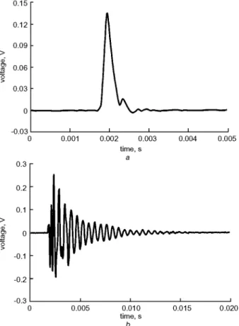
Fig. 2a is a representation of a typical input force used in triggering a central incisor to vibrate. Subsequent to the test teeth having been excited to vibration, the vibration signal was recorded and appears as shown in Fig. 2b. When the vibration signal was converted from a time-domain to a frequency-domain format, the natural frequency of the teeth was recorded in the position reflecting maximum tooth mobility. Fig. 3 illustrates the frequency response of a vibrating central incisor and also a first molar from the same volunteer.
According to Table 3, the natural frequencies corresponding to each type of tooth revealed no significant difference between the four quadrants, although the natural frequency of molars was noted to be significantly lower than that for the other three tooth types, no matter what quadrant was tested ( p < 0.01 ). According to the data presented in Table 3, it can clearly be seen that the mean natural frequency of molar teeth, regardless of the tooth's
0.15 0.12 0.09 > d 0.06 0.03 -0.03 0 0.3 m i 0.001 I I I I 0.002 0.003 0.004 0.005 time, s a 0.2 0.1 > o o -0.1 -0.2
.U
-0.3 i i i i 0 0.005 0.010 0.015 0.020 time, s bFig. 2 ~,'pical spectra, (a) impulse force and (b) the vibrational response o f the test tooth as detected bv the microphone
300 600 900 1200 1500 1800 frequency, Hz
Frequeno; response o f a vibrating central incisor ( ) and also a first molar (---) for one vohmwel: The resonance fi'equency can be obtained through the fi'equency response
spectrum with the highest amplitude o f vibration
location quadrant, is 1.2 kHz, and the corresponding figure for the other three remaining tooth types lies between 1.3 kHz and 1.35 kHz. As it would appear that no obvious relationship exists between the natural frequency of the teeth and the tooth's location quadrant, the following analyses consequently do not use the specific quadrant location o f the tooth as a parameter. After combining all the computations, the mean natural frequency of the molar teeth is 1.20 4-0.15 kHz, which is still significantly lower (p < 0.01 ) than the mean natural frequency of central incisors (1.34 4- 0.2 kHz), canines ( 1.35 4- 0.17 kHz) and the first premolars ( 1.32 4- 0.18 kHz).
With regard to the influence of gender, as indicated in Table 4, it would appear that regardless of the tooth type, males demon- strate an obviously lower mean natural frequency than females (p<0.01). For the central incisor, canine and premolar, the mean value for the natural frequency for males lies between 1.27 kHz and 1.30 kHz, and the corresponding value for females lies between 1.37 kHz and 1.40 kHz. For molars, the mean value for the natural frequency for males is 1.16 kHz, again signifi- cantly lower than the analogous figure for females (1.25 kHz).
Tables 5a and b depict the mean values for the natural frequency of male and female teeth based upon different tooth type and age categories. According to data presented in Table 5a,
different dynamic characteristics of the teeth were noted for different age categories for male volunteers. Prior to the age of 50 years, the natural frequency of central incisors, canines and the first premolars of male volunteers increases significantly as age increases ( p < 0 . 0 5 ) . The mean natural frequency of the same teeth, however, decreased after 50 years. With regard to female volunteers (Table 5b), no significant difference in natural
frequency was found between the different age groups for central incisors, canines and first premolars. For molars, based upon different age categories, regardless of gender, there is a significant increase in the mean natural-frequency value (p < 0.05). in addition, the mean natural frequency of molars for volunteers aged below 50 years is obviously lower than the corresponding mean value for the three other teeth types (p < 0.01 ) for both male and female groups. For the age category of 50 years and over, there would appear to be no significant resonant frequency difference between molars and the three other teeth types.
4 Discussion
An accelerometer is one of the most common devices used for vibrational signal detection (NONES, 1999); however, consid- ering the size of this apparatus, it is not suitable for use in the
Table 3 Resonance fi'equencies (kHz) o f teswd weth (mean ± SD) with different anatomical shapes at different location quadrants within oral cavity. Frequencies corresponding to each type o f tooth revealed no significant differences between four quadrants. Howevel: fi'equencies o f molars were noted to be significantly lower than those for other three tooth types. One- way ANOVA was used for statistical examination
UR UL LR LL p CI 1.35 ± 0.20 1.34 ± 0.19 1.32 ± 0.20 1.34 ± 0.20 0.93 CN 1.35 ± 0.18 1.32 ± 0.17 1.32± 0.18 1.38± 0.16 0.12 FP 1.32 ± 0.17 1.30 ± 0.15 1.30 ± 0.16 1.34 ± 0.21 0.42 FM 1.19 ± 0.13 1.21 ± 0.16 1.18± 0.16 1.22± 0.15 0.50 p <0.01 <0.01 <0.01 <0.01
UR -- upper right; UL -- upper left; LR -- lower right; LL -- lower left; CI -- central incisor; CN -- canine; FP -- first premolar;
FM -- First molar
Table 4 Difference in resonance fi'equencies (kHz) of four different weth O.'pes (mean ± SD) for male and female wst populations. Natural fi'equency o f Jbmale teeth is" significantly greater than corresponding figure for male teeth
Central incisor Canine First premolar First molar p
Male 1.27±0.15 1.30±0.15 1.27±0.15 1.16±0.12 <0.05*
Female 1.41 ± 0.21 1.40 ± 0.18 1.37 ± 0.20 1.25 ± 0.16 < 0.05"
p <0.01t <0.01 t <0.01 t <0.01 t
*Statistical examination by one-way ANOVA tStatistical examination by Student t-test
Table 5 (a) Relationship between value o f natural fi'equencies (kHz) and differing age categories among male vohmteers. Mean fi'equencv o f teeth for male subjects is" influenced bv increasing age. One-way ANOVA was used for statistical examination (b) Relationship bent'een value o f natural fi'equencies and differing age categories among female vohmteers. Frequencies seem to be almost independent o f increasing age. One-way ANOVA was used for statistical examination
20-29 years 30-39 years 40-49 years ~> 50 years p
CI 1.24 ± 0.10 1.25 ± 0.15 1.38 ± 0.23 1.32 ± 0.17 < 0.05 CN 1.24±0.12 1.32±0.13 1.41±0.15 1.34±0.17 <0.05 FP 1.23±0.11 1.28±0.16 1.39±0.17 1.28±0.17 <0.05 FM 1.13 ± 0.11 1.16 ± 0.10 1.19± 0.12 1.24 ± 0.15 < 0.05 p <0.01 <0.01 <0.01 0.15 a
20-29 years 30-39 years 40-49 years ~> 50 years p
CI 1.44 ± 0.22 1.34 ± 0.18 1.40 ± 0.23 1.43 ± 0.17 0.35
CN 1.41 ± 0.20 1.33 ± 0.16 1.40± 0.19 1.41 ± 0.12 0.34
FP 1.41 ± 0.25 1.35 ± 0.12 1.35 ± 0.18 1.36 ± 0.13 0.58
FM 1.23 ± 0.16 1.21 ± 0.09 1.26 ± 0.12 1.39 ± 0.28 < 0.05
p < 0.01 < 0.01 < 0.01 0.65
limited space in the oral cavity. To examine the dynamic behaviour o f the periodontal membrane, OKAZAKI et al. (1996) used an accelerometer attached with dental utility wax to the surface o f test incisors to receive the vibration signal. However, analogous assessment was difficult to perform for teeth in the posterior region. In addition, when the test tooth was excited, the accelerometer and the tooth vibrated together, owing to their physical attachment. Thus, when the overall vibration response is measured, the signal includes a regulatory contribu- tion based upon the construction materials o f the accelerometer. KITAZAKI and GRIFFIN (1995), when assessing the vibration characteristics o f lumbar vertebrae, found that the weight o f the accelerometer could not be ignored with regard to the natural- frequency assessment o f individual vertebra. To eliminate the mass loading effect o f the accelerometer, LOWET et al. (1993) used a microphone instead o f an accelerometer for measuring the vibrational response of long bones.
i n our study, to overcome the problem of the difficulty associated with measuring the vibration signals from posterior teeth, we, like LOWET et al., used a microphone sensor as a
transducer to measure the vibrational response of teeth. As illustrated in Figs 2 and 3, the spectrum o f frequency response of the test teeth was both clear and sensitive.
In the oral cavity, one end of a tooth is exposed to air, whereas the other end, under normal conditions, is constrained firmly within the alveolar bone; thus we can evaluate the tooth' s natural frequency b y applying the formula for a cantilevered beam, as follows (LOWET et al., 1993; MEREDITH et al., 1996):
f;,
V)-g
(1)where f,, is the natural frequency o f the beam, l is the effective vibrational length of the beam, E is Y o u n g ' s modulus, I is the m o m e n t o f inertia, p is the mass per unit effective vibrational length, and e is a constant related to its boundary conditions. Clearly, from the formula, if an increase in the effective length o f vibration l is noted, the natural frequency o f the beam will decrease.
A previous study has noted that variation in the attachment level of tooth periodontal ligament is associated with variation in the natural frequency of the tooth. A diseased tooth possessing a lower periodontal attachment level exhibits a lower natural frequency than a healthy tooth, owing to a larger value for l for the former (LEE et al., 2000). To diminish the effects of a variable periodontal condition upon the natural frequency of teeth, the periodontal conditions of all the test teeth selected in this study were tested, and their healthy status was confirmed by means of probing depth and X-ray image examinations before modal analysis was performed.
According to the literature concerning living tissue, the value of the first natural frequency lies, mostly, below 1000Hz; for example, the value for the femur is approxi- mately 250Hz (KHALIL et al., 1981), that for the tibia lies in the range of 260-430Hz (VAN DER PERRE et al., 1983; CORNELISSEN et al., 1986; THOMSEN, 1990; HOBATHO et al.,
1991), the mean natural frequency of the third lumbar vertebra is approximately 30Hz (JURIST and KIANIAN,
1973), and the figure for the human head lies between 300 and 600Hz (STALNAKER et al., 1971). In Our study, the average value of the natural frequency of various tooth groups is around 1000Hz, or slightly above, this figure being obviously larger than the corresponding value reported for other hard tissue. We suggest that the main reason for this difference is that the size of the tooth is small in comparison with other tested hard-tissue structures, and thus the effective length of vibration is likely to be lower than the analogous value for long bones.
As demonstrated in Table 3, owing to the fact that a tooth possesses a characteristic of symmetry to some degree, the natural frequency of a healthy tooth inside the oral cavity reveals no significant difference between the four different oral cavity quadrants. The OKAZAKI et al. (1996) results also demonstrated that there is no difference in the characteristics of anterior tooth vibration between the left and fight sides of the oral cavity. Although the results of NOYES and SOLT (1973) did suggest that there is an obvious difference between the resonant frequency for upper and lower central incisors, their conclusions must remain questionable given the small sample size adopted in their analysis.
From the mobility study of OIKARINEN et al. (1992), after a tooth has been impacted, it will have the motion of a harmo- nically rigid body and a structural resonant vibration at the same time. These authors reported that no difference in vibrational response exists between central incisors, lateral incisors and canines. Data illustrated in Table 3 also reveal a similar result, with no significant difference in tooth natural frequency existing between central incisors, canines and first premolars, whereas the opposite was the case for comparison between the first molar and the three other tooth type groups. This would appear to be the case because molar teeth expose a proportion- ally larger component of their overall volume external to the alveolar bone compared with the other three tooth groups. Thus the mass per unit effective vibrational length p is undoubtedly larger for molars than for any of the other tooth types. From the formula above (eqn 1), it is apparent that an increase in the value o f p will lead to a decrease in the natural frequency of the tooth.
OIKARINEN et al. (1992) noted that the mean attenuation duration of percussion sound was lower for women than for men; they reasoned that this was because the degree of mobility of female teeth is lower than that of men's teeth, in the context of general vibration theory, those objects with a lower degree of mobility will reveal a higher frequency of vibration. Our results also suggest that the natural frequency of female teeth is significantly greater than the corresponding figure for male teeth (Table 4).
The effect of aging upon the periodontal ligament (PDL) results in a progressive decrease in the relative width of the periodontal membrane (SICHER and BHASKAR, 1972). This phenomenon will decrease the degree of mobility of the tooth, resulting in an increase in the natural frequency of teeth, in addition, however, aging will also cause the alveolar bone to be resorbed and cause the natural frequency of teeth to decrease. Our results indicated that these aging effects on natural frequen- cies of teeth in males and females are quite different. The mean frequency of teeth for the male subjects is influenced by increasing age. However, the frequencies in females seem to be almost independent of increasing age.
Based upon the results depicted in Table 5a, the natural frequencies of a central incisor, canine and first premolar from our male population of volunteers aged less than 50 years tend to increase with increasing age. This arises because the width of the periodontal ligament decreases with age and results in a decrease in tooth mobility. For males who have reached the age of 50 years or greater, the bone-loss effect is more substantial than that which occurs at a younger age (HILDEBOLT, 1997); hence, a tooth's natural frequency will tend to decrease with increasing age. As females reveal a more severe bone loss with normal aging than is the case for males, such bone loss and the aging of the periodontal ligament will, at the same time, elicit an effect upon the natural frequency of teeth. Such a combined effect will result in a slight change in the natural frequency of a female's central incisor, canine and first premolar (Table 5b). For molars, an increasing natural frequency with increasing age, indepen- dent of gender, was found in Tables 5a and b. As the overall surface area of posterior teeth is larger than that of the other three tooth types, the aging effect upon the periodontium will be larger than the bone loss effect with regard to the value of a tooth's natural frequency.
Because only healthy teeth were selected to be tested in this study, the numerical differences in all the data of the natural frequencies in Tables 3-5 are very small. According to the finite element analysis of LEE et al. (2000), the natural frequency of a central incisor exhibiting periodontal disease decreases linearly at a rate of 188 Hz m m 1. However, the way in which natural frequencies change with the degree of disease in vivo is still unknown, it should be an important subject for future study. In summary, our results have indicated that the natural frequency of each different anatomical form of tooth within the different quadrants of the oral cavity did not show any obvious difference with regard to tooth siting. The natural frequency of teeth is, however, related to age, gender and tooth type. The results of this study can thus be a useful reference for future research into the dynamic characteristic assessment of teeth and associated dental-trauma analysis.
Ac~Tlowledgments--This study was supported by a grant (TMC 88-Y05- A132) from the Taipei Medical University and by Grant NSC 89-2815- C-038-003-R from the National Science Council, Taipei, Taiwaxl. The authors also wish to thank Prowave Engineering, Inc., for their assistance and for providing instrumentation for use in this study.
References
ANDREASEN, J. O., and ANDREASEN, E M. (1994): 'Classification,
etiology and epidemiology' in ANDREASEN, J. O., and
ANDREASEN, E M. (Eds): 'Textbook and color atlas of traumatic injuries to the teeth' (Mosby, St Louis), pp. 151-180
CORNELISSEN, P., CORNELISSEN, M., VAN DER PERRE, G., CHRISTENSEN, A. B., AMMITZBOLL, E, and DYRBYE, C. (1986): 'Assessment of tibial stiffness by vibration testing in situ - II. Influence of soft tissue, Joints and fibula', J. Biomech., 19, pp. 551- 561
COUTEAU, B., HOBATHO, M. C., DARMANA, R., BRIGNOLA, J. C., and ARLAUD, J. Y. (1998): 'Finite element modelling of the vibrational behaviour of the human femur using CT-based individualized geometrical and material properties', J Biomech., 31, pp. 383-386 ELIAS, J. J., BRUNSKI, J. B., and SCARTON, H. A. (1996): 'A dynamic modal testing technique for noninvasive assessment of bone-dental implant interfaces', Int. J OralMaxilloJac. Implants, 11, pp. 728-734 GURDJIAN, E. S., HODGSON, V R., and THOMAS, L. M. (1970): 'Studies on mechanical impedance of the human skull: preliminary report', J. Biornech., 3, pp. 239-247
HILDEBOLT, C. F. (1997): 'Osteoporosis and oral bone loss', Dento- rnaxilloJac. Radiol., 26, pp. 3-15
HOBATHO, M. C., DARMANA, R., PASTOR, P., BARRAU, J. J., LAROZE, S., and MORUCCI, J. R (1991): 'Development of three- dimensional finite element model of a human tibia using experi- mental modal analysis', J. Biomech., 24, pp. 371-383
JURIST, J. M., and KianiaxL K. (1973): 'Three models of the vibrating ulna', J Biomech., 6, pp. 331-342
KHALIL, T. B., VIANO, D. C., and SMITH, D. L. (1979): 'Experimental analysis of the vibrational characteristics of the human skull', J. Sound Vib., 63, pp. 351-376
KHALIL, T. B., VIANO, D. C., and TABER, L. A. (1981): 'Vibrational characteristics of the embalmed human femur', J Sound Vib., 75, pp. 417-436
KITAZAKI, S., and GRIFFIN, M. J. (1995): 'A data correction method for surface measurement of vibration on the human body', J. Biomech., 28, pp. 885-890
KUMARESAN, S., and RADHAKRISHNAN, S. (1996): 'Importance of partitioning membranes of the brain and the influence of the neck in head injury modelling', Med. Biol. Eng. Comput., 34, pp. 27-32 LEE, S. Y., HUANG, H. M., and LIN, C. Y. (2000): 'ln vivo mad in vitro
natural frequency analysis of periodontal conditions, an innovative method', J Periodont., 71, pp. 632-640
LOWET, G., VAN AUDEKERCKE, R., VAN DER PERRE, G., GEUSENS, P, DEQUEKER, J., and LAMMENS, J. (1993): 'The relation between resonant frequencies and torsional stiffness of long bones in vitro validation of a simple beam model', J Biomech., 26, pp. 689-696 MEREDITH, N., ALLEYNE, D., and CAWLEY, P. (1996): 'Quantitative
determination of the stability of the implaxlt-tissue interface using resonance frequency analysis', Clin. Oral Impl. Res., 7, pp. 261-267
MEREDITH, N., BOOK, K., FRIBERG, B., JEMT, T., and SENNERBY, L. (1997): 'Resonance frequency measurements of implant stability in vivo', Clin. Oral Irnpl. Res., 8, pp. 226-233
MOAVENI, S. (1999): 'Verification of results', in MOAVENI, S. (Ed.): 'Finite element analysis: theory and application with ANSYS' (Prentice-Hall, New Jersey), pp. 1-45
NOKES, L. D. M. (1999): 'The use of low-frequency vibration measurement in orthopaedics', Proc. hist. Mech. Eng, 213, pp. 271-290
NOYES, D. H., and SOLT, C. W. (1973): 'Measurement of mechanical mobility of human incisors with sinusoidal forces', J. Biomech., 6, pp. 439-442
OIKARINEN, K., KAUPPINEN, P., and HERRALA, E. (1992): 'Mobility and percussion sound of healthy upper incisors and canines', Endod. Dent. Traumatol., 8, pp. 21-25
OKAZAKI, M., FUKUMOTO, M., and TAKAHASHI, J. (1996): 'Damped oscillation analysis of natural and artificial periodontal membranes', Ann. Biornech. Eng., 24, pp. 234-240
RAMFJORD, S. P. (1959): 'Indices for prevalence and incidence of periodontal disease', J. Periodontol., 30, pp. 51-59
RASMUSSON, L., MEREDITH, N., CHO, I. H., and SENNERBY, L. (1999): 'The influence of simultaneous versus delayed placement on the stability of titanium implants in onlay bone grafts', Int. J. Oral MaxilloJac. Surg., 28, pp. 224-231
SENNERBY, L., and MEREDITH, N. (1998): 'Resonance frequency analysis: measuring implant stability and osseointegration', Corn- pendiurn, 19, pp. 493-502
SICHER, H., and BHASKAR, S. N. (1972): 'Periodontal ligament', in SICHER, H., and BHASKAR, S. N. (Eds): 'OrbaxFs oral histology and embryology' (Mosby, St Louis), pp. 180-200
STALNAKER, R. L., FOGLE, J. L., and MCELHANEY, J. H. (1971): 'Driving point impedance characteristics of the head', J Biomech., 4, pp. 127-139
THOMSEN, J. J. (1990): 'Modelling human tibia structural vibrations', J Biomech., 23, pp. 215-228
VAN DER PERRE, G., VAN AUDEKERCKE, R., MARTENS, M., and MULIER, J. C. (1983): 'Indentification of in vivo vibration modes of human tibiae by modal analysis', ASME J Biomed. Eng., 105, pp. 244-248
WILLINGER, R., TALEB, L., and KOPR C. M. (1995): 'Modal and temporal analysis of head mathematical models', J. Neurotrauma, 12, pp. 743-754
Author's biography
. . . . ~ ~ ~ . . HAW-MING HUANG received his MSc in Biome- dical Engineering from Yaxlg-Ming Medical University, Taipei, TaiwaxL in 1993. He is cur- rently a lecturer in the Department of Medical Technology in Taipei Medical University, Taipei, TaiwaxL At present, he is researching for his PhD at the Graduate Institute of Medical ~ - - Sciences in Taipei Medical University. His main areas of research are those concerning traumatic biomechanics, including neurotrauma and dental trauma. He is also particularly interested in non-destructive and non-invasive diagnosis methods in dentistry.