Anti-inflammatory Effects of Scoparia dulcis L. and
Betulinic Acid
Jen-Chieh Tsaia, †, Wen-Huang Pengb, †, Tai-Hui Chiua, Shang-Chih Laic, and
Chao-Ying Leea,*
a
School of Pharmacy, College of Pharmacy, China Medical University, Taichung, Taiwan, R.O.C.
b
School of Chinese Pharmaceutical Sciences and Chinese Medicine Resources, College of Pharmacy, China Medical University, Taichung, Taiwan, R.O.C.
c
Department of Health and Nutrition Biotechnology, College of Health Science, Asia University, Taichung, Taiwan, R.O.C.
Running title: Anti-inflammatory Effects of Scoparia dulcis L. and Betulinic Acid
†These two authors contributed equally to this work
*Corresponding author:Dr. Chao-Ying, Lee
91, Hsueh-Shih Road, Taichung, Taiwan, R.O.C.
School of Pharmacy, College of Pharmacy, China Medical University.
Tel: (+886) 4-2205-3366 (ext 5116)
Fax: (+886) 4-2203-1075
Abstract
The aims of this study intended to investigate the anti-inflammatory activity of
the 70% ethanol extract from Scoparia dulcis (SDE) and betulinic acid based on
λ-carrageenan-induced paw edema in mice. The anti-inflammatory mechanisms of
SDE and betulinic acid were examined by detecting the levels of cyclooxygenase-2
(COX-2), nitric oxide (NO), tumor necrosis factor (TNF-α), interleukin-1β (IL-1β)
and malondialdehyde (MDA) in the edema paw tissue and the activities of superoxide
dismutase (SOD), glutathione peroxidase (GPx) and glutathione reductase (GRd) in
the liver. The betulinic acid content of SDE was detected by high performance liquid
chromatography (HPLC). In the anti-inflammatory model, the results showed that
SDE (0.5 and 1.0 g/kg) and betulinic acid (20 and 40 mg/kg) reduced the paw edema
at 3, 4 and 5 hours after λ-carrageenan administration. Moreover, SDE and betulinic
acid affected the levels of COX-2, NO, TNF-α and IL1-β in the
λ-carrageenan-induced edema paws. The activities of SOD, GPx and GRd in liver
tissues were increased and the MDA levels in the edema paws were decreased. It is
suggested that SDE and betulinic acid possessed anti-inflammatory activities and the
anti-inflammatory mechanisms appear to be related to the reduction of the levels of
COX-2, NO, TNF-α and IL1-β in inflamed tissues, as well as the inhibition of MDA
that the content of betulinic acid was 6.25 mg/g extract.
Keywords: Scoparia dulcis; Betulinic acid; Anti-Inflammation; Cyclooxygenase-2;
Introduction
Scoparia dulcis L. (Scrophulariaceae), a small, much branched, glabrous leafy
annual herb, is a well-known folk medicine used for hypertension in Taiwan (Chow et
al., 1974). In India, it is used to treat diabetes, toothache and gastric disorders
(Satyanarayana, 1969). In recent years, some dietary supplements containing S. dulcis
were used as health foods and drinks. Phytochemical investigations on S. dulcis have
been reported to contain steroids, diterpenoids, triterpenoids, flavonoids and
benzenoids (Kawasaki et al., 1988; Hayashi et al., 1993). Betulinic acid is one of the
constituents isolated from S. dulcis (Mahato et al., 1981), and has been reported to
possess anti-HIV, anti-bacterial, antimalarial, anticancer, analgesic and
anti-inflammatory activities (Yogeeswari and Sriram, 2005). There have been a
number of studies that indicated the pharmacological activities of S. dulcis, such as
anti-viral (Hayashi et al., 1988), analgesic, anti-inflammatory (Freire et al., 1993),
anti-ulcerative (Sonia et al., 2007) and anti-hyperglycemia effects (Pari and
Venkateswaran, 2002). In our previous study, the hepatoprotective effect of S. dulcis
was proved (Tsai et al., 2010). Furthermore, even though the anti-inflammatory effect
of S. dulcis have been reported, the mechanism was not clear enough.
Inflammation is a series of processes involving cytokines and various mediators,
and IL1-β) excite the production of many cytokines during the response to
inflammation, including prostaglandins (PGs) and NO (Chao et al., 2009). Previous
studies indicated that the inflammatory response induced by λ-carrageenan was also
proved to be associated with antioxidant enzyme activities (Lu et al., 2007). Therefore,
the aim of this study is to investigate the effects of the 70% ethanol extract from S.
dulcis (SDE) and betulinic acid by the inflammation induced by λ-carrageenan in
mice. We also examined the levels of cyclooxygenase-2 (COX-2), nitric oxide (NO),
tumor necrosis factor (TNF-α), interleukin-1β (IL-1β) and malondialdehyde (MDA)
in the edema paw and the activities of superoxide dismutase (SOD), glutathione
peroxidase (GPx) and glutathione reductase (GRd) in the liver to investigate the
anti-inflammatory mechanisms of SDE and betulinic acid. Additionally, the content of
betulinic acid was analyzed by HPLC.
Materials and Methods
Plant Materials
Whole plants of S. dulcis were collected from Taichung County, Taiwan as
described by Flora of Taiwan. A voucher specimen (Number: TNM S100389) was
deposited in the National Museum of Natural Science (TNM), Taichung, Taiwan. The
Museum of Natural Science, Taichung, Taiwan. It was authenticated in many aspects,
including morphology (flowers, fruits, seeds and pollens), histological microscopic
examination (leaves and stems) and ITS (Internal Transcribed Spacer) region of
rDNA. The sequence data was submitted to the National Center for Biotechnology
Information (NCBI) GenBank.
Chemicals
The following chemicals and reagents, betulinic acid, λ-carrageenan,
indomethacin, Griess reagent, etc., were purchased from Sigma-Aldrich Chemical Co.
The SOD, GPx, GRd and MDA activity assay kits were purchased from Randox
Laboratory Ltd. The NO and COX-2 assay kits were purchased from Cayman
Chemicals Co. Chemicals and enzyme immunometric assay kits for mouse IL-1β and
TNF-α were obtained from eBioscience Inc. All of the other reagents used were
analytical grade.
Preparation of Plant Extracts
Whole plants of S. dulcis were cut into small pieces and air dried, then crushed
into coarse powder. The coarse powder (1.5 kg) was extracted with 2L of 70% ethanol
rotary evaporator. The remaining solution was lyophilized and 108 g (7.2 % net gain)
of crude extract was yielded. The extract was stored in a refrigerator before the
experiment.
Experimental Animals
Male ICR mice (20~25 g) were purchased from BioLasco Charles River
Technology, Taipei, Taiwan. They were raised in the animal center of China Medical
University at 22 ± 1 ℃, relative humidity 55 ± 5 %, with a light and dark cycle of 12
hours for at least one week before the experiment. Animals were provided with a
rodent diet and clean water ad libitum. Animal tests used in this study were conducted
in accordance with the NIH Guide for the Care and Use of Laboratory Animals. The
experimental protocol was approved by the Committee on Animal Research, China
Medical University, under the code 99-134.
λ-Carrageenan-Induced Mice Paw Edema
The anti-inflammatory activities of SDE and betulinic acid were determined by
the λ-carrageenan-induced edema test in the hind paws of mice.Male ICR mice (10
per each group) were fasted for 24 hours before the experiment with free access to
side of right hind paws of the mice (Posadas et al., 2004). Paw volume was measured
at 1, 2, 3, 4 and 5 hours after the administration of the λ-carrageenan using a
plethysmometer. The degree of swelling was evaluated by the delta volume (a–b),
where a and b were the volume of the right hind paw after and before the
λ-carrageenan treatment, respectively. Indomethacin (20 mg/kg, p.o.), SDE (0.1, 0.5
and 1.0 g/kg, p.o.) and betulinic acid (10, 20 and 40 mg/kg, p.o.) were administered at
2 hours after λ-carrageenan injection. The control group was given an equal volume
of saline.
In the secondary experiment, the whole right hind paw tissues and liver tissues
were taken at the third hour. The right hind paw tissue was rinsed in ice-cold normal
saline, and immediately placed in its four volumes of cold normal saline and
homogenized at 4℃. Then the homogenate was centrifuged at 12,000 rpm for 5
minutes. The supernatant was stored at -80℃ for the COX-2, NO, TNF-α, IL-1β and
MDA assays. Additionally, the whole liver tissue was rinsed in ice-cold normal saline,
and immediately placed in an equal volume of cold normal saline and finally
homogenized at 4℃. Then the homogenate was centrifuged at 12,000 rpm for 5
minutes. The supernatant was obtained and stored at -80℃ for the antioxidant
COX-2 assay
COX-2 was examined according to manufacturer’s instructions. The peroxidase
activity of COX-2 was assayed colorimetrically by monitoring the appearance of
oxidized N, N, N’, N’- tetramethyl-p-phenylenediamine (TMPD) at 590 nm. The
COX-2 activity was expressed as U/ml per mg protein. One unit was equal to
nmol/min.
NO assay
NO was measured based on the method of Moshage et al. (1995). For nitrite
determination, nitrate was converted into nitrite utilizing nitrate reductase; NO2- was
measured by using the Griess reaction (Green et al., 1982). The absorbance of the
final product (purplish red) was determined at 540 nm. Values obtained by this
procedure represent the sum of nitrite and nitrate.
TNF-α and IL-1β assays
TNF-α and IL-1β assays were measured by enzyme-linked immunosorbent
assays (ELISA). Assays were performed according to manufacturer’s instructions.
(0-1000 pg/ml) constructed in each assay. The concentration of TNF-α and IL-1β in
each sample were expressed as picogram per milligram protein (pg/mg) for cytokine
concentration.
MDA assay
MDA was evaluated by the thiobarbituric acid reacting substance (TBARS)
method (Draper and Hadley, 1990). Briefly, MDA reacted with thiobarbituric acid in
an acidic condition with high temperature (above 90℃) and formed a red-complex
TBARS. The absorbance of TBARS was determined at 532 nm.
Antioxidant Enzymatic Activity Measurements
The following biochemical parameters were analyzed to evaluate the antioxidant
activities of SDE and betulinic acid by the methods given below. SOD enzymatic
activity was determined in accordance with the method of Misra and Fridovich (1972)
at room temperature. 100 µl of liver homogenate supernatant was added to 880 µl
(0.05 M, pH 10.2, 0.1 mM EDTA) carbonate buffer. 20 µl of 30 mM epinephrine (in
0.05% acetic acid) was added to the mixture at 480 nm for 4 minutes on a Hitachi U
2000 Spectrophotometer. The enzymatic activity was expressed as the amount of
GPx enzyme activity was determined according to the method of Flohe and
Gunzler (1984) at 37℃. A reaction mixture consisted of 500 µl phosphate buffer, 100
µl 0.01 M GSH (reduced form), 100 µl 1.5 mM NADPH and 100 µl GRd (0.24 units).
100 µl of supernatant was added to the reaction mixture and incubated at 37℃ for 10
minutes. Then 50 µl of 12 mM t-butyl hydroperoxide was added to 450 µl of the
tissue reaction mixture and measured at 340 nm for 180 seconds. The molar extinction
coefficient of 6.22 × 10⁻³ was used to determine the enzymatic activity. One unit of
activity was equal to the mM of NADPH oxidized/min per mg protein.
GRd enzyme activity was determined by the method of Carlberg and Mannervik
(1985) at 37℃. 50 µl of NADPH (2 mM) in 10 mM Tris buffer (pH 7.0) was added in
a cuvette containing 50 µl of GSSG (20 mM) in phosphate buffer. 100 µl of
supernatant was added to the NADPH-GSSG buffered solution and measured at 340
nm for 3 minutes. The molar extinction coefficient of 6.22 × 10⁻³ was used to
determine the GRd enzyme activity. One unit of activity was equal to the mM of
NADPH oxidized /min per mg protein.
Phytochemical Analysis of SDE by HPLC
The HPLC profile was established for betulinic acid and SDE. The HPLC
L-7100 HPLC solvent delivery pump, and a Hitachi L-7455 diode array detector.
Chromatographic separation was performed with a LiChroCART RP-18 endcapped
column (250 × 4.6 mm, i.d., 5 µm pore size, Merck, Germany). The mobile phase
consisted of 0.2 % formic acid and acetonitrile (25:75, v/v), under isocratic conditions.
The sample injection volume was 20 µl. The flow rate was 1.0 ml/min and the
detection wavelength was 205 nm. Three injections were performed for each sample.
Statistical Analysis
All data were represented as mean ± SE. Statistical analyses were performed
with SPSS software. Statistical analyses were carried out using one-way ANOVA
followed by Scheffe’s multiple range test.
Results
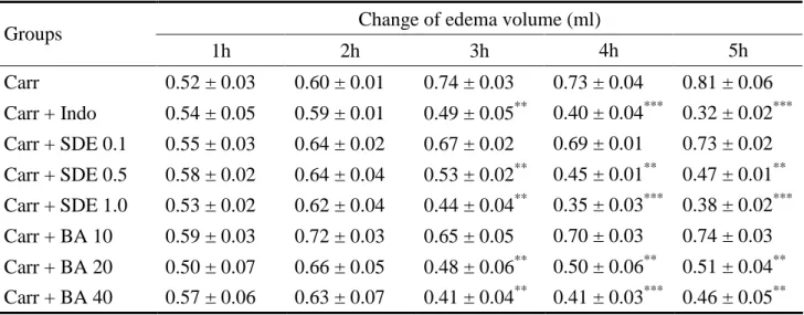
Effects of SDE and Betulinic Acid on λ-carrageenan-induced Mice Paw Edema
The results of λ-carrageenan-induced mice paw edema were represented in Table
1, it was observed that SDE (0.5 and 1.0 g/kg) and betulinic acid (20 and 40 mg/kg)
significantly inhibited (p < 0.01-0.001) the development of paw edema induced by
carrageenan after 3, 4 and 5 hours of treatment. A similar result was observed by
Effects of SDE and Betulinic Acid on COX-2 level Measurements
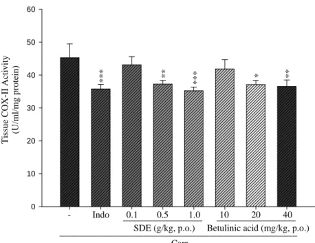
As shown in Fig. 1, the activity of COX-2 increased significantly in the edema
paw of mice after carrageenan administration on the third hour. However, COX-2
activities were decreased significantly by treatments with SDE (0.5 and 1.0 g/kg) and
betulinic acid (20 and 40 mg/kg), as well as indomethacin at 20 mg/kg (p <
0.05-0.001).
Effects of SDE and Betulinic Acid on NO level Measurements
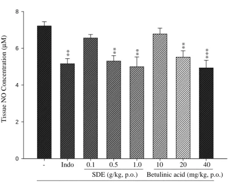
The results of NO level were shown in Fig. 2. The NO level in the edema paw
induced by λ-carrageenan was significantly increased. There was a significant effect
in the NO level when treating with SDE (0.5 and 1.0 g/kg) and betulinic acid (20 and
40 mg/kg) (P < 0.01-0.001).
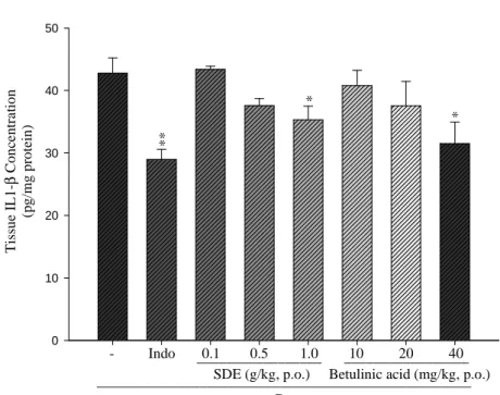
Effects of SDE and Betulinic Acid on TNF-α and IL-1β Levels
As the results shown, TNF-α level in the λ-carrageenan induced edema paws was
raised significantly. The increased TNF-α levels were reduced by treatment with SDE
(0.5 and 1.0 g/kg) and betulinic acid (20 and 40 mg/kg) (P < 0.01-0.001, Fig. 3). The
increased IL-1β levels were decreased by treatment with SDE at dose of 1.0 g/kg and
Effects of SDE and Betulinic Acid on MDA level Measurements
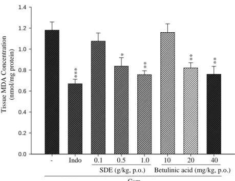
As shown in Fig. 5, the levels of MDA in the edema paw induced by
λ-carrageenan were significantly elevated. However, MDA levels were reduced by
pretreatment with SDE 0.5 g/kg (P<0.05) and 1.0 g/kg (P<0.01), as well as betulinic
acid (20 and 40 mg/kg) (P<0.01) and indomethacin (20 mg/kg) (P<0.001).
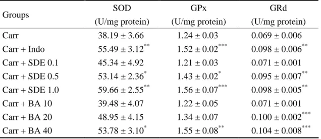
Measurements of Antioxidant Enzymatic Activities
The results of antioxidant enzymes such as SOD, GPx and GRd at the 3rd hour
following the intrapaw injection of λ-carrageenan in mice are presented in Table 2.
SOD, GPx and GRd activities in liver tissue were decreased significantly after
λ-carrageenan administration at the 3rd hour. Treatment with SDE at doses of 0.5 g/kg
and 1.0 g/kg, betulinic acid at a dose of 40 mg/kg and indomethacin at a dose of 20
mg/kg increased the levels of SOD, GPx and GRd activities significantly.
Phytochemical Analysis of SDE by HPLC
The HPLC chromatographic profiles of betulinic acid and SDE are shown in Fig.
6. In the chromatogram of the standard, a peak of betulinic acid at the retention time
chromatogram. According to the calibration curve, the content of betulinic acid in
SDE was 6.25 mg/g of extract.
Discussion
The anti-inflammatory activities of SDE and betulinic acid were evaluated in
λ-carrageenan-induced paw edema, an in vivo animal model of acute inflammation
commonly employed for assessing the anti-edematous effect of various natural
products and experimental compounds (Lai et al., 2010). The cellular and molecular
mechanism of the λ-carrageenan-induced inflammation is well characterized. In our
study, SDE, betulinic acid and indomethacin revealed anti-inflammatory effects in
λ-carrageenan-induced mice paw edema. These findings demonstrated that SDE and
betulinic acid have in vivo anti-inflammatory effects in λ-carrageenan-induced acute
inflammation.
It is well-known that the degree of the edema induced by λ-carrageenan was
maximal 3 hours after injection (Kirkova et al., 1992). Inflammation response induced
by λ-carrageenan immediately induced the release of several inflammatory mediators
such as histamine, serotonin and bradykinin, and then further the biosynthesis of
prostaglandins (PGs) and nitric oxide (NO), which are produced by inducible
al., 1994). COX-2 is responsible for the biosynthesis of PGs under acute
inflammatory conditions (Xie et al., 1991). NO, produced byinducible nitric oxide
synthase (iNOS) during conversion of l-arginine to l-citrulline, is an important
pro-inflammatory mediator in the pathogenesis of inflammation (Salvemini et al.,
1996). The COX-2 and NO levels in the edema paw tissues of mice were significantly
diminished by treatment with SDE and betulinic acid. These findings demonstrated
that the mechanisms of anti-inflammatory activities of SDE and betulinic acid in the
model of λ-carrageenan-induced paw edema of mice might act through the inhibitions
of COX-2 and NO levels.
Some inflammatory mediators, including TNF-α and IL-1β, are involved in the
development of many inflammatory disorders (Dinarello, 1997). TNF-α is a major
pro-inflammatory cytokine which can induce immune responses by activating T cells
and macrophages and can stimulate secretion of other inflammatory cytokines
(Beutler and Cerami, 1989). IL-1β is another pro-inflammatory cytokine, primarily
released by monocytes, macrophages, fibroblasts and endothelial cells (Dung et al.,
2009). Because cytokines are critical to the inflammatory responses, modulation of
their production can improve therapeutic benefits. In the present work, treatment of
SDE and betulinic acid significantly decreased the TNF-α and IL-1β levels in the
possess anti-inflammatory activities.
Previous studies indicated that among the several models of acute inflammation,
λ-carrageenan-induced inflammation was concerned with free radical and has been
applied to research the free radical generation in liver tissue after inflammatory states
(Lai et al., 2009). The λ-carrageenan-induced inflammatory response has been linked
to neutrophil infiltration and the production of neutrophil derived free radicals, such
as hydrogen peroxide, superoxide and hydroxyl radicals, as well as the release of
other neutrophil-derived mediators (Dawson et al., 1991). MDA formation is a key
event of oxidative stress and widely used as a marker of free radical mediated lipid
peroxidation injury (Flemming et al., 1997). Thus, inflammatory effect would result
in the accumulation of MDA. Our results indicated that the production of MDA was
reduced by treatment of SDE and betulinic acid. Glutathione is a known oxyradical
scavenger and enhancement of the level of Glutathione is conducive to reduction of
the production of MDA (Ko et al., 2010). GPx and GRd are GSH-related enzymes
and have anti-oxidative roles in cellular defense against reactive free radicals
(Cuzzocrea et al., 1999). SOD is evidenced as an effective anti-oxidative enzyme. The
reaction of NO with superoxide anion forms peroxynitrite, a strong cytotoxic oxidant
causing lipid peroxidation and cellular damage, which increases the production of
activities with SDE and betulinic acid treatment. Furthermore, we assume the
suppression of MDA production was likely due to the increases of SOD, GPx and
GRd activities. Also, the increase of SOD not only enhances the superoxide anion
scavenging capacity but also prevents the peroxynitrite-mediated tissue inflammatory
response.
Phytochemical investigations have shown that S. dulcis contains diterpenoids,
triterpenoids and flavonoids (Kawasaki et al., 1988; Hayashi et al., 1993). Betulinic
acid is one of the triterpenoids isolated from S. dulcis. In our laboratory, we also have
isolated this compound from S. dulcis. Betulinic acid has been confirmed to express
anti-HIV, anti-bacterial, antimalarial, anticancer, analgesic and anti-inflammatory
activities (Yogeeswari and Sriram, 2005). Our results of anti-inflammatory activity
were in agreement with the previous reports. As mentioned above, the
anti-inflammatory mechanism of betulinic acid in the paw edema mice induced by
λ-carrageenan was determined in this study. Previous studies demonstrated that two
triterpenoids in S. dulcis, glutinol and scoparinol, have analgesic and
anti-inflammatory activities (Amhed et al., 2001; Freire et al., 1993). The content
analysis of betulinic acid by HPLC also showed the presence of 6.25 mg/g in SDE.
Therefore, betulinic acid may be another important active constituent with
In conclusion, these results suggested that SDE and betulinic acid exhibited
anti-inflammatory activities against λ-carrageenan-induced paw edema. The
anti-inflammatory mechanisms of SDE and betulinic acid are considered to be closely
related to the inhibition of the formation of PGs by suppressing TNF-α, IL-1β and
COX-2 levels and decreasing the levels of MDA and NO via increasing the activities
of SOD, GPx and GRd activities. Betulinic acid may be one of biomarkers in SDE.
Therefore, SDE has been shown to possess the potential to be developed into a
pharmacological agent for the prevention or treatment of inflammatory disorders.
Acknowledgements
We would like to thank Mr. Derek Lewis for revising the English language in
this manuscript.
References
Ahmed, M., H.A. Shikha, S.K. Sadhu, M.T. Rahman and B.K. Datta. Analgesic,
diuretic, and anti-inflammatory principle from Scoparia dulcis. Pharmazie. 56:
657-660, 2001.
Beutler, B. and A. Cerami. The biology of cachectin/TNF—a primary mediator of the
Carlberg, I. and B. Mannervik. Glutathione reductase. Methods Enzymol. 113:
484-499, 1985.
Chao, J., T.C. Lu, J.W. Liao, T.H. Huang, M.S. Lee, H.Y. Cheng, L.K. Ho, C.L. Kuo
and W.H. Peng. Analgesic and anti-inflammatory activities of ethanol root
extract of Mahonia oiwakensis in mice. J. Ethnopharmacol. 125: 297-303,
2009.
Chow, S.Y., S.M. Chen, C.M. Yang and H. Hsu. Pharmacological studies on China
herbs. (I) Hypotensive effect of 30 Chinese herbs. J. Formosan Med. Assoc. 73:
729-739. 1974.
Cuzzocrea, S., G. Costantino, B. Zingarelli, E. Mazzon, A. Micali and A.P. Caputi.
The protective role of endogenous glutathione in carrageenan induced pleurisy
in the rat. Eur. J. Pharmacol. 372: 187-197, 1999.
Daniela, S., Z.Q. Wang, P.S. Wyatt, D.M. Bourdon, M.H. Marino, P.T. Manning and
M.G. Currie. Nitric oxide: a key mediator in the early and late phase of
carrageenan-induced rat paw inflammation. Br. J. Pharmacol. 118: 829-838,
1996.
Dawson, J., A.D. Sedgwick, J.C. Edwards and P. Lees. A comparative study of the
cellular, exudative and histological responses to carrageenan, dextran and
Dinarello, C.A. Proinflammatory and anti-inflammatory cytokines as mediators in the
pathogenesis of septic shock. Chest. 112: 321-329, 1997.
Draper, H.H. and M. Hadley. Malondialdehyde determination as index of lipid
peroxidation. Methods Enzymol. 186: 421-431, 1990.
Dung, N.T., V.K. Bajpai, J.I. Yoon and S.C. Kang. Anti-inflammatory effects of
essential oil isolated from the buds of Cleistocalyx operculatus (Roxb.) Merr
and Perry. Food Chem. Toxicol. 47: 449-453, 2009.
Flemming, N., B.B. Mikkelsen, J.B. Nielsen, H.R. Andersen and P. Grandjean.
Plasma malondialdehyde as biomarker for oxidative stress: reference interval
and effects of life-style factors. Clin. Chem. 43: 1209-1214, 1997.
Flohe, L. and W.A. Gunzler. Glutathione peroxidase. Methods Enzymol. 105: 115-121,
1984.
Freire, S.M.F., J.A.S. Emim, A.J. Lapa, C. Souccar and L.M.B. Torres. Analgesic and
anti inflammatory properties of Scoparia dulcis L. extracts and glutinol in
rodents. Phytother. Res. 7: 408-414, 1993.
Green, L.C., D.A. Vagner, J. Glogowski, P.L. Skipper, J.S. Wishhnok and S.R.
Tannenbaum. Analysis of nitrate, nitrite, and (15N) nitrate in biological fluids.
Anal. Biochem. 26: 131-138, 1982.
and in vivo antiviral activity of Scopadulcic acid B from Scoparia dulcis,
Scrophulariaceae, against Herpes simplex virus type 1. Antiviral Res. 9:
345-354, 1988.
Hayashi, T., K. Okamura, Y. Tamada, A. Lida, T. Fujita and N. Morita. A new
chemotype of Scoparia dulcis. Phytochemistry. 32: 349-352, 1993.
Kawasaki, M., T. Hayashi, M. Arisawa, N. Morita and L.H. Berganza.
8-Hydroxytricetin 7-glucuronide, a beta-glucuronidase inhibitor from Scoparia
dulcis. Phytochemistry. 27: 3709-3711, 1988.
Kirkova, M., T. Kassabova and E. Russanov. In vivo effects of indomethacin. I.
Activity of antioxidant enzymes and lipid peroxidation. Gen. Pharmacol. 23:
503-507, 1992.
Ko, Y.J., T.C. Lu, S. Kitanaka, C.Y. Liu, J.B. Wu, C.L. Kuo, H.Y. Cheng, Y.C. Lin
and W.H. Peng. Analgesic and anti-inflammatory activities of the aqueous
extracts from three Flemingia species. Am. J. Chin. Med. 38: 625-638, 2010.
Lai, S.C., W.H. Peng, S.C. Huang, Y.L. Ho, T.H. Huang, Z.R. Lai and Y.S. Chang.
Analgesic and anti-inflammatory activities of methanol extract from
Desmodium triflorum DC in Mice. Am. J. Chin. Med. 37: 573-588, 2009.
Lai, Z.R., W.H. Peng, Y.L. Ho, S.C. Huang, T.H. Huang, S.C. Lai, Y.R. Ku, J.C. Tsai,
methanol extract of Kalanchoe gracilis (L.) DC stem in Mice. Am. J. Chin. Med.
38: 529-546, 2010.
Lu, T.C., Y.Z. Ko, H.W. Huang, Y.C. Hung, Y.C. Lin and W.H. Peng. Analgesic and
anti-inflammatory activities of aqueous extract from Glycine tomentella root in
mice. J. Ethnopharmacol. 113: 142-148, 2007.
Mahato, S.B., M.C. Das and N.P. Sahu. Triterpenoids of Scoparia dulcis.
Phytochemistry. 20: 171-173, 1981.
Misra, H.P. and I. Fridovich. The role of superoxide anion in the autoxidation of
epinephrine and a simple assay for superoxide dismutase. J. Biol. Chem. 247:
3170-3175, 1972.
Moshage, H., B. Kok and J.R. Huizenga. Nitrite and nitrate determination in plasma: a
critical evaluation. Clin. Chem. 41: 892-896, 1995.
Pari, L. and S. Venkateswaran. Hypoglycemic activity of Scoparia dulcis L. extract in
alloxan induced hyperglycemic rats. Phytother. Res. 16: 662-664, 2002.
Posadas, I., M. Bucci, F. Roviezzo, A. Rossi, L. Parente, L. Sautebin and G. Cirino.
Carrageenan-induced mouse paw oedema is biphasic, age-weight dependent
and displays differential nitric oxide cyclooxygenase-2 expression. Br. J.
Pharmacol. 142: 331-338, 2004.
Evidence of peroxynitrite involvement in the carrageenan induced rat paw
edema. Eur. J. Pharmacol. 303: 217-220, 1996.
Satyanarayana, K. Chemical examination of Scoparia dulcis (Linn). J. Indian Chem.
Soc. 46: 765-766, 1969.
Seibert, K., Y. Zhang, K. Leahy, S. Hauser, J. Masferrer, W. Perkins, L. Lee and P.
Isakson. Pharmacological and biochemical demonstration of the role of
cyclooxygenase 2 in inflammation and pain. Proc. Natl. Acad. Sci. USA. 91:
12013-12017, 1994.
Sonia, M., M. Bielavsky, L.M.B. Torres, S.M. Freire, M.T.R. Lima-Landman, C.
Souccar and A.J. Lapa. In vivo inhibition of gastric acid secretion by the
aqueous extract of Scoparia dulcis L. in rodents. J. Ethnopharmacol. 111:
403-408, 2007.
Tsai, J.H., W.H. Peng, T.H. Chiu, S.C. Huang, T.H. Huang, S.C. Lai, Z.R. Lai and
C.Y. Lee. Hepatoprotective effect of Scoparia dulcis on carbon tetrachloride
induced acute liver injury in mice. Am. J. Chin. Med. 38: 761-775, 2010.
Xie, W., J.G. Chipman, D.L. Robertson, R.L. Erikson, and D.L. Simmons. Expression
of a mitogen-responsive gene encoding prostaglandin synthase is regulated by
mRNA splicing. Proc. Natl. Acad. Sci. USA. 88: 2692-2696, 1991.
Figure Legend
Figure 1. Effects of SDE, betulinic acid and indomethacin (Indo) on tissue COX-2
concentration of edema paw in mice. Each value represents as mean ± S.E.M. *p <
0.05, **p < 0.01, ***p < 0.001 as compared with the λ-carrageenan (Carr) group
(one-way ANOVA followed by Scheffe’s multiple range test).
Figure 2. Effects of SDE, betulinic acid and indomethacin (Indo) on nitrate/nitrite
concentration of edema paw in mice. Each value represents as mean ± S.E.M. **p <
0.01, ***p < 0.001 as compared with the λ-carrageenan (Carr) group (one-way
ANOVA followed by Scheffe’s multiple range test).
Figure 3. Effects of SDE, betulinic acid and indomethacin (Indo) on the tissue TNF-α
concentration of edema paw in mice. Each value was represented as mean ± SEM.
**p < 0.01, ***p < 0.001 as compared to the λ-carrageenan (Carr) group (one-way
ANOVA followed by Scheffe’s multiple range test).
Figure 4. Effects of SDE, betulinic acid and indomethacin (Indo) on the tissue IL-1β
< 0.05, **p < 0.01 as compared to the λ-carrageenan (Carr) group (one-way ANOVA
followed by Scheffe’s multiple range test).
Figure 5. Effects of SDE, betulinic acid and indomethacin (Indo) on the tissue MDA
concentration of edema paw in mice. Each value represents as mean ± S.E.M. *p <
0.05, **p < 0.01, ***p < 0.001 as compared with the λ-carrageenan (Carr) group
(one-way ANOVA followed by Scheffe’s multiple range test).
Fig. 1 0 10 20 30 40 50 60 * * * * ** - Indo _________________ 10 20 40 SDE (g/kg, p.o.) * * * * * 0.1 0.5 1.0 _________________
Betulinic acid (mg/kg, p.o.)
T is su e C O X -I I A ct iv it y (U /m l/ m g p ro te in ) ___________________________________________________ Carr
Fig. 2 0 2 4 6 8 * * * * * * * - Indo _________________ 10 20 40 SDE (g/kg, p.o.) * * * * 0.1 0.5 1.0 _________________
Betulinic acid (mg/kg, p.o.)
T is su e N O C o n c en tr at io n ( µ M ) ___________________________________________________ Carr
Fig. 3 0 10 20 30 40 50 60 * * * * * * - Indo _________________ 10 20 40 SDE (g/kg, p.o.) * * * * * 0.1 0.5 1.0 _________________
Betulinic acid (mg/kg, p.o.)
T is su e T N F -α C o n ce n tr a ti o n (p g /m g p ro te in ) ___________________________________________________ Carr
Fig. 4 0 10 20 30 40 50 - Indo _________________ 10 20 40 SDE (g/kg, p.o.) * 0.1 0.5 1.0 _________________
Betulinic acid (mg/kg, p.o.)
T is su e IL 1 -β C o n ce n tr a ti o n (p g /m g p ro te in ) ___________________________________________________ Carr * * *
Fig. 5 0.0 0.2 0.4 0.6 0.8 1.0 1.2 1.4 * * * * * * * - Indo _________________ 10 20 40 SDE (g/kg, p.o.) * * * 0.1 0.5 1.0 _________________
Betulinic acid (mg/kg, p.o.)
T is su e M D A C o n ce n tra ti o n (n m o l/ m g p ro te in ) ___________________________________________________ Carr
Fig. 6
(a)
Table 1. Effects of the 70% ethanol extract of S.dulcis (SDE), betulinic acid (BA) and indomethacin (Indo) on the hind paw edema induced by λ-carrageenan in mice.
Each value represents as mean ± S.E.M. **p < 0.01, ***p < 0.001 as compared with the Carr (λ-carrageenan) group (one-way ANOVA followed by Scheffe’s multiple range test).
Change of edema volume (ml) Groups 1h 2h 3h 4h 5h Carr 0.52 ± 0.03 0.60 ± 0.01 0.74 ± 0.03 0.73 ± 0.04 0.81 ± 0.06 Carr + Indo 0.54 ± 0.05 0.59 ± 0.01 0.49 ± 0.05** 0.40 ± 0.04*** 0.32 ± 0.02*** Carr + SDE 0.1 0.55 ± 0.03 0.64 ± 0.02 0.67 ± 0.02 0.69 ± 0.01 0.73 ± 0.02 Carr + SDE 0.5 0.58 ± 0.02 0.64 ± 0.04 0.53 ± 0.02** 0.45 ± 0.01** 0.47 ± 0.01** Carr + SDE 1.0 0.53 ± 0.02 0.62 ± 0.04 0.44 ± 0.04** 0.35 ± 0.03*** 0.38 ± 0.02*** Carr + BA 10 0.59 ± 0.03 0.72 ± 0.03 0.65 ± 0.05 0.70 ± 0.03 0.74 ± 0.03 Carr + BA 20 0.50 ± 0.07 0.66 ± 0.05 0.48 ± 0.06** 0.50 ± 0.06** 0.51 ± 0.04** Carr + BA 40 0.57 ± 0.06 0.63 ± 0.07 0.41 ± 0.04** 0.41 ± 0.03*** 0.46 ± 0.05**
Table 2. Effects of the 70% ethanol extract of S.dulcis (SDE), betulinic acid (BA) and indomethacin (Indo) on the liver SOD, GPx, and GRd activities in mice
Each value represents as mean ± S.E.M. *p < 0.05, **p < 0.01, ***p < 0.001 as compared with the Carr (λ-carrageenan) group (one-way ANOVA followed by Scheffe’s multiple range test).
Groups SOD (U/mg protein) GPx (U/mg protein) GRd (U/mg protein) Carr 38.19 ± 3.66 1.24 ± 0.03 0.069 ± 0.006 Carr + Indo 55.49 ± 3.12** 1.52 ± 0.02*** 0.098 ± 0.006** Carr + SDE 0.1 45.34 ± 4.92 1.21 ± 0.03 0.071 ± 0.001 Carr + SDE 0.5 53.14 ± 2.36* 1.43 ± 0.02* 0.095 ± 0.007** Carr + SDE 1.0 59.66 ± 2.55** 1.56 ± 0.07*** 0.098 ± 0.005** Carr + BA 10 39.48 ± 4.07 1.22 ± 0.05 0.071 ± 0.001 Carr + BA 20 48.95 ± 4.15 1.34 ± 0.07 0.100 ± 0.002*** Carr + BA 40 53.78 ± 3.10* 1.55 ± 0.08** 0.104 ± 0.008***