Biosensor Technologies for
Augmented Brain–Computer
Interfaces in the Next Decades
This paper focuses on recent and projected advances of a wide range of sensor and
acquisition neurotechnologies enabling online brain–signal processing in everyday,
real-life environments, and highlights current and future approaches to
address the challenges in this field.
By Lun-De Liao,
Member IEEE
, Chin-Teng Lin,
Fellow IEEE
,
K a l e b M c D o w e l l ,
Senior Member IEEE
, Alma E. Wickenden,
Member IEEE
,
K l a u s G r a m a n n
, Tzyy-Ping Jung,
Senior Member IEEE
, Li-Wei Ko,
Member IEEE
, a n d
J y h - Y e o n g C h a n g ,
Member IEEE
ABSTRACT
|
The study of brain–computer interfaces (BCIs) has undergone 30 years of intense development and has grown into a rich and diverse field. BCIs are technologies that enable direct communication between the human brain and externaldevices. Conventionally, wet electrodes have been employed to obtain unprecedented sensitivity to high-temporal-resolution brain activity; recently, the growing availability of various sen-sors that can be used to detect high-quality brain signals in a wide range of clinical and everyday environments is being exploited. This development of biosensing neurotechnologies and the desire to implement them in real-world applications have led to the opportunity to develop augmented BCIs (ABCIs) in the upcoming decades. An ABCI is similar to a BCI in that it relies on biosensors that record signals from the brain in everyday environments; the signals are then processed in real time to monitor the behavior of the human. To use an ABCI as a mobile brain imaging technique for everyday, real-life applica-tions, the sensors and the corresponding device must be light-weight and the equipment response time must be short. This study presents an overview of the wide range of biosensor approaches currently being applied to ABCIs, from their use in the laboratory to their application in clinical and everyday use. The basic principles of each technique are described along with examples of current applications of cutting-edge neuroscience research. In summary, we show that ABCI techniques continue to grow and evolve, incorporating new technologies and ad-vances to address ever more complex and important neuro-science issues, with advancements that are envisioned to lead to a wide range of real-life applications.
KEYWORDS
|
Augmented brain–computer interface (ABCI); biosensor; cognitive-state monitoring; electroencephalogram (EEG); human brain imagingManuscript received November 28, 2011; accepted December 20, 2011. Date of publication March 12, 2012; date of current version May 10, 2012. L.-D. Liao and C.-T. Lin contributed equally to this work. This work was supported in part by the UST-UCSD International Center of Excellence in Advanced Bio-engineering sponsored by the Taiwan National Science Council I-RiCE Program under Contracts NSC-99-2911-I-010-101 and NSC-100-2911-I-010-101, in part by the National Science Council, Taiwan, under Contract NSC 100-2321-B-009-003, and in part by the Aiming for the Top University Plan of National Chiao Tung University, the Ministry of Education, Taiwan, under Contract 100W9633. Research was also sponsored in part by the Army Research Laboratory and was accomplished under Cooperative Agreement Number W911NF-10-2-0022. The views and the conclusions contained in this document are those of the authors and should not be interpreted as representing the official policies, either expressed or implied, of the Army Research Laboratory or the U.S Government. The U.S Government is authorized to reproduce and distribute reprints for Government purposes notwithstanding any copyright notation herein.
L.-D. Liao is with the Department of Electrical Engineering, National Chiao Tung University, Hsinchu, Taiwan and also with the Brain Research Center, National Chiao Tung University, Hsinchu 300, Taiwan.
C.-T. Lin is with the Department of Electrical Engineering, National Chiao Tung University, Hsinchu, Taiwan, the Brain Research Center, National Chiao Tung University, Hsinchu, Taiwan, and the CS/EE Departments, National Chiao Tung University, Hsinchu 300, Taiwan (e-mail: [email protected]).
K. McDowell is with the Translational Neuroscience Branch, Army Research Laboratory, Aberdeen Proving Ground, MD 21005 USA.
A. E. Wickenden is with the Sensors & Electron Devices Directorate, Army Research Laboratory, Adelphi, MD 20783 USA.
K. Gramann and T.-P. Jung are with the Swartz Center for Computational Neuroscience, Institute for Neural Computation, University of California at San Diego, La Jolla, CA 92093 USA.
L.-W. Ko is with the Brain Research Center, National Chiao Tung University, Hsinchu, Taiwan and also with Department of Biological Science and Technology, National Chiao Tung University, Hsinchu 300, Taiwan.
J.-Y. Chang is with the Department of Electrical Engineering, National Chiao Tung University, Hsinchu 300, Taiwan.
I .
I N T R O D U C T I O N
As the proliferation of technology dramatically infiltrates all aspects of social life, the development of strategies and techniques to enhance human–computer interfaces is be-coming increasingly important. Recent developments in neurotechnologies are addressing these issues through novel concepts that directly link brain activity to compu-ters. Major forerunners in this area are brain–computer interfaces (BCIs), which are based on a direct communi-cation pathway between the human brain and an external device and have been primarily applied in laboratory and clinical settings. As biosensing technologies continue to progress in the upcoming decades, the ability to image brain activity will move away from traditional BCI settings and into everyday environments. Such capabilities will enable the development of potentially revolutionary ap-proaches that will alter the nature of how people interact with technology in their everyday environments through novel augmented BCIs (ABCIs), which are BCIs that can be used by individuals for everyday use.
Several existing imaging technologies are currently used to sense brain activity for both basic research and clinical applications, including functional magnetic reso-nance imaging (fMRI) [1], positron emission tomography (PET) [2], electroencephalograms (EEGs) and optical brain imaging techniques (i.e., near-infrared spectroscopy (NIRS) [3], laser speckle imaging [4], and functional photo-acoustic imaging (fPAM) [5], [6]). fMRI offers mul-tiparametric and noninvasive measurements of blood oxy-genation [7], blood flow [8], and oxygen metabolism, making this approach especially useful in imaging the brain, muscles, the heart, and cancers as compared with other medical imaging techniques, such as computed tomography (CT) or X-rays [1], [9]. The PET imaging technique is most useful in combination with anatomical imaging methods, such as CT. Modern PET scanners are now available with integrated high-end multidetector-row CT scanners, which can perform sequential scans during the same session [2]. These imaging techniques are pow-erful tools for human brain imaging; however, the machines used for these techniques are bulky and thus difficult to use for measuring human brain activity in real-life applications [3]. In contrast, the machines used for optical brain-imaging techniques are relatively smaller and are capable of assessing hemodynamic changes associated with brain activity [5], [6]; however, the reconstructed images from NIR suffer from poor spatial resolution due to the diffusive nature of light in biological tissue [10]–[12]. A magnetic or optically based system for measuring brain activity in everyday applications remains to be discovered. Of all the current imaging modalities, EEG offers the most near-term potential for applications in everyday en-vironments. EEG is a powerful technique that can provide high temporal resolution to directly reflect the dynamics of brain activity [13], [14] and has been widely used for both
medical diagnoses and neurobiological research [15]–[19]. EEG-based BCI techniques acquire, process, and then translate signals from brain activity into machine codes or commands to provide a direct communication pathway between the brain and the external world [18], [20], [21]. The acquisition of brain activity by EEG-based BCIs can be divided into two categories [21]: invasive [22], [23] and noninvasive [24], [25]. Invasive BCIs use sensors inside the human/animal brain to obtain high-quality brain-activity signals or to send external signals into the brain [22]. Invasive systems provide a reliable manner for con-necting neurons and devices based on appropriate surgical techniques; however, the relative increase in signal quality as compared with scalp-based sensors has been disputed [26]. Furthermore, for everyday applications in healthy popula-tions, any potential benefit based on increased signal quality must be balanced against the potential risks asso-ciated with both the surgery and the long-term implanta-tion of these devices.
Noninvasive EEG-based BCIs, which measure the elec-trical activity of the brain using electrodes placed along the scalp skin, have been shown to provide a feasible method for communication of the human brain with external devices [27]–[29]. As compared with other mobile imaging approaches, the usability and strong reliability of these noninvasive devices, which are worn on the outside of the head and are removable, have made the use of EEG signals the most common approach for BCIs [30]. In recent years, EEG sensors and sensing circuit designs have enabled the integration of sensors into portable multimodal acquisition devices to measure a wide variety of physiological signals. Conventional wet Ag/AgCl electrodes are most frequently used to measure EEG signals [19]. The characteristics of conventional wet electrodes, including their applications [31], have been widely studied and discussed in detail. With proper skin preparation and the use of conductive gels, the quality of the EEG signal measured by wet elec-trodes is excellent. However, the use of wet elecelec-trodes, which require an extended setup and preparation time and are based on an electrolyte being applied to the user’s hair, greatly limit the applicability of BCIs for everyday use. Recent efforts in sensing technologies have shown prog-ress in the development of dry sensors that have the po-tential to dramatically improve system usability [32]–[34]. This progress coupled with advancements in signal pro-cessing techniques is enabling mobile brain imaging and novel BCIconcepts that will influence many aspects of everyday life and a broad population of users. At the cur-rent pace of research, the development of new EEG-based ABCI devices with novel, highly usable, comfortable, ro-bust and noninvasive sensors for everyday applications is expected in the upcoming decades.
In this paper, we review a variety of biosensing techniques and ABCI approaches that are based on the associated devices. The basic principles of each technique are summarized, and examples of their use in cognitive
applications are also provided. Makeig et al. [35] and Lance et al. [36] have conducted a thorough review of the advances in signal processing and ABCI applications. Developments in biosensors for ABCI applications are described in Section II. Section III discusses the recent development of a noninvasive biosensing device for human brain imaging and an advanced device for mobile brain imaging in everyday applications. Finally, in Section IV, we briefly discuss the expected trends for ABCI applications in the upcoming decades.
I I .
T H E D E V E L O P M E N T O F B I O S E N S I N G
T E C H N I Q U E S F O R A B C I A P P L I C A T I O N S
A. Wet Sensors
Conventional wet electrodes are the most frequently used sensors for measuring EEG signals. Many types of wet electrodes are available, and their individual characteristics and clinical applications have been widely studied and discussed in detail [31], [37], [38]. The various types in-clude the following: 1) disposable electrodes (pre-gelled types); 2) reusable disc electrodes (gold, silver, stainless steel or tin); 3) saline-based electrodes; and 4) needle electrodes. For noninvasive multichannel measurements, electrode caps are preferred, which are placed on the sur-face of the user’s scalp. The most common wet electrodes are coated with Ag–AgCl and have a diameter of 1 to 3 mm with long, flexible leads that can be easily plugged into the readout circuit device. Ag–AgCl electrodes can accurately record small potential changes over relatively short dura-tions [39]. In contrast, needle electrodes are preferred for long recordings and are invasively inserted under the scalp. The development of effective and comfortable EEG sensors for everyday use requires the consideration of sev-eral factors, including 1) the ability to acquire high-quality signals from a wide range of individuals with different head shapes and sizes, hair types and lengths, and scalp properties (e.g., scalp toughening due to ultraviolet light exposure of balding areas or different chemical or soap residues associated with hygiene practices); 2) long-duration inter-application sensor stability, sensor attach-ment, and user comfort issues; 3) the effects of long-term use (multiple acquisitions) on sensor stability/durability and the head/scalp; and 4) other practical considerations such as simplicity of application and cost. Additionally, the type and design of the electrode can have a significant impact on artifact signals (see [37] for a discussion on the developments in signal processing and machine-learning algorithms).
Novel Wet Approaches: Recently, Alba et al. explored the benefits of a cross-linked polyacrylate gel at the electrode/ skin interface. As a superabsorbent hydrogel, polyacrylate can absorb an electrolyte solution and swell to a degree far beyond typical contemporary electrode materials,
provid-ing a strong hydratprovid-ing effect to the skin surface [40]. This hydrating power allows the material to increase the effective skin contact surface area through wetting and noninvasively decreasing or bypassing the highly resistive barrier of the stratum corneum. Cross-linked sodium poly-acrylate gel was synthesized using a method proposed by Sohn et al. [41]. The dimension of the polyacrylate gel electrode is illustrated in Fig. 1(a). The development of water-based sensors for EEG-based BCI applications was studied by Volosyak et al. [42] [see Fig. 1(b)]. This group has shown that water-based sensors can measure EEG activity using tap water as the interface to the scalp. How-ever, movement artifacts, primarily influenced by the shape of the electrode, remain one of the major problems with such electrodes. This group has also concluded that optimal designs of the electrode and the electrode mate-rials for maintaining low impedance still require future improvement.
B. Dry Biosensors
With proper skin preparation and the use of conductive gels, the EEG signal quality from wet sensors is excellent [19]. However, the skin preparation processes used to reduce the skin-electrode contact interface impedance can be time-consuming and uncomfortable for the user [43], making them impractical for everyday use. Furthermore, as the EEG signal quality may degrade over time as the skin regenerates and/or the conductive gel dries [43], these electrodes require repeated skin preparations and gel ap-plications, which may also cause allergic reactions or in-fections. Issues also arise when measuring a location of interest that is covered with hair, which can lead to insuf-ficient skin-electrode contact area, especially for long-term applications.
To overcome these problems, dry-contact- and non-contact-type EEG sensors have been developed to improve EEG measurements [31], [44]–[46]. Dry contact sensors
Fig. 1.Several types of EEG sensors: (a) wet sensors; (b) water-based EEG sensors proposed by Volosyak et al. [42]; (c)–(g) dry EEG sensors developed by Yu et al. [46], Liao et al. [34], Matthews et al. [54], Grozea et al. [56], and Liao et al. [33]; and (h) noncontact EEG sensors.
can be separated into the following types: 1) dry micro-electromechanical system (MEMS) sensors [see Fig. 1(c)]; 2) dry fabric-based sensors [see Fig. 1(d)]; 3) hybrid dry sensors [see Fig. 1(e)–(g)]. Dry MEMS EEG sensors use Bmicroneedles[ to reach the skin to acquire the EEG sig-nals without the use of conductive gels on the forehead or at other hairless sites [45]. The characteristics of dry MEMS EEG sensors have been discussed in detail [47], including the dependency of the electrode-skin-electrode impedance (ESEI) on electrode size [47]. However, de-pending on their design, these sensors can be slightly inva-sive as the electrodes (microneedles) penetrate the stratum
corneum and sometimes live skin layers, possibly resulting in pain or infection. These dry MEMS sensors can perform well in measuring EEG signals when applied to the forehead or other hairless sites; however, evidence regarding the quality of the EEG signals at sites covered with hair using dry MEMS-based EEG sensors is less convincing.
Recently, fabric-based sensors were proposed for mea-suring biopotential signals [48]–[52]. Beckmann et al. have conducted detailed investigations of the characterization of fabric materials with different fabric specifications for electrocardiography (ECG) measurements [51]. Baek et al.
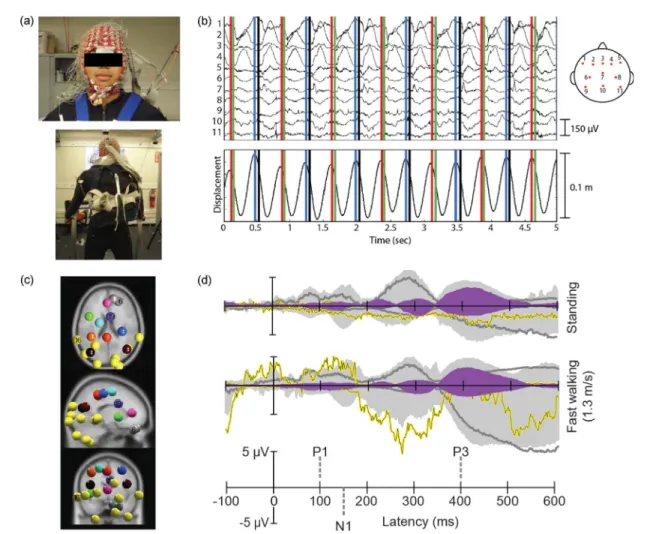
Fig. 2.(a) Photograph of a participant wearing a 256-channel EEG cap with motion capture emitters placed on the cap and along standard positions in a full-body motion capture suit; the same participant seen from behind wearing a backpack containing the EEG amplifier system. (b) Spontaneous EEG activity recorded for selected channels (as indicated on the figure’s head to the right), showing single-channel activity during several stride cycles and (below) vertical center-of-mass displacement during the gait cycle. The gait cycle begins and ends with the left toe raised (red vertical lines). The other vertical lines indicate the timing of the left heel strike (black vertical lines), the right toe lift (blue vertical lines), and the right heel strike (green vertical lines). (c) Centroids of independent component (IC) clusters (colored spheres) visualized in the Montreal Neurological Institute brain volume in horizontal, sagittal, and coronal views. The gray and yellow spheres represent eye and neck muscle activity clusters, respectively. The spheres of other colors mark the centroids of brain IC clusters. (d) Grand-mean event-related potential (ERP) envelopes (maximum and minimum channel ERP values for each latency) time-locked to target stimulus onset for two different movement conditions. The light gray shaded area shows the summed back-projection of all IC clusters. The purple shaded area shows the back-projected contribution of the IC cluster with equivalent dipoles in or near the anterior cingulate cortex [19 in (d)]. The yellow and dark gray traces show the back-projected contribution of a representative neck muscle cluster [16 in (d)] and a representative eye movement cluster [6 in (d)], respectively.
fabricated a polydimethylsiloxane (PDMS)-based dry elec-trode for long-term ECG measurements [50]. Both studies indicate that long-term biopotential signal monitoring is possible using fabric-based electrodes [48]. The test results also indicate that the performance of fabric-based electro-des is comparable to that of commercial Ag/AgCl electroelectro-des for ECG applications. More recently, Lin et al. developed foam-based sensors wrapped in conductive fabric materials to acquire forehead EEG signals [34] [see Fig. 1(d)]. They also showed that the foam-based sensors have the potential to reduce motion artifacts when obtaining measurements from a freely moving user using conventional wet electrodes. Compared with dry MEMS-based EEG sensors, fabric-based sensors are relatively comfortable and nonin-vasive. However, fabric-based sensors for acquiring EEG signals at sites covered with hair require further improve-ment because the contact area of the skin-electrode interface is significantly reduced by the presence of hair.
In addition to the previously described types of dry sensors, Matthews et al. proposed a hybrid dry sensor for EEG measurements [53]–[55] [see Fig. 1(e)]. This sensor, which combines high-impedance resistive and capacitive characteristics, contacts the scalp surface without any skin preparation and is dependent on the high contact impe-dance between the scalp and the electrode. However, these sensors possess hard substrates, which led to discomfort or even pain on the scalp surface when force was applied for attachment. The hard substrate also led to EEG signal dis-tortions caused by motion effects [31]. Moreover, the fabrication cost for a high contact impedance electrode may be higher than for other types of dry EEG electrodes, such as fabric-based electrodes. In response to this con-cern, Cristian et al. presented a new, low-cost dry EEG sensor made of flexible metal-coated polymer bristles [56] [see Fig. 1(f)]. They suggested that the dry bristle sensors produce high-quality EEG recordings and are thus suitable for a wide range of EEG studies and BCI applications. Moreover, Liao et al. proposed a novel dry spring-loaded
contact probe EEG sensor for measuring EEG signals, es-pecially at sites covered with hair [33] [see Fig. 1(g)]. Each of the spring-loaded probes is used to attach the sensors tightly to the scalp surface. These probes were designed to be inserted into a thin plate for additional conductivity. Most importantly, this thin plate is flexible so that it will fit the scalp surface well when applying force to the sensor. The spring-loaded probes and thin plate serve as a buffer to avoid causing pain when force is applied to the sensor and to improve the skelectrode contact impedance. An in-jection molding process is used to package the sensors, which can decrease the fabrication cost of the entire acquisition system, depending on the cost of the electro-des. Test results have demonstrated the feasibility of using dry spring-loaded probe electrodes for measuring EEG signals at sites covered with hair.
Noncontact (capacitive) sensors with spaces between the electrode and the body and without skin preparation also have the potential to acquire EEG signals [57] [see Fig. 1(h)]. However, dry capacitive sensors are sensitive to motion artifacts [31], and Gert et al. indicated that design-ing an amplifier to acquire signals with such high source impedance remains a challenging issue [31]. Because of these issues, dry capacitive sensors require further improvement.
C. Nano- and Microtechnology Sensors
Nanoelectronic device technology holds promise for the next generation of electronics, leading to advancement through the development of novel sensors, flexible, trans-parent, and wearable high-performance electronics, smart bandages, optoelectronics, on-chip electronic-optical cou-pling, radiation hard electronics, and communications and processing electronics for deployable sensor platforms. For example, researchers in Spain and the United Kingdom have developed a new method for measuring electrical activity in the brain that uses sensors constructed from carbon nanotubes (CNTs) [58]. Ruffini et al. also demon-strated the use of carbon-nanotube-based dry sensors in biopotential signal studies [44], [58]. In the future, active, short-range communication of information between body-worn sensors may be enabled by spin-torque nano-oscillators (STNOs). These devices are being actively studied as a technology for magnetic memory applications, and may also be used as miniature frequency-agile radio frequency (RF) sources and sensitive magnetic field detec-tors [59]. For example, the extremely low-power (250 pW) transmission of microwave radiation through air has been demonstrated from a discrete 50-nm device, with broad-band frequency agility over at least four octaves of fre-quency without conjugate matching [60], enabling a new class of low-power wireless communications for wearable sensor technologies. Bio-inspired nanotechnologies mim-icking gecko foot structures are being developed as engineered reversible adhesive devices to enable mm- to cm-scale robotic platforms to crawl on surfaces [61] and
Fig. 3.Wearable EEG devices: (a) Emotiv, (b) NeuroSky, (c) Zeo, (d) StarLab, (e) EmSense, (f) nia Game Controller, (g) Mindo 4 with dry foam electrodes, and (h) Mindo 16 with dry spring-loaded probe sensors.
may be applicable to future biocompatible dry electrode adhesives for EEG sensors. Maturing micro- and nano-electromechanical system (MEMS/NEMS) technologies also hold promise for novel actuation devices, tactors and state-measurement devices. In the future, carbon-based or other biocompatible nanoscale sensing technologies may be envisioned that could be injected into blood vessels, cross the blood-brain barrier, attach to specific neurons or cells, sense the desired signals and transmit to an external receiver though the intact skull. While a very high spatial-temporal resolution of the EEG signals could potentially be provided in this manner, the resolution of many significant technical and ethical considerations will be required to facilitate the use of such technologies, similar to the exist-ing drug-development protocols.
D. Multimodality Sensors
In addition to those sensors that are only used to measure EEG signals, the simultaneous recording of hemo-dynamic responses using NIRS and neural activity using EEG through multimodality sensors while users receive stimulation is also a critical issue in the neuroscience do-main. NIRS and EEG techniques are based upon different imaging principles, and therefore, cross-validation can improve our understanding of both the relationship be-tween hemodynamic responses and neural activity under-lying cortical activation and the biophysics behind the measurement techniques themselves. Furthermore and critical to ABCIs, simultaneous NIRS and EEG imaging can provide novel insight into the phenomenon of neurovas-cular coupling changes for studying human brain mapping in everyday environments. Takeuchi et al. developed a head cap for both NIRS and EEG whole-brain imaging [62], and neurohemodynamic changes have been ad-dressed in detail. Cooper et al. also proposed a novel probe design for simultaneous EEG and NIRS imaging of cortical activation in the human brain [63]. To accomplish this imaging, anBopto-electrode probe[ was designed to house both an EEG electrode and an optical fiber bundle. This probe illustrates the potential applications of simultaneous NIR and EEG imaging. Although such novel ABCIs could provide simultaneous EEG and NIRS imaging, conductive gels and proper skin preparation are still required on the scalp skin surface at the electrode sites. In the future, we envision that dry EEG sensors will be integrated into simultaneous EEG and NIRS imaging.
I I I .
I N T E G R A T I O N O F B I O S E N S O R S A N D
S E N S I N G D E V I C E S I N T O A B C I S Y S T E M S
A. Current Developments in Biosensing Device Technology
Basic System Design: Traditionally, EEG systems have used relatively bulky, wired laboratory or clinically
oriented equipment to measure EEG signals. Analog EEG signals are directly transmitted to a sensing device through wires that limit the routine activity of users. Recently, small wearable EEG acquisition devices have become available that are capable of recording EEG signals without hindering the user in the performance of routine tasks in everyday operational environments (for a detailed over-view of wearable EEG-based BCI technology, refer to [64], [65]). Wearable EEG systems have been developed using headbands [66]–[68], headphones [69], caps [70], helmets [55], headsets [71], [72] and even cat ears [73]. Their shape determines the potential positions of the electrodes, which limits their functionality; however, the design of wearable EEG devices must consider both functionality and appearance. For example, a forehead-based system would not effectively utilize evoked potentials from the visual cortex, which are typically measured from electrodes located on the posterior portion of the head.
Analog Front-End: An instrumentation amplifier is used to augment the small signals received from electrodes and requires careful design to achieve a high signal-to-noise ratio (SNR). The common-mode rejection ratio (CMRR) is usually used to evaluate amplifier instruments, i.e., an amplifier with a higher CMRR reduces the common-mode noise in measurements. The traditional implementation of amplifiers uses a three op-amp configuration, which re-quires precise matching of the resistors used in the feed-back network to achieve a high CMRR. Such matching usually requires expensive laser-trimmed resistors that consume a significant amount of chip area. One technique for overcoming this problem is to use current feedback instrumentation amplifiers [74]–[76], which requires two resistors to adjust the gain, but the resistors are not re-quired to be matched. The advantage of a current feedback instrumentation amplifier is that it could be implemented using CMOS techniques and could reduce the chip area due to a lower resistor count, which is critical for amplifier miniaturization.
During the acquisition of EEG signals, which contain low frequencies, flicker noise (1/f noise) and input-referred dc offsets are undesirable signals that must be suppressed to achieve a high SNR. Flicker noises are signals in which the spectrum is inversely proportional to the noise. Input-referred dc offsets are caused by electrode polarization. Depending on the electrode placement, these dc offsets can be large and can easily saturate high-gain instrumentation amplifiers. Chopper amplifiers are com-monly used for reducing the noise associated with op-amp imperfections and input-referred dc offset voltage. This modulation transposes the signal to the odd harmonic frequencies of the chopper frequency. After modulation, the signals are amplified and demodulated back to the original frequency band. The input offset voltage and noise are demodulated to the higher harmonics of the chopper frequency in the second multiplier. Another technique for
reducing input-referred dc offsets is autozeroing. Auto-zeroing amplifiers typically operate in one of two phases: the sampling phase or the output phase. Autozeroing can meet low-offset requirements but may not be suitable for low-power applications [77] because it requires the offset to be stored on a capacitor that may be relatively large and may thus increase power consumption. This issue should be resolved before it is used in EEG-monitoring appli-cations. In [78], the authors apply autozeroing to an instrument amplifier and use a low-pass filter for noise-cancelling feedback.
Transmission Medium: EEG acquisition devices are usually designed for low-power consumption and low cost. The acquired signals are transmitted to another device with a high-end processor through a transmission medium. Currently, most projects use conventional wireless tech-nology such as Bluetooth [66]–[68], 802.15.4/Zigbees [70], or RF transmitters [55], [69], [71], [72]. Bluetooth was developed as a replacement for cables and is a common feature in laptops and cellular phones. However, it also has a heavy protocol stack and high power consumption. In contrast, the protocol stacks of RF transmitters and 820.15.4/ Zigbees are light and more efficient in terms of their power use, but they are less common in laptops.
Multichannel EEG transmission should have a high data rate and high power consumption. Compression tech-nology can be used to reduce the data rate. Lossless com-pression schemes are reliable but have lower comcom-pression ratios [79], [80]. Lossy compression schemes [81], [82] have high compression ratios but introduce artifact issues.
Table 1 summarizes the EEG acquisition devices men-tioned above [55], [66]–[71], which are responsible for wirelessly acquiring EEG signals and transmission data. Signal processing requires high computation power and is handled by hosts such as computers and cellular phones. Therefore, acquisition devices could be implemented with low-cost microcontrollers. Microcontrollers acquire digi-tized EEG signals from an ADC through a serial interface. According to the Nyquist-Shannon sampling theorem, the sampling rate should be greater than twice the maximum frequency of the acquired EEG signals. For example, SSVEP signals with a maximum frequency of 50 Hz are acquired, using a sampling rate of 128 Hz. A higher sam-pling rate and a higher resolution provide better accuracy for EEG signals but also increase the data rate and power consumption.
B. Technologies That Will Change Future Biosensing Techniques for ABCIs
Miniaturization of Power Sources: The miniaturization of power sources will enable the development of lightweight, portable sensor technologies. Li-ion battery technology, in which the energy density is proportional to the voltage of the cell, is being advanced through the development of high-voltage cathodes and electrolytes capable of operating at those higher voltages. Researchers at the U.S. Army Research Laboratory (ARL) have developed breakthrough electrolytes that allow for the operation of high-voltage lithium-nickel-manganese spinel oxides at 4.7 V against Li with 99% Coulombic efficiency and 85% capacity
retention after 200 cycles [83]. ARL researchers have also developed a stabilized 4.8-V lithium-cobalt-phosphate high-voltage cathode. Such a stabilized cathode can be cycled against Li with only 12% capacity loss compared with 33% capacity loss for the same cathode without stabi-lization after 10 cycles. This increased capacity retention has not been previously achieved [84]. Power sources using energetic materials based on nuclear isomers are being investigated for future power generation. Radio-isotopes possess intrinsic energy storage that is more than 100 times greater than that of fuel cells and batteries, and they have the potential ability to release that energy upon demand. Recent results have demonstrated an induced energy release from isomers using neutrons [85], and alpha-based isotope batteries are being studied as a long-lived power source with an anticipated 50-year lifetime [86], which exceeds current battery lifetimes. Power con-version systems are being investigated at the cubic-millimeter, milligram scale to create high-efficiency, high-power-density actuation systems. CMOS design tech-niques, such as specific guard-rings and floating wells, that enable on-chip handling of more than 20 V in a 130-nm 1.2-V CMOS process have been demonstrated [85] and represent a significant step towards the high voltages re-quired for novel actuators. MEMS-based ultraminiature air-core inductors with high-quality (Q) factors ðQ > 20Þ and inductance densities ð> 100 nanohenries/mm2Þ have been shown to outperform most inductors fabricated with thin magnetic films, forming the basis for high-efficiency, high-frequency (> 20 MHz) boost converters [88]. Novel techniques using capillary wicking are being used to shape magnetic or high-k dielectric nanoparticles into complex 3-D microstructures [89], resulting in power conversion device components that show significant improvements in capacitance or inductance with no parasitic effect on the quality factor (i.e., efficiency) up to 200 MHz. Nano-electronic devices are also being explored, including super-capacitors for energy storage [90] and graphene-based metal-insulator-metal (MIM) diode structures for harvest-ing energy and rectifyharvest-ing antenna structures.
The design of power-efficient control theory is another approach to achieving reduced power requirements for sensor array information processing. Bio-inspired and bio-plausible control theory, in which the control laws could, in principle, be implemented on the neural substrate of simple insects, is being investigated for applications in which computational power is extremely limited. Research groups, such as Richard Murray’s group at Caltech, are developing control theories that focus on tight coupling between the temporal dynamics of the system and the temporal properties of the control algorithm [91]. These bioplausible algorithms use approaches that involve parallel, asynchronous information processing and emerg-ing circuit technologies, such as those beemerg-ing developed in the DARPA-sponsored SyNAPSE (Systems of Neuromor-phic Adaptive Plastic Scalable Electronics) project. These
technologies could, for example, facilitate the fusion of inputs from different sensor modes that have distinctly different time constants, such as EEG and fMRI, enhanc-ing localization of the brain response for improved BCI interactions.
Mobile Human Brain Imaging for Real-Life Applications: Traditional neuroscientific approaches to measuring and imaging the hemodynamics coupled to neural activity that accompany cognitive processing (e.g., fMRI, PET) permit only minimal movement of the participants’ hands or feet. This restriction of the participants’ movement range is necessary to avoid movement of the signal-generating volume, i.e., the participants’ brain, which cannot be fol-lowed by the heavy sensor arrays [92]. In addition, brain imaging techniques that directly measure electrical activity originating from the neural populations underlying cogni-tive processing further restrict the movement of the participants’ head and eyes to avoid contamination of the signal of interest with current source activity originating from the neck, supracranial, and facial muscles as well as strong current source activity stemming from horizontal and vertical eye movement. From the standpoint of em-bodied cognition, however, cognitive processes are tightly coupled to physical activity and make use of our physical structure to reach behavioral goals [92], [93].
Consequently, results from traditional imaging ap-proaches can only explain the neural basis of cognitive processes that are restricted to specific recording environ-ments and do not necessarily reflect natural cognitive processing. Thus far, no investigation has compared hu-man brain dynamics during free movement in the world with the brain dynamics of restricted behavior during traditional laboratory experiments. New results from ani-mals in motion, however, demonstrate a direct coupling of behavioral states with brain states [94], [95]. These studies support the assumption that changes in behavioral states are accompanied by changes in brain dynamic states to adapt to differences in incoming sensory information. While the restrictions of traditional brain imaging ap-proaches do not invalidate theoretical views formed on the basis of such experiments, several recent investigations in human participants point to a tight coupling of motor behavior and, for example, attention and spatial cognition. An example of such coupling was described by Wykotska and colleagues [96], demonstrating a direct influence of (planned) movement on target detection when targets were defined by features relevant to the movement. The authors showed that shape-defined targets were detected faster when participants planned to grasp a target object while luminance-defined targets were detected faster when participants planned on pointing towards the target object. An even more pronounced coupling of active be-havior with human brain dynamics could be expected for spatial cognitive processes that are tightly coupled to and make efficient use of idiothetic information derived from
the vestibular and proprioceptive system [97]. Spatial up-dating of one’s own position and orientation is a prere-quisite of successful spatial orientation but relies heavily on idiothetic information that is absent in traditional brain imaging approaches. As a consequence, the brain dynamics accompanying active spatial orientation including physical rotation will provide novel insights into the brain dynamics associated with idiothetic information processing and will thus help us better understand the brain dynamics asso-ciated with spatial orientation. To overcome the restric-tions of traditional imaging approaches and to investigate the brain dynamics accompanying active behavior in hu-mans, our group, recently developed and demonstrated a mobile brain/body imaging (MoBI) approach [91], [98]–[100]. Through various experiments, we demonstrat-ed the feasibility of this concept and revealdemonstrat-ed interesting insights into the cognitive dynamics of attention during treadmill walking [98] and into the coupling of cortical activity with gait-cycle phases [100]. However, these experiments were clearly restricted because of the hard-wiring of the EEG system. In [99], the analysis of human brain dynamics for subjects who were standing and walking at different speeds was demonstrated. However, the cable sway associated with the displacement of the participants’ heads while running required extensive template-based artifact rejection (see Fig. 2). In another experiment [Gramann et al., unpublished manuscript], the use of traditional hard-wired EEG sensors restricted the movement range of the participants’ heads when they attempted to actively orient towards laterally placed objects in their environment. In general, in the field of motor control and physical behavior, numerous attempts to study active cognitive processing have been hampered due to these types of issues associated with the tethered nature of standard EEG equipment. Classic examples include EEG studies of shooting that have been largely limited to static targets as opposed to reactive, dynamic targets; studies of golf that have been largely limited to putting as opposed to driving and studies of basketball that have been limited to free-throw shooting as opposed to any other aspect of game-play. These examples demonstrate the need for wireless sensor systems to allow for future investigation and deeper insights into the architecture of active and mobile cognition.
Advanced Flexible Electronics and Display Technologies: Future ABCIs are envisioned to be moving towards seam-less integration with users’ clothing and the environment; advancements in technologies such as flexible electronics and displays, which are maturing to provide lightweight, rugged, and ultralow-power flexible imaging systems for high-yield manufacturing processes, will enable such im-provements [see, for example, http://flexdisplay.asu.edu/]. Full-color organic light-emitting-diode (OLED) displays have been demonstrated on plastic substrates, and charge-transfer complex molecular OLED systems have been integrated into emissive flexible displays to dramatically
enhance the charge injection and transport properties of organic devices [100]. Flexible reflective displays for system-level integration have transitioned in industry to meet emerging information technology needs for the mobile soldier. Large-area distributions of flexible electro-nic circuits, discrete elements, and sensors are being in-tegrated with flexible solar cells, antennas, and solid-state lighting to enhance next-generation displays. Enhance-ments in carrier transport, brightness, and speed are being investigated with the use of flexible and stretchable silicon or metal technologies [102]. These technologies will enable the creation of high-performance conformable circuits that are needed for more seamless integration of ABCIs into clothing as well as advanced concepts for smart clothes and body armor. Stretchable silicon-integrated circuits that can be applied directly to the skin have recently been demon-strated [103]. These tattoo-like structures are an example of the new epidermal electronics technology that can be used to directly measure vital signs. Such advancements will permit a range of advancements from novel approaches to sensor design to the collection and integration of high-density signals across numerous behavioral and physiolog-ical sources for enhanced ABCI performance.
I V .
T R E N D S I N A B C I s
A. Current Applications of ABCIs
Wet electrodes have their own readout circuit sys-tems and are reliable for clinical applications. For dry/ noncontact electrodes, developing the proper readout de-vice for everyday use is important. Dede-vices with dry elec-trodes are more convenient and comfortable than traditional EEG systems with wet electrodes [31] and are, thus, more practical for use in everyday applications. Although dry/noncontact EEG devices have not been pro-posed or used for clinical applications, many commercial devices use EEG measurements for entertainment (Neu-rosky, Emotiv, StarLab, EmSense, and nia Game Control-ler) [104], [105] and for monitoring personal sleeping status (MyZeo) [106], as shown in Fig. 3(a)–(f). These figures demonstrate that the development of portable EEG devices with dry electrodes has become an important goal for mobile human brain imaging.
Recently, Lin et al. proposed a wearable, wireless EEG device (Mindo) for everyday use [32]. The Mindo 4 EEG device with 4-channel foam electrodes has proven to be reliable for controlling games according to the user’s mental focusing state based on signals from forehead sensor sites [34], as shown in Fig. 3(g). It also has the potential to acquire the EEG status during sleep. Another multichannel EEG device, Mindo 16, which has spring-loaded probe electrodes, was designed by Lin et al. for wirelessly measuring EEG signals, especially at sites with hair, as the corresponding dry sensors have the potential to properly reach the scalp skin through the hair [see Fig. 3(h)]. In addition to wireless EEG
devices with dry contact electrodes, Gert et al. designed a wireless device with non-contact electrodes for measuring both EEG and ECG [107]. There is no doubt that developing a truly wearable, wireless EEG device using dry/noncontact electrodes and extending the limitations of this technique from basic research to clinical applications are important goals. Highly desirable characteristics of future devices include a minimized readout circuit size and easy prepara-tion when using dry electrodes.
B. Future ABCI Applications Based on Advanced Biosensing Technology
Gaming control, homecare, and rehabilitation engi-neering applications are potential future applications of ABCIs in the coming decades. ABCI applications for gam-ing are one of the major focuses of this technology, and existing prototypes demonstrate the feasibility of games controlled by an ABCI [40]–[42]. It is possible that an EEG-based BCI device with novel EEG sensors that is capable of interpreting the cognitive relevance of neuron interactions in the brain will become available and reliable in the near future [108]. Another feasible future trend for ABCIs is remote monitoring, which can be used in homecare and rehabilitation engineering applications [109]. The elderly and ill often prefer living in their own houses to being in a hospital, but living alone can be dangerous because of unpredictable accidents such as falling and epileptic seizures [110]. Remote-sensing and monitoring would enable the remote monitoring of a user’s EEG signals. EEG-based ABCIs may be able to assist with depression and many other psychological and cranial nerve diseases, such as schizophrenia, Parkinson’s disease and seizures, in the
near future. We refer the reader to another paper written by Lance et al. [36] for details of future ABCI applications.
V .
S U M M A R Y
We have studied a wide range of approaches to ABCIs and explored their applications to neuroscientific questions and cognitive engineering. We have provided insights into the fundamental basis of many ABCI techniques and highlighted important considerations for their practical implementation. The miniaturization of sensors, elec-tronics, and power sources; the design of power-efficient information processing; and the emergence of flexible electronics and display technologies have the potential to radically enhance future ABCI capabilities. We hope that these details will help those who are interested in using or developing biosensing techniques for ABCIs to understand the key aspects that should be considered when acquiring measurements or analyzing data.
We have surveyed the large body of literature that discusses studies in which biosensing technologies and devices have been successfully used for ground-breaking and important research on ABCIs and their applications. The development of ABCIs is a rapidly expanding field that is continually evolving to embrace new technologies and real-life applications.h
A c k n o w l e d g m e n t
The authors would like to thank S.-Y. Li for his help in discussing part of the context in this paper.
R E F E R E N C E S
[1] Y.-Y. I. Shih, C.-C. V. Chen, B.-C. Shyu, Z.-J. Lin, Y.-C. Chiang, F.-S. Jaw, Y.-Y. Chen, and C. Chang,BA new scenario for negative functional magnetic resonance imaging signals: Endogenous neu-rotransmission,[ J. Neurosci., vol. 29, pp. 3036–3044, 2009.
[2] Y.-Y. Chen, Y.-Y. I. Shih, Y.-C. Lo, P.-L. Lu, S. Tsang, F.-S. Jaw, and R.-S. Liu,BMicroPET imaging of noxious thermal stimuli in the conscious rat brain,[ Somatosens. Motor Res., vol. 27, pp. 69–81, 2010, 2011/04/13.
[3] E. M. C. Hillman,BOptical brain imaging in vivo: Techniques and applications from animal to man,[ J. Biomed. Opt., vol. 12,
pp. 051402–051428, 2007. [4] N. Li, X. Jia, K. Murari, R. Parlapalli,
A. Rege, and N. V. Thakor,BHigh spatiotemporal resolution imaging of the neurovascular response to electrical stimulation of rat peripheral trigeminal nerve as revealed by in vivo temporal laser speckle contrast,[ J. Neurosci. Methods, vol. 176, pp. 230–236, 2009.
[5] L.-D. Liao, Y.-Y. Chen, C.-T. Lin, J.-Y. Chang, and M.-L. Li,BFunctional transcranial photoacoustic micro-imaging of mouse cerebrovascular cross-section and
hemoglobin oxygenation changes during forepaw electrical stimulation,[ in Proc. Photons Plus Ultrasound: Imaging and Sensing 2011, San Francisco, CA, 2011, vol. 7899, pp. 78 992–78 997.
[6] L.-D. Liao, M.-L. Li, H.-Y. Lai, Y.-Y. I. Shih, Y.-C. Lo, S. Tsang, P. C.-P. Chao, C.-T. Lin, F.-S. Jaw, and Y.-Y. Chen,BImaging brain hemodynamic changes during rat forepaw electrical stimulation using functional photoacoustic microscopy,[ NeuroImage, vol. 52, pp. 562–570, 2010.
[7] S. Ogawa, D. W. Tank, R. Menon, J. M. Ellermann, S. G. Kim, H. Merkle, and K. Ugurbil,BIntrinsic signal changes accompanying sensory stimulation: Functional brain mapping with magnetic resonance imaging,[ Proc. Nat. Acad. Sci., vol. 89, pp. 5951–5955, 1992.
[8] J. A. Detre, W. Zhang, D. A. Roberts, A. C. Silva, D. S. Williams, D. J. Grandis, A. P. Koretsky, and J. S. Leigh,BTissue specific perfusion imaging using arterial spin labeling,[ NMR Biomed, vol. 7, pp. 75–82, Mar. 1994.
[9] B. P. Chugh, J. P. Lerch, L. X. Yu, M. Pienkowski, R. V. Harrison, R. M. Henkelman, and J. G. Sled, BMeasurement of cerebral blood volume in mouse brain regions using micro-computed tomography,[ NeuroImage, vol. 47, pp. 1312–1318, 2009.
[10] H. Dehghani, S. Srinivasan, B. W. Pogue, and A. Gibson,BNumerical modelling and image reconstruction in diffuse optical tomography,[ Philosoph. Trans. Royal Soc. A: Math., Phys. Eng. Sci., vol. 367, pp. 3073–3093, 2009.
[11] L. V. Wang and H.-I. Wu, Biomedical Optics: Principles and Imaging. New York: Wiley, 2007.
[12] G. Gratton and M. Fabiani,BDynamic brain imaging: Event-related optical signals (EROS) measures of the time course and localization of cognitive-related activity,[ Psychonom. Bull. Rev., vol. 5, pp. 535–563, 1998.
[13] E. Moser, M. Meyerspeer, F. P. S. Fischmeister, G. Grabner, H. Bauer, and S. Trattnig,BWindows on the human body in vivo high-field magnetic resonance research and applications in medicine and psychology,[ Sensors, vol. 10, pp. 5724–5757, 2010. [14] A. Gevins, J. Le, N. K. Martin, P. Brickett,
J. Desmond, and B. Reutter,BHigh resolution EEG: 124-channel recording, spatial deblurring and MRI integration methods,[ Electroencephalogr. Clin. Neurophys., vol. 90, pp. 337–358, 1994. [15] C.-T. Lin, R.-C. Wu, S.-F. Liang, W.-H. Chao,
Y.-J. Chen, and T.-P. Jung,BEEG-based drowsiness estimation for safety driving using independent component analysis,[
IEEE Trans. Circuits Syst. Part I: Regular Papers, vol. 52, pp. 2726–2738, 2005. [16] G. K. Wenning, I. Litvan, M. Verny,
K. Ray-Chaudhuri, R. Granata, W. Poewe, and K. Jellinger,BIs EEG useful in the differential diagnosis of Parkinsonism?[ Parkinsonism Rel. Disorders, vol. 4, pp. 79–80, 1998.
[17] S. Sanders, S. Rowlinson, I. Manidakis, C. D. Ferrie, and M. Koutroumanidis, BThe contribution of the EEG technologists in the diagnosis of Panayiotopoulos syndrome (susceptibility to early onset benign childhood autonomic seizures),[ Seizure, vol. 13, pp. 565–573, 2004. [18] N. Thakor,BIn the spotlight:
Neuroengineering,[ IEEE Rev. Biomed. Eng., vol. 2, pp. 18–20, 2009.
[19] N. V. Thakor,BBiopotentials and electro-physiology measurement,[ in The Measurement, Instrumentation, and Sensors Handbook. Boca Raton, FL: CRC, 1999, pp. 74–71.
[20] S. G. Mason and G. E. Birch,BA general framework for brain computer interface design,[ IEEE Trans. Neural Syst. Rehab. Eng., vol. 11, pp. 70–85, 2003.
[21] R. Rao and R. Scherer, BBrain-computer interfacing [in the spotlight],[ IEEE Signal Process. Mag., vol. 27, pp. 152–150, 2010.
[22] Y.-Y. Chen, H.-Y. Lai, S.-H. Lin, C.-W. Cho, W.-H. Chao, C.-H. Liao, S. Tsang, Y.-F. Chen, and S.-Y. Lin,BDesign and fabrication of a polyimide-based microelectrode array: Application in neural recording and repeatable electrolytic lesion in rat brain,[ J. Neurosci. Meth., vol. 182, pp. 6–16, 2009.
[23] T. Yamakawa, T. Yamakawa, T. Inoue, S. Aou, S. Ishizuka, M. Fujii, and M. Suzuki, BMinimally invasive ecog recording using the novel subdural electrodes manipulated by a shape memory alloy guidewire,[ Epilepsia, vol. 52, pp. 201–201, Aug. 2011.
[24] G. Xiaorong, X. Dingfeng, C. Ming, and G. Shangkai,BA BCI-based environmental controller for the motion-disabled,[ IEEE Trans. Neural Syst. Rehab. Eng., vol. 11, pp. 137–140, 2003.
[25] A. S. Royer, A. J. Doud, M. L. Rose, and B. He,BEEG control of a virtual helicopter in 3-dimensional space using intelligent control strategies,[ IEEE Trans. Neural Syst. Rehab. Eng., vol. 18, pp. 581–589, 2010.
[26] J. R. Wolpaw,BBrain-computer interfaces as new brain output pathways,[ J. Physiol.-London, vol. 579, pp. 613–619, Mar. 15, 2007.
[27] A. Nijholt and D. Tan,BBrain-computer interfacing for intelligent systems,[ IEEE Intell. Syst., vol. 23, pp. 72–79, 2008. [28] K. D. Nielsen, A. F. Cabrera, and
N. Omar Feix Do,BEEG based BCI-towards a better control. Brain-computer interface research at aalborg university,[ IEEE Trans. Neural Syst. Rehab. Eng., vol. 14, pp. 202–204, 2006.
[29] G. R. Muller-Putz, R. Scherer, C. Neuper, and G. Pfurtscheller,BSteady-state somatosensory evoked potentials: Suitable brain signals for brain-computer interfaces?[ IEEE Trans. Neural Syst. Rehab. Eng., vol. 14, pp. 30–37, 2006. [30] S. Mason, M. Jackson, and G. Birch,
BA general framework for characterizing studies of brain interface technology,[
Ann. Biomed. Eng., vol. 33, pp. 1653–1670, 2005.
[31] Y. M. Chi, T. P. Jung, and G. Cauwenberghs, BDry-contact and noncontact biopotential electrodes: Methodological review,[ IEEE Rev. Biomed. Eng., vol. 3, pp. 106–119, 2010.
[32] C.-T. Lin, L.-W. Ko, J.-C. Chiou, J.-R. Duann, R.-S. Huang, T.-W. Chiu, S.-F. Liang, and T.-P. Jung,BNoninvasive neural prostheses using mobile and wireless EEG,[ Proc. IEEE, vol. 96, pp. 1167–1183, 2008. [33] L.-D. Liao, I. J. Wang, S.-F. Chen,
J.-Y. Chang, and C.-T. Lin,BDesign, fabrication and experimental validation of a novel dry-contact sensor for measuring electroencephalography signals without skin preparation,[ Sensors, vol. 11, pp. 5819–5834, 2011.
[34] C. T. Lin, L. D. Liao, Y. H. Liu, I. J. Wang, B. S. Lin, and J. Y. Chang,BNovel dry polymer foam electrodes for long-term EEG measurement,[ IEEE Trans. Biomed. Eng., vol. 58, pp. 1200–1207, 2011. [35] S. Makeig, C. Kothe, T. Mullen,
N. Bigdely-Shamlo, Z. Zhang, and K. Kreutz-Delgado,BEvolving signal processing for brain-computer interfaces,[ Proc. IEEE, 2012, DOI: 10.1109/JPROC.2012. 2185009.
[36] B. J. Lance, S. E. Kerick, A. J. Ries, K. S. Oie, and K. McDowell*,BBrain-computer interface technologies in the coming decades,[ Proc. IEEE, 2012, DOI: 10.1109/JPROC.2012.2184830. [37] M. Roberto,BThe electrode-skin interface
and optimal detection of bioelectric signals,[ Physiologic. Measur., vol. 31, 2010.
[38] A. Searle and J. Kirkup,BA direct comparison of wet, dry and insulating biolelectric recording electrodes,[ Physiologic. Measur., vol. 21, pp. 271–283, 2000.
[39] T. W. Picton, S. Bentin, P. Berg, E. Donchin, S. A. Hillyard, R. Johnson, G. A. Miller, W. Ritter, D. S. Ruchkin, M. D. Rugg, and M. J. Taylor,BGuidelines for using human event-related potentials to study cognition: Recording standards and publication criteria,[ Psychophysiology, vol. 37, pp. 127–152, Mar. 2000.
[40] N. A. Alba, R. J. Sclabassi, M. Sun, and X. T. Cui,BNovel hydrogel-based preparation-free EEG electrode,[ IEEE Trans. Neural Syst. Rehab. Eng., vol. 18, pp. 415–423, Aug. 2010.
[41] O. Sohn and D. Kim,BTheoretical and experimental investigation of the swelling behavior of sodium polyacrylate superabsorbent particles,[ J. Appl. Polymer Sci., vol. 87, pp. 252–257, Jan. 10, 2003. [42] I. Volosyak, D. Valbuena, T. Malechka,
J. Peuscher, and A. Gra¨ser,BBrain-computer interface using water-based electrodes,[ J. Neural Eng., vol. 7, p. 066007, 2010. [43] T. C. Ferree, P. Luu, G. S. Russell, and
D. M. Tucker,BScalp electrode impedance, infection risk, and EEG data quality,[ Clin. Neurophys., vol. 112, pp. 536–544, 2001.
[44] G. Ruffini, S. Dunne, E. Farre´s, J. Marco-Pallare´s, C. Ray, E. Mendoza, R. Silva, and C. Grau,BA dry electrophysiology electrode using CNT arrays,[ Sens. Actuators A: Phys., vol. 132, pp. 34–41, 2006.
[45] P. Griss, P. Enoksson, H. K. Tolvanen-Laakso, P. Merila¨inen, S. Ollmar, and G. Stemme, BMicromachined electrodes for biopotential
measurement,[ IEEE J. Microelectromech. Syst., vol. 10, pp. 10–15, 2001.
[46] Y. Wang, K. Guo, W.-H. Pei, Q. Gui, X.-Q. Li, H.-D. Chen, and J.-H. Yang,BFabrication of dry electrode for recording bio-potentials,[ Chinese Phys. Lett., vol. 28, p. 010701, 2011. [47] P. Griss, H. K. Tolvanen-Laakso,
P. Merila¨inen, and G. Stemme, BCharacterization of micromachined spiked biopotentials electrodes,[ IEEE Trans. Biomed. Eng., vol. 49, pp. 597–604, 2002.
[48] G. Anna, S. Hansen, and J. Mu¨ller, BNovel dry electrodes for ECG monitoring,[ Physiologic. Measur., vol. 28, p. 1375, 2007. [49] K. P. Hoffmann and R. Ruff,BFlexible
dry surface-electrodes for ECG long-term monitoring,[ in Proc. IEEE 29th Annu. Int. Conf. Eng. Med. Biol. Soc., EMBS 2007, 2007, pp. 5739–5742.
[50] J.-Y. Baek, J.-H. An, J.-M. Choi, K.-S. Park, and S.-H. Lee,BFlexible polymeric dry electrodes for the long-term monitoring of ECG,[ Sens. Actuators A: Phys., vol. 143, pp. 423–429, 2008.
[51] L. Beckmann, C. Neuhaus, G. Medrano, N. Jungbecker, M. Walter, T. Gries, and S. Leonhardt,BCharacterization of textile electrodes and conductors using standardized measurement setups,[ Physiologic. Measur., vol. 31, p. 233, 2010. [52] P. J. Xu, H. Zhang, and X. M. Tao,
BTextile-structured electrodes for electrocardiogram,[ Textile Progr., vol. 40, pp. 183–213, 2008.
[53] W. S. Eric, T. Peter, A. S. William, M. Tobin, M. V. Theresa, and M. Robert,BA novel dry electrode for brain-computer interface,[ in Proc. 13th Int. Conf. Human-Computer Interaction. Part II: Novel Interaction Methods and Techniques, San Diego, CA, 2009. [54] R. Matthews, N. J. McDonald, H. Anumula, J. Woodward, P. J. Turner, M. A. Steindorf, K. Chang, and J. M. Pendleton,BNovel hybrid bioelectrodes for ambulatory zero-prep EEG measurements using multi-channel wireless EEG system,[ Lecture Notes in Computer Science, vol. 4565, pp. 137–146, 2007.
[55] R. Matthews, P. J. Turner, N. J. McDonald, K. Ermolaev, T. M. Manus, R. A. Shelby, and M. Steindorf,BReal time workload classification from an ambulatory wireless EEG system using hybrid EEG electrodes,[ in Proc. IEEE 30th Annu. Int. Conf., Eng. Med. Biol. Soc., EMBS 2008, 2008, pp. 5871– 5875.
[56] G. Cristian, C. D. Voinescu, and S. Fazli, BBristle-sensors-low-cost flexible passive dry EEG electrodes for neurofeedback and BCI applications,[ J. Neural Eng., vol. 8, p. 025008, 2011.
[57] R. Matthews, N. J. McDonald, I. Fridman, P. Hervieux, and T. Nielsen,BThe invisible electrodeVZero prep time, ultra low capacitive sensing,[ in Proc. 11th Int. Conf. Human Computer Int., Las Vegas, NV, 2005, pp. 22–27.
[58] G. Ruffini, S. Dunne, L. Fuentemilla, C. Grau, E. Farr, J. Marco-Pallar, P. C. P. Watts, and S. R. P. Silva,BFirst human trials of a dry electrophysiology sensor using a carbon nanotube array interface,[ Sens. Actuators A: Phys., vol. 144, pp. 275–279, 2008.
[59] J. A. Katine and E. E. Fullerton,BDevice implications of spin-transfer torques,[ J. Magn. Magn. Mater., vol. 320, pp. 1217–1226, Apr. 2008.
[60] A. E. Wickenden, C. Fazi, B. Huebschman, R. Kaul, A. C. Perrella, W. H. Rippard, and M. R. Pufall,BSpin Torque Nano Oscillators as Potential Terahertz (THz) Communications Devices,[ Army Research Laboratory Tech. Rep., 2009, vol. ARL-TR-4807.
[61] H. Lee, B. P. Lee, and P. B. Messersmith, BA reversible wet/dry adhesive inspired by mussels and geckos,[ Nature, vol. 448, pp. 338–U334, Jul. 19, 2007.
[62] M. Takeuchi, E. Hori, K. Takamoto, A. Tran, K. Satoru, A. Ishikawa, T. Ono, S. Endo, and H. Nishijo,BBrain cortical mapping by simultaneous recording of functional near infrared spectroscopy and
electroencephalograms from the whole brain during right median nerve stimulation,[ Brain Topogr., vol. 22, pp. 197–214, 2009.
[63] R. J. Cooper, N. L. Everdell, L. C. Enfield, A. P. Gibson, A. Worley, and J. C. Hebden, BDesign and evaluation of a probe for simultaneous EEG and near-infrared imaging of cortical activation,[ Phys. Med. Biol., vol. 54, pp. 2093–2102, Apr. 7, 2009. [64] C.-T. Lin, L.-W. Ko, M.-H. Chang,
J.-R. Duann, J.-Y. Chen, T.-P. Su, and T.-P. Jung,BReview of wireless and wearable electroencephalogram systems and brain-computer interfacesVA mini-review,[ Gerontology, 2009. [65] A. Casson, D. Yates, S. Smith, J. Duncan,
and E. Rodriguez-Villegas,BWearable electroencephalography,[ IEEE Eng. Med. Biol. Mag., vol. 29, pp. 44–56, 2010. [66] C.-T. Lin, L.-W. Ko, C.-J. Chang, Y.-T. Wang,
C.-H. Chung, F.-S. Yang, J.-R. Duann, T.-P. Jung, and J.-C. Chiou,BWearable and wireless brain-computer interface and its applications,[ in Foundations of Augmented Cognition. Neuroergonomics and Operational Neuroscience, vol. 5638, D. Schmorrow, I. Estabrooke, and M. Grootjen, Eds. Berlin, Germany: Springer, 2009, pp. 741–748.
[67] Y. M. Chi, P. Ng, E. Kang, J. Kang, J. Fang, and G. Cauwenberghs,BWireless non-contact cardiac and neural monitoring,[ in Proc. Wireless Health 2010, San Diego, California, 2010, pp. 15–23.
[68] Y.-T. Wang et al.,BA cell-phone-based brain-computer interface for communication in daily life,[ J. Neural Eng., vol. 8, p. 025018, 2011.
[69] T. Torfs, V. Leonov, R. F. Yazicioglu, P. Merken, C. V. Hoof, R. J. M. Vullers, and B. Gyselinckx,BWearable autonomous wireless electro-encephalography system fully powered by human body heat,[ in Proc. 2008 IEEE Sensors, 2008, pp. 1269–1272. [70] A. Riera, S. Dunne, I. Cester, and G. Ruffini,
BSTARFAST: A wireless wearable EEG/ECG biometric system based on the ENOBIO sensor,[ in Proc. 5th pHealth Workshop Wearable Micro Nanosyst. Pers. Health (pHealth’08), 2008.
[71] L. Brown, J. V. D. Molengraft, R. F. Yazicioglu, T. Torfs, J. Penders, and C. V. Hoof,BA low-power, wireless, 8-channel EEG monitoring headset,[ in Proc. IEEE 2010 Annu. Int. Conf. Eng. Med. Biol. Soc. (EMBC), 2010, pp. 4197–4200. [72] M. K. Mukerjee,BNeuroPhone:
Brain-mobile phone interface using a wireless EEG headset,[ MobiHeld, 2010 [73] Neurowear, Necomimi, 2011. [Online].
Available: http://neurowear.net/?p=28 [74] R. Martins, S. Selberherr, and F. A. Vaz,
BA CMOS IC for portable EEG acquisition
systems,[ IEEE Trans. Instrum. Measur., vol. 47, pp. 1191–1196, 1998. [75] S.-B. Maryam, K. L. Rakesh, and
K. S. Dinesh,BA low-power and compact analog CMOS processing chip for portable ECG recorders,[ in Proc. 2005 Asian Solid-State Circuits Conf., 2005, pp. 473–476. [76] H. Chun-Chieh, H. Shao-Hang, C. Jen-Feng,
L. D. Van, and L. Chin-Teng,BFront-end amplifier of low-noise and tunable BW/gain for portable biomedical signal acquisition,[ in Proc. 2008 IEEE Int. Symp. Circuits Syst, ISCAS 2008, 2008, pp. 2717–2720. [77] M. Belloni, E. Bonizzoni, A. Fornasari, and
F. Maloberti,BA micropower chopperVCDS operational amplifier,[ IEEE J. Solid-State Circuits, vol. 45, pp. 2521–2529, 2010. [78] G. Costa, A. Arnaud, and M. Miguez,
BA precision autozero amplifier for EEG signals,[ in Proc. 23rd Symp. Integr. Circuits Syst. Design, Sa˜o Paulo, Brazil, 2010, pp. 28–32.
[79] E. Chua and W. C. Fang,BMixed bio-signal lossless data compressor for portable brain-heart monitoring systems,[ IEEE Trans. Consumer Electron., vol. 57, pp. 267–273, Feb. 2011.
[80] N. Sriraam and C. Eswaran,BAn adaptive error modeling scheme for the lossless compression of EEG signals,[ IEEE Trans. Inf. Technol. Biomed., vol. 12, pp. 587–594, Sep. 2008.
[81] J. R. Tolbert, P. Kabali, S. Brar, and S. Mukhopadhyay,BAn accuracy aware low power wireless EEG unit with information content based adaptive data compression,[ in Proc. 2009 IEEE Annu. Int. Conf. Eng. Med. Biol. Soc., EMBC 2009, 2009, pp. 5417–5420.
[82] G. Higgins, S. Faul, R. P. McEvoy, B. McGinley, M. Glavin, W. P. Marnane, and E. Jones,BEEG compression using JPEG2000: How much loss is too much?[ in Proc. 2010 IEEE Annu. Int. Conf. Eng. Med. Biol. Soc. (EMBC 2010), 2010, pp. 614–617.
[83] A. von Cresce and K. Xu,BElectrolyte additive in support of 5 V Li ion chemistry,[ J. Electrochem. Soc., vol. 158, pp. A337–A342, 2011.
[84] J. L. Allen, T. R. Jow, and J. Wolfenstine, BImproved cycle life of Fe-substituted LiCoPO(4),[ J. Power Sources, vol. 196, pp. 8656–8661, Oct. 15, 2011. [85] S. A. Karamian and J. J. Carroll,BCross
section for inelastic neutronBacceleration[ by (178)Hf(m2),[ Phys. Rev. C, vol. 83, Feb. 9, 2011.
[86] M. S. Litz, G. Merkel, N. R. Pereira, C. N. Boyer, G. E. Holland, J. W. Schumer, J. F. Seely, L. T. Hudson, and J. J. Carroll, BAnomalous fluorescence line intensity in megavoltage bremsstrahlung,[ Phys. Plasmas, vol. 17, Apr. 2010. [87] X. Lin, C. Meyer, C. M. Dougherty,
S. Bedair, B. Morgan, D. P. Arnold, and R. Bashirullah,BTowards miniature step-up power converters for mobile microsystems,[ in Proc. IEEE 2011 Twenty-Sixth Annu. Appl. Power Electron. Conf. Expo., 2011, pp. 1451–1455.
[88] C. D. Meyer, S. S. Bedair, B. C. Morgan, and D. P. Arnold,BHigh-inductance-density, air-core, power inductors, and transformers designed for operation at 100–500 MHz,[ IEEE Trans. Magn., vol. 46, pp. 2236–2239, Jun. 2010.
[89] S. S. Bedair, C. D. Meyer, and B. Morgan, BClosed core inductor and high-K dielectric capacitor fabrication through evaporation
driven nanoparticle assembly in capillaries,[ J. Appl. Phys., vol. 109, Apr. 1, 2011. [90] L. T. Le, M. H. Ervin, H. W. Qiu, B. E. Fuchs,
and W. Y. Lee,BGraphene supercapacitor electrodes fabricated by inkjet printing and thermal reduction of graphene oxide,[ Electrochem. Commun., vol. 13,
pp. 355–358, Apr. 2011. [91] S. Han, A. Censi, A. D. Straw, and
R. M. Murray,BA bio-plausible design for visual pose stabilization,[ in Proc. Int. Conf. Intell. Robots Syst., 2010, pp. 5679–5686.
[92] S. Makeig, K. Gramann, T. P. Jung, T. J. Sejnowski, and H. Poizner, BLinking brain, mind and behavior,[ Int. J. Psychophys., vol. 73, pp. 95–100, Aug. 2009.
[93] A. Clark,BAn embodied cognitive science?[ Trends in Cogn. Sci., vol. 3, pp. 345–351, Sep. 1999.
[94] G. Maimon, A. D. Straw, and
M. H. Dickinson,BActive flight increases the gain of visual motion processing in Drosophila,[ Nature Neurosci., vol. 13, pp. 393–399, Mar. 2010.
[95] C. M. Niell and M. P. Stryker,BModulation of visual responses by behavioral state in mouse visual cortex,[ Neuron, vol. 65, pp. 472–479, Feb. 25, 2010.
[96] A. Wykowska, A. Schubo, and B. Hommel, BHow you move is what you see: Action planning biases selection in visual search,[ J. Exper. Psychology-Human Perception Perform., vol. 35, pp. 1755–1769, Dec. 2009. [97] K. Gramann,BEmbodiment of spatial
reference frames and individual differences in reference frame proclivity,[ Spatial Cogn. Comput., 2011.
[98] K. Gramann, J. T. Gwin, N. Bigdely-Shamlo, D. P. Ferris, and S. Makeig,BVisual evoked responses during standing and walking,[ Frontiers Human Neurosci., vol. 5, p. 12, Oct. 29, 2010.
[99] J. T. Gwin, K. Gramann, S. Makeig, and D. P. Ferris,BRemoval of movement artifact from high-density EEG recorded during walking and running,[ J. Neurophys., vol. 103, pp. 3526–3534, Jun. 2010. [100] J. T. Gwin, K. Gramann, S. Makeig, and
D. P. Ferris,BElectrocortical activity is coupled to gait cycle phase during treadmill walking,[ Neuroimage, vol. 54, pp. 1289–1296, Jan. 15, 2011. [101] S. C. Liu, J. M. Shi, E. W. Forsythe,
S. M. Blomquist, and D. Chiu, BPolymer charge-transfer complexes for opto-electronic applications,[ Synth. Metals, vol. 159, pp. 1438–1442, Jul. 2009. [102] Y. G. Sun and J. A. Rogers,BInorganic
semiconductors for flexible electronics,[ Adv. Mater., vol. 19, pp. 1897–1916, Aug. 3, 2007.
[103] D. H. Kim, N. S. Lu, R. Ma, Y. S. Kim, R. H. Kim, S. D. Wang, J. Wu, S. M. Won, H. Tao, A. Islam, K. J. Yu, T. I. Kim, R. Chowdhury, M. Ying, L. Z. Xu, M. Li, H. J. Chung, H. Keum, M. McCormick, P. Liu, Y. W. Zhang, F. G. Omenetto, Y. G. Huang, T. Coleman, and J. A. Rogers, BEpidermal electronics,[ Science, vol. 333, pp. 838–843, Aug. 12, 2011.
[104] K. Crowley, A. Sliney, I. Pitt, and D. Murphy, BEvaluating a brain-computer interface to categorise human emotional response,[ in Proc. IEEE 10th Int. Conf. Adv. Learning Technol., 2010, pp. 276–278.
[105] Emotiv. [Online]. Available: http://www. emotiv.com


![Table 1 summarizes the EEG acquisition devices men- men-tioned above [55], [66]–[71], which are responsible for wirelessly acquiring EEG signals and transmission data](https://thumb-ap.123doks.com/thumbv2/9libinfo/7511833.117797/7.864.80.795.137.519/summarizes-acquisition-devices-responsible-wirelessly-acquiring-signals-transmission.webp)
