行政院國家科學委員會專題研究計畫 成果報告
核心設施-微陣列基因體醫學核心實驗室 III
計畫類別: 個別型計畫 計畫編號: NSC93-3112-B-002-026-Y 執行期間: 93 年 01 月 01 日至 94 年 04 月 30 日 執行單位: 國立臺灣大學醫學院內科 計畫主持人: 楊泮池 共同主持人: 陳健尉 報告類型: 完整報告 報告附件: 出席國際會議研究心得報告及發表論文 處理方式: 本計畫可公開查詢中 華 民 國 94 年 8 月 8 日
一. ABSTRACT:
Microarray technology is the central platform for functional genomics and translational research in the post-genomic era. Our main goal is to establish an up-to-date microarray core facility, dedicated to provide high quality microarrays, technical supports and services to researchers in genomic medicine nationwide. Access to this high quality microarray will allow our researchers to be on the cutting edge of genetic research, focus on microarray-based gene expression profiling to explore the pathogenic mechanism of human diseases. The microarray technology is particularly important to clinical medicine in post-genomic era. It will provide high throughput and valuable insights into differences in an individual’s disease as compared with constitutional mRNA expression. The microarray information can provide tissue-specific disease signatures and therefore, can improve disease diagnosis, disease classification, prognosis evaluation, identify biomarkers, individualized patient treatment and improve treatment outcome. Although the cDNA microarray is extremely powerful, the current application is still limited. The major obstacles are its high cost and difficulty in maintaining good quality microarray and experiment. The purpose of this project presented here is to establish the Core Lab and libraries for high quality service of microarray under the reasonable price. The core facility will provide services including target labeling, microarray hybridization, colorimetric and fluorescence detections, image digitization, and cluster analysis. In addition, the services of custom-made chips including membrane- and glass-format microarray are also provided. According to the work capacity, microarray core facility will open for the investigators of academic and research institution participated in National Research Program of Genomic Medicine to apply. The core facility has been established in National Taiwan University College of Medicine for more than four years. Also, the Core Lab offered related research consultation and education training to medical campus. National Science Council supports our core facility from 2002 to set up the “Microarray Core Facility for Genomic Medicine”, which offers nation-wide microarray services right away. The microarray core facility has provided complete microarray services for dozens of institutions, including the researchers of Taiwan University, Chung-Shan, Yang-Ming University, Chang-Geng University, Chung-Shan Medical University, and so on, for 2,853 pieces of microarray membranes including human, mouse, and custom-made chips. There are still hundreds of chips waiting for preparation. We had published 7 papers on SCI journals and submitted 3 papers. The users of this core facility also had published 5 papers on SCI journals and submitted 2 papers (see publication). We will provide services by the enrolled order and work capacity. To provide a large number and swift microarray services and develop new microarray format such as cell-based siRNA and promoter screening microarrays, the additional equipment and personnel are essential. The Web address of Microarray Core Facility is http://microarray.mc.ntu.edu.tw in which containing application procedure and working flow path, applicants can apply for services on-line and download the application form.
微陣列是發展及應用較為成熟的生物晶片技術之一,是後基因時代基因體醫學研究之重要 平台,此晶片技術已廣泛的應用於各領域的基礎研究,亦將直接或間接的應用於疾病之診斷、
分類、疾病之預後評估,並改善疾病之治療。由於基因晶片的設備及成本高昂,導致此技術無 法廣為應用於各領域之研究。本計劃之目的為設立微陣列核心實驗室及基因庫,將以合理價格 提供高品質之人類及老鼠基因微陣列晶片全套性且完整性實驗服務,包括核酸標記、雜交反 應、呈色偵測、影像分析、數值擷取及叢集分析,另外並代為製作各式晶片。核心實驗室將依 最大工作能量,提供參與基因體醫學國家型科技計畫之各學術及研究機構研究人員申請使用。 本核心實驗室已於台大醫學院設置四年有餘,並提供醫學校區相關的研究諮詢與教育訓 練。九十一年度受國科會補助成立「微陣列基因體醫學核心實驗室」,並立即提供全國性的微 陣列服務,為國內能完整提供基因微陣列服務的核心實驗室之一。自從九十一年八月受國科會 補助開始對外服務後,目前已提供台大、陽明、中山、長庚、中山醫學等大學超過 2,853 片人 類、老鼠及代製晶片,後續登記要求服務的更高達數百片,將依登記先後及實驗室製作容量提 供服務。我們已在國際期刊發表七篇 SCI 文章,另有三篇正在審查中;核心設施使用者也發表 了五篇 SCI 文章並有兩篇審查中(如 Publication 段落)。為能提供高品質、大量且快速的微陣 列服務並發展新的微陣列格式,如細胞 siRNA 及啟動子微陣列等,增購相關設備及增聘研究人 員則是必要的。為提昇基因微陣列之教學及人員操作訓練,我們編撰基因微陣列操作儀使用手 冊及基因微陣列資料分析實務手冊〈請見附件〉。提供使用者深入瞭解基因微陣列儀器之操作 及資料分析。我們並建立基因體醫學核心實驗室網站〈http://microarray.mc.ntu.edu.tw〉, 內含申請程序及作業流程,可供使用者上網申請並下載表格。研究同仁可直接上網申請服務, 找尋資訊或查詢資料
二. Preliminary Studies and Progress Report
Preliminary Studies
1. Discovery of tumorigenesis-related genes a. HLJ-1
We have identified a novel candidate gene HLJ-1 involving tumor metastasis using microarray technology. Immuno-fluorescent staining indicated that HLJ-1 protein was located in nucleolus and focused on chromosomes at anaphase. The transfectants with HLJ-1 possessed the less ability of in vitro invasion, migration, and colony formation. Tumorigenic analyses in SCID mice showed that the expression of HLJ-1 could suppress the tumor growth and reduce the tumor mass volume. The results of real-time quantitative reverse-transcription polymerase chain reaction (RTQ-RT-PCR) also revealed that HLJ-1 was expressed more in normal tissues (80 %) than in tumor ones (20 %), furthermore, significantly correlated with survival and relapse in lung adenocarcinoma patients. All the results suggested that HLJ-1 is a potential invasion/tumor-suppressor gene. HLJ1 inhibits cell proliferation by blocking G1/S transition. To synchronize, the cells were serum-starved and 0.5ug/ml of aphidicolin was added to block the cells at G0/G1for 24hr. Next, cells were stimulated with 10% FBS and the same times, 40ng/ml Nocodazole was added to block the cells at G2/M. The cells were harvested at various time points, fixed with 70% cold ethanol and stained with propidium iodide (PI) for DNA content analysis by flow cytometry. Results showed that the CL1-5 and PCC10 control cells entered S phase 2 hr after being released from the G1/S block, while HLJ1 tranfected cells had just started DNA synthesis at 4 hr after the removal of aphidicolin. These data suggest that HLJ1 may block or retard initiation of DNA synthesis. In further studies, we investigated in more responsible for this growth suppression. We found that HLJ1 suppresses the growth of non- small lung cancer cells prominently up-requlated expression of cyr61 and p21.
We identify tumorgenesis-associated genes using cDNA microarrays containing 31104 nonredundant expressed sequence tag clones. Among identified targets, microphthalamia-associated transcription factor (MITF) was noticed, whose expression was negatively correlated with cell line invasiveness and tumorgenesis. MITF encodes a transcription factor that is essential for normal melanocyte development and appears to regulate expression of several pigmentation genes. The transcriptional expression analysis of MITF using quantitative RT-PCR demonstrated a tight correlation between its expression level and clinical characteristics in lung cancer specimen. MITF was lower expressed in cancer tissues compared to the counterparts of normal tissues and its expression level also significantly corrected to survival and disease free of cancer patients. 2. Development of cell-based microarray and viral chip
a. Cell-based microarray
Cell-based microarray performed by reverse transfection of plasmid DNAs in glass slides
layered with HEK293 cells.
First, we used a recently developed new technique so-called reverse-transfection to develop cell-based microarray. The EGFP-expressed vector in 0.2%gelatin solution printed onto a glass slide with varied
concentration (100pg to 1000pg) by a solid pin arrayer about 500 um apart. The fluorescence expression levels of cell clusters in a microarray were proportional to the amount of vector DNA printed onto the slide. When the cells were reverse transfected with pEGFP-expressed plasmid and pDSRED-expressed plasmid at the same spot printed with both plasmids, the fluorescence microscopy image showing green and red signals indicated that the co-transfection is possible in cell-based microarray.
b. Viral chip
Flavivirus; Dengue virus type 2
F F F F E E E E E F~ FlavivirusE~ Enterovirus co nse rved p rob es unique probes
Layout of the test virus chip
Flavivirus; Dengue virus type 3 Flavivirus; Dengue virus type 4
Flavivirus; Japanese encephalitis virus Coronavirus; Transmissible gastroenteritis virus
Coronavirus; SARS-CoV Enterovirus; Enterovirus 71
Dengue 2 probes Flavivirus genus probes C C C C C~ Coronavirus C C
Flavivirus genus probes Flavivirus genus probes
Dengue 4 probes Dengue 3 probes
Flavivirus genus probes JEV probes
Coronavirus genus probes TGEV probes
Coronavirus genus probes SARS-CoV probes
Enterovirus genus probes Dengue 2 probes
Flavivirus genus probes C C C C C~ Coronavirus C C
Flavivirus genus probes Flavivirus genus probes
Dengue 4 probes Dengue 3 probes
Flavivirus genus probes JEV probes
Coronavirus genus probes TGEV probes
Coronavirus genus probes SARS-CoV probes
Enterovirus genus probes
Experimental results for the virus chip of seven viral samples (left panel) and illustration of viral
genome amplification protocol (right panel).
To verify the approach of probe design, 128 70-mer oligonucleotides covering three viral genera (Coronavirus, Flavivirus, and Enterovirus) were synthesized and spotted on glass slides. Seven viral genomes (SARS-CoV, transmissible gastroenteritis virus, Japanese encephalitis virus, Dengue virus II/III/IV, and Enterovirus 71) extracted from infected cell cultures were individually labeled with fluorescent dye and hybridized to the oligonucleotide microarray. The experimental results indicate that only the correct spot/target pairs yield detectable signals (left panel). We have also established a standard operating procedure (SOP) to amplify and detect unknown viruses in clinical specimens. The SOP applies to detection of viruses of either DNA or RNA genomes (right panel). The current detection limit of the virus chip is less than 100 virus particles for SARS-CoV.
Simulation result of PathBLAST using the published diauxic shift experiments.
We tested the utility of PathBLAST initially with the data from the diauxic shift experiments (38),which
have become the "gold standard" for the application of expressionarrays to the study of metabolism. The
experiment investigatesthe temporal program of gene expression accompanying the metabolicshift from
fermentation to respiration that occurs when fermentingyeast cells, inoculated into a rich medium containing
glucose (20 g/L), turn to aerobic utilization of the ethanol produced during the fermentation after the
fermentable sugar is exhausted.The authors made whole-genome hybridization experimentscomparing gene
expression at seven timepoints (T1-T7) to characterizethe changes in gene expression that take place during
the diauxicshift.
We used PathBLAST to rank the statistical significance of the changes observed in the genes organized
accordingto the logic of the 92 KEGG metabolic pathways during the diauxicshift. PathBLASTemploys the
fold-change weighted scoring test to measure the probability thata pathway is significantly altered, for any
specified threshold (usually the cutoff is <= 0.05). The results from PathBLAST indicate that the main
positively affectedpathways during the diauxic shift are oxidative phosphorylation, the citrate cycle, the
electrontransport system complexes II and IV, and pyruvate metabolism.The negative values of the genes for
ribosomal proteins and RNApolymerase are also in agreement with the progressivereduction in cellular
metabolism, DNA and RNA synthesis, and entry into stationary phase, which are expected with the
exhaustionof the sugars and alternative carbon sources (see above Figure). The above in-silicon simulation
has fully demonstrates the validation of this approach on the diauxic shift experiments and its potential to help
interpretthe results from one or more experiments, by examining differentialexpression.
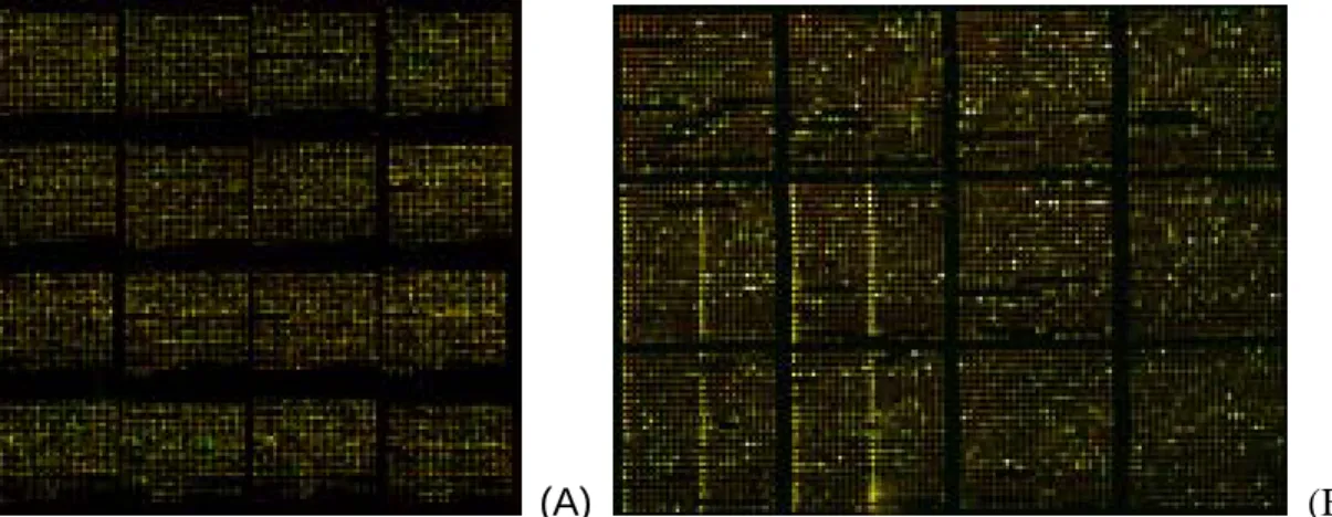
(A) (B)
Figure 1. (A) A hybridization image of 150mer gene-specific oligonucleotide microarray. (B) A part of hybridization image of new 20k oligo microarray.
Figure 2. The homepage of UniProbe database. Home-made types of oligonucleotide microarrays
With the aid of liquid handing robotics and integrated data management system established in our laboratory, we successfully developed a high throughput platform for PCR mass-production. About ~14,000 human gene-specific 150-mer probes have been generated by PCR with genomic DNA and spotted on surface-activated glass slides. An image of the oligonucleotide microarray hybridized with Cy3- and Cy5-labeled samples is shown in Figure1. At the same time a web-based database called UniProbe (http://140.109.43.20/genestamp/uniprobe.htm) is constructed for large scale gene expression analysis. UniProbe is implemented on a MySQL relational database management system. The Web application to query and manage the database is based on PHP. The complete application runs on an Apache web server in a Linux 9 environment. Currently the database contains about 14,000 human named genes, and each gene includes information on empirically validated gene-specific DNA probe and PCR primer sequences for microarray fabrication and for real-time qPCR assays. In addition to the internally generated data, UniProbe provides external links to the National Center for Biotechnology Information (NCBI) LocusLink for more detailed
gene function description, Kyoto Encyclopedia of Genes and Genomes (KEGG) and BioCarta databases for pathway information via UniGene Cluster ID, GenBank accession number, or gene symbol hyperlinks. Figure 2 displays the homepage of UniProbe database. All the gene-specific PCR primer pairs in the database have been empirically verified and can be readily employed in quantitative real-time QPCR assays.
Progress Report
Microarray service
The core facility has been established in National Taiwan University Hospital, National Taiwan University College of Medicine for more than four years and manufactured more than 1,800 pieces of microarray membranes including human, mouse, and custom-made chips. Also, the core Lab offered related research consultation and education training to medical campus. National Science Council supports our core facility two years ago to set up the “Microarray Core Facility for Genomic Medicine”, which offers countrywide microarray services right away.
Our microarray core facility has provided complete microarray services for dozens of institutions, including the researchers of Taiwan University, Yang-Ming University, Chang-Geng University, Chung-Shan Medical University, and so on, for more than 1,000 pieces of microarray membranes including human, mouse, and custom-made chips. There are still hundreds of chips waiting for preparation. We will provide services by the enrolled order and work capacity. To provide a large number and swift microarray services and develop new microarray format such as home-made oligonucleotide chips, viral chips, cell-based, promoter screening microarrays and SiRNA microarrays, and SNP analysis system, the additional equipment and personnel are essential.
The statistical analysis for service is provided as follows to show the working status of core facility, including user list, institution, chip type, number, service items, and so on, in the past three years. Briefly, we have provided 842, 145, and 48 chips of complete microarray services at 2002, 2003 and half a year of 2004, respectively. And we have also provided 1422, 299 and 175 chips at 2002, 2003 and half a year of 2004, respectively. The semi-annual income of 2004 is NT 832,702.
Training and Education
1. Our core facility has given a 50-hour microarray training course including lectures and experimental practices every year in the past three years. The total number of trainees is more than fifty. The schedule of course is provided as followed.
2. We also offer related research consultation and education training anytime if applicants request. We have trained more than 100 persons for microarray experiments and they can successfully carry out microarrays themselves in the past three years.
三. Related publications by using this core facilities:
1. Hong TM, Yang PC, Peck K, Chen JJW, Yang SC, Chen YC, and Wu CW. (2000). Profiling the Down Stream Genes of Tumor Suppressor PTEN in Lung Cancer Cells by cDNA Microarray. Am. J. Respir. Cell Mol. Biol. 23:355-363.
2. Hong TM, Chen JJW, Peck K, Yang PC, and Wu CW (2001). P53 amino acids 339-346 represent the minimal p53 repression domain. J. Biol. Chem. 276: 1510-1515.
3. Chen JJW, Peck K, Hong TM, Yang SC, Sher YP, Shih JY, Wu R, Wu CW, and Yang PC. (2001). Global analysis of gene expression in invasion by a lung cancer model. Cancer Res. 61:
5223-5230.
4. Shih JY, Yang SC, Hong TM, Yuan A, Chen JJW, Yu CJ, Chang YL, Lee YC, Peck K, Wu CW, and Yang PC. (2001). Collapsin response mediator protein-1 and the invasion and metastasis of cancer cells. J. Natl. Cancer I. 93: 1392-1400.
5. Chen HW, Chen JJW, Tzeng CR, Li HN, Chang SJ, Cheng YF, Chang CW, Wang RS, Yang PC, and Lee YT. (2002). Global Analysis of Differentially Expressed Genes in Early Gestational Decidua and Chorionic Villi using a 9,600 Human cDNA Microarray. Mol. Hum. Reprod. 8: 475-484. (Cover story)
6. Shih JY, Lee YC, Yang SC, Hong TM, Huang CY, and Yang PC. (2003). Collapsin response mediator protein-1: a novel invasion-suppressor gene. Clin Exp Metastasis. 20(1): 69-76.
7. Chen JJW, Yao PL, Yuan A, Hong TM, Shun CT, Kuo ML, Lee YC, and Yang PC. (2003). Up-regulation of Tumoral Interleukin-8 Expression by Infiltrating Macrophages: Its Correlation with Tumor Angiogenesis and Patient Survival in Non-Small Cell Lung Cancer. Clin. Cancer Res. 9: 729-737.
8. Chen HW, Yu SL, Chen JJW, Li HN, Lin YC, Yao PL, Chou HY, Chien CT, Chen WJ, Lee YT, and Yang PC. (2004). Anti-Invasive Gene Expression Profile of Curcumin in Lung Adenocarcinoma Based on a High Throughput Microarray Analysis. Mol. Pharmacol. 65: 99-110.
9. Yang WC, Chen HW, Au HK, Chang CW, Huang CT, Yen YH, and Tzeng CR. (2004). Serum and endometrial markers. Best Pract Res Clin Obstet Gynaecol. 18: 305-318.
10. Yu SL, Chen HW, Yang PC, Peck K, Chen JJW, and Lin FY. (2004). Differential Gene Expression in Gram-Negative and Gram-positive Sepsis. Am. J. Resp. Crit. Care Med. 169:(10) 1135-1143.
11. Chen HW, Yu SL, Chen WJ, Yang PC, Chien CT, Chou HY, Li HN, Peck K, Huang CH, Lin FY, Chen JJW and Lee YT. (2004). Dynamic Changes of Gene Expression Profiles during Postnatal Heart Development in Mice. Heart 90: 927-934.
12. Kuo Yuan, Tse-Ming Hong, Jeremy Chen, Wan-Hua Tsai, and Ming T Lin (2004). The Impact of Low-dose Busulfan on Clonal Dynamics in Non-Human Primates Blood 104: 1025-1033.
13. Yuan A, Chen JJW, Yao PL, and Yang PC. (2005) The role of interleukin- 8 in cancer cells and microenvironment interaction. Frontier in Bioscience. 10: 853-865.
14. Chen JJW, Lin YC, Yao PL, Yuan A, Chen HY, Tsai MF, Chen CH, and Yang PC. (2005). Tumor-Associated Macrophages: The Double Edge Sword in Cancer Progression. J Clin Oncol 23: 953-964.
15. Yao PL, Yi-Chen Lin YC, Chen JJW, Wang SS, and Yang PC. (2005). Autocrine and Paracrine Regulation of IL-8 Expression in Lung Cancer Cells. Am J Respir Cell Mol (in press).
16. Huei-Wen Chen , Jeremy J.W. Chen, Sung-Liang Yu, Han-Ni Li, Pan-Chyr Yang, Ching-Mao Su, Heng-Kien Au, Ching-Wen Chang, Li-Wei Chien, Chieh-Sheng Chen, and Chii-Ruey Tzeng (2005). Transcriptomic analysis during blastocyst hatching by cDNA microarrray. Human
Reproduction (in press).
17. Chen WJ, Chen HW, Yu SL, et al. (2005). Gene Expression Profiles in Hypoxic Preconditioning using cDNA Microarray Analysis: Altered Expression of an Angiogenic Factor, CEACAM-1. Shock (in press).
18. Wang CC, Tsai MF, Hong TM, Chang GC, Chen CY, Yang WM, Chen JJW*, and Yang PC. (2005). The transcriptional factor YY1 upregulates the novel invasion suppressor HLJ1 expression andinhibits cancer cell invasion. Oncogene (in press).
19. Wen-Chien Chou, Hsuan-Yu Chen, Sung-Liang Yu , Linzhao Cheng, Pan-Chyr Yang, Chi V. Dang. (2005) Arsenic suppresses gene expression in promyelocytic leukemia cells partly through Sp1 oxidation. Blood (in press).