國
立
交
通
大
學
材料科學與工程學系
博 士 論 文
2-羥基乙基丙烯酸甲酯複合物奈米結構與
控制釋放行為之研究
Nanostructural evolution and controlled
release behavior of SiO
2
/pHEMA and
Cu/pHEMA hybrids
研究生:劉彥妤
指導教授:陳三元 博士
中文摘要
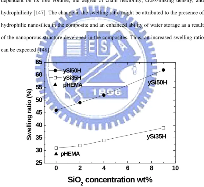
就許多醫療器材而言,改善其與血液接觸所產生之相容性至今日仍是相當重要的 議題。高分子是目前最被廣泛應用於製造醫療元件的材料,通常需藉由改質來改善其 血液相容性。許多文獻指出,表面改質方法如電漿表面改質或是肝素接枝法都已被廣 泛且成功的運用在與血液接觸的元件上;然而,材料表面為較容易損壞之部分,對於 需要植入體內之醫療元件,其可能因腐蝕、與血液接觸或因個人生活型態迥異而造成 不同物理環境,產生非預期之化學作用,其更須具備長期穩定之特性。因此,考量醫 療元件整體之功用及其在臨床治療上所面臨之問題,材料表面改質及材料整體改質仍 具有許多研究及發展的空間。 本論文之研究主旨,即在於利用奈米級無機粒子與金屬粒子之混摻,探討其對於 高分子水膠微結構以及整體性質之改變,與此複合材料於藥物釋放以及血液相容性上 之討論。論文主體分為兩部分: 第一部分探討奈米級矽分散粒子與矽醇水合前驅物,對於2-羥基乙基丙烯酸甲酯 高分子載體其微結構以及其在血液相容性之影響。我們利用即時(in-situ)光聚合法,成 功製備出矽/2-羥基乙基丙烯酸甲酯 (SiO2/pHEMA and Silanol/pHEMA) 複合材。從電子顯微鏡可知,複合材料孔洞隨無機物含量遞增,含有4%奈米 SiO2粒子之複合材有 最佳機械性質。此外,水膠複合材之膨潤度隨無機物含量遞增,當無機物含量由2% 增 加至9%,藥物擴散速率提高約 100 倍。經由血小板貼覆測試可知 SiO2/pHEMA 複合 材具有相當良好之血液相容性。 第二部分探討奈米級金屬銅粒子,對於2-羥基乙基丙烯酸甲酯高分子載體其微結 構以及其在血液相容性之影響。我們利用即時(in-situ)光聚合法,將銅離子均勻分散於 2-羥基乙基丙烯酸甲酯高分子載體中,其後並以化學還原法,製備得到 Cu(0)-pHEMA 複合材。由穿透電子顯微鏡可知,隨溶劑與單體莫耳比增加,可得奈米級銅顆粒均勻 分散於高分子載體,顆粒大小約為5~10nm。此外,並探討 Cu(0)-pHEMA 複合材中, 銅溶解擴散模式以及其對於內皮細胞成長之影響。由結果可知,銅離子可經由
Cu(0)-pHEMA 複合材中以穩定緩慢的模式釋放,可經由複合材料組成之變化,控制銅 離子釋放濃度。於體外實驗,與控制組相較,當每日銅釋放量約為10ppm 時,可提升 內皮細胞增長量至120%;若每日銅釋放濃度高於 20ppm,則會產生毒化情形。 最後,探討Cu(0)-pHEMA 複合材之電化學特性,與其行氧化作用使 GSNO (Nitrosoglutathione)還原產生一氧化氮(NO)之反應探討。由循環伏安測試我們可得一氧 化電流(-320mV vs. Ag/AgCl),Cu(0)-pHEMA 複合材具有氧化能力。由交流阻抗分析 得知,隨Cu(0)-pHEMA 複合材中之銅含量之增加至 1%, 阻抗可降低至 6438 ohm。在 定電位測試中可知,Cu(0)-pHEMA 複合材可將 GSNO (Nitrosoglutathione)還原產生一 氧化氮(NO)。 這個以奈米複合材微結構來控制藥物釋放之血液相容元件的研究具有很好的前 瞻性。在本研究中,與血液接觸之相容性以及藥物釋放之性質同等重要,而了解複合 材中無機奈米顆粒在微結構變化、表面組織及生物相容特性所扮演的角色,將有助於 我們發現新的科學現象並設計更新穎的材料以應用在材料科學、生物醫學以及藥物釋 放學上。 關鍵詞: 2-羥基乙基丙烯酸甲酯、矽、銅、複合材料、藥物控制釋放、血液相容、表面 電性
Abstract
Use of medical devices with clinically acceptable blood compatibility has gained increasing attention over the years. This has been consciously alerted due to a current understanding that the devices used to contact with human blood has been criticized to having insufficient anti-blood clotting surface for short-to-long term invasion medication. Therefore, development of new biomaterials with improved blood compatibility has continuously attracted great attention.
In this thesis, the incorporation of inorganic (silica) and metal (Cu) nanoparticles into the poly(2-hydroxyethyl methacrylate) (pHEMA) matrix to form inorganic/organic nanocomposites for drug controlled release and blood compatibility was achieved.
The first part in this thesis, an in-situ method is developed successfully to mix well-dispersed silica colloidal suspension and silica sol-gel solution with HEMA monomers following photopolymerization to form a nanocomposite. The incorporation of SiO2 nanoparticles and Silanol into pHEMA matrix revealed a significant effect on the reaction rate of crosslinking during polymerization, resulting in composites with varying nanoporous structures. The nanocomposites showed improved tensile strength, and the platelet adhesion property remained as excellent as that of neat pHEMA, which encourages the use of such composites for antithrombotic applications. Drug diffusion characteristics in the composites can be well modulated by controlling the concentrations of SiO2 nanoparticles and silanol and water in the starting stage of synthesis.
In the second part, a novel in-situ synthesis method is developed where a hybrid system based on HEMA monomers that were photopolymerized in the presence of Cu2+ precursor was prepared through an in-situ synthesis, following an in-situ chemical reduction of the Cu2+ precursor to form resulting metallic Cu-containing hybrid. The cell proliferation, surface potential, and interaction with blood of the Cu-pHEMA hybrid nanocomposites was
systemically discussed. From the results, coordinated interaction between Cu(II) with the hydroxyl groups within the pHEMA matrix was confirmed by the infrared spectral analysis and considerable improvement of the thermal stability of the Cu(0)-pHEMA hybrids. Localization of the metallic copper particles within pHEMA network structure as a result of those intermolecular interactions gives rise to the formation of discretely distributed nanocrystallites with a particle size ranging from 10 to 25 nm in diameter. A relatively slow and sustained release of the Cu (in form of cupric ion) from the hybrids for a time period over 10 days was measured, which also illustrated a Cu(II)-induced proliferation of the endothelial cells. The hybrids also showed negative surface charge and considerable improvement in blood compatibility compared to neat pHEMA.
Finally, the electrochemical properties of the Cu-pHEMA hybrid nanocomposites were investigated by cyclic voltammetry (CV) and alternating current (AC) impedance measurements. The generation of nitric oxide in aqueous by Cu(0)-pHEMA hybrid was also tested. From the results, the Cu(0)-pHEMA hybrid exhibits an oxidation current at -310mV. The charge transfer resistances (RCT) of Cu(0)-pHEMA hybrids are estimated to be reduced from 11743 to 6438 ohm, respectively as the content of nano copper particle in polymer matrix increased to 1 wt%. The reduction currents of Cu(0)-pHEMA hybrid varied with the concentration of nitrosoglutathione. With increasing levels of nitrosoglutathione (GSNO), the amount of NO generation increased.
Keywords: 2-hydroxyethyl methacrylate, silica, copper, hybrid, drug controlled release, blood compatibility, surface charge, and electrochemical properties
Contents
Abstract ...III Figure captions... VII Table captions ...IX
Chapter 1 ... 1
Introduction ... 1
1.1 Silica-base pHEMA nanocomposites (Part 1) ... 3
1.2 Copper-base pHEMA nanocomposites (Part2)... 4
Chapter 2 ... 6
Literature Review and Theory ... 6
2.1 pHEMA nanocomposites hydrogel... 6
2.1.1 Silica/pHEMA nanocomposites ... 7
2.1.2 Metal/pHEMA nanocomposites ... 10
2.2 pHEMA nanocomposites for drug delivery system ... 13
2.3 Blood compatibility of pHEMA nanocomposites... 17
2.4 Effect of copper particles on blood compatibility... 19
Chapter 3 ... 22
Experimental... 22
3.1 Flowchart of Experiment Process...22
3.2 Materials and Equipment ... 23
3.3 Characteristics Analysis... 26
3.4 Methods ... 28
Chapter 4 ... 34
Inorganic/organic Hybrid – SiO2/pHEMA –Synthesis and Characterization of Nanoporous SiO2/pHEMA Biocomposites... 34
4.1 Introduction ... 34
4.2 Fabrication of pHEMA and SiO2/pHEMA Composite ... 34
4.3 Microstructure and Physical Characterization ... 36
4.4 Platelet Adhesion ... 41
4.5 Drug Release Characterization ... 44
4.6 Summary ... 48
Chapter 5 ... 50
Inorganic/organic Hybrid – Silanol/pHEMA – Synthesis and Characterization of Silanol/pHEMA Biocomposites for Drug Controlled Release ... 50
5.1 Introduction ... 50
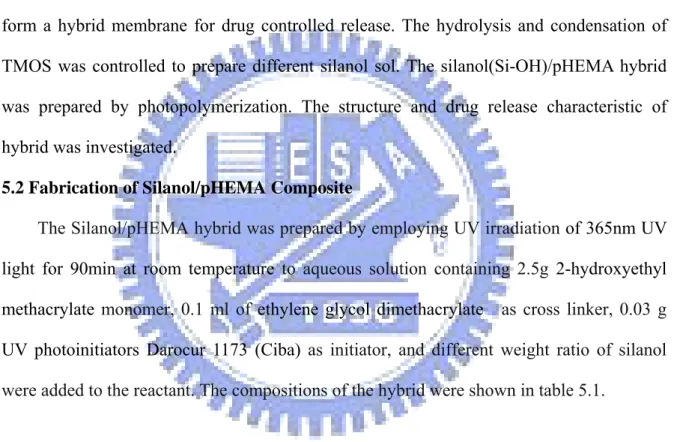
5.2 Fabrication of Silanol/pHEMA Composite ... 50
5.3 Chemical and Physical Characterization ... 52
5.4 Drug diffusion Characterization ... 55
5.5 Summary ... 57
Chapter 6 ... 58
Metal/organic Hybrid – Cu/pHEMA - Structural Evolution of Cu-pHEMA Hybrid and Cu Release Behavior on the Proliferation of Endothelial Cells... 58
6.1. Introduction ... 58
6.2 Fabrication of Cu-pHEMA Hybrid synthesis ... 59
6.3 Hybrid Characterization... 61
6.4 Nanostructural evolution of the Hybrid ... 63
6.5 Cu(Ⅱ) release... 67
6.7 Summary ... 72
Chapter 7 ... 74
Metal/organic Hybrid – Cu/pHEMA –In-Situ Synthesis of Hybrid Nanocomposite with Highly-Order Arranged Amorphous Metallic Copper Nanoparticle in Poly(2-hydroxyethyl methacrylate) and Its Potential for Blood-contact Uses... 74
7.1 Introduction ... 74
7.2 Fabrication of Cu-pHEMA Hybrid ... 75
7.3 Structural Analysis ... 76
7.4 Zeta potential measurement ... 81
7.5 Blood and platelet test ... 82
7.6 Summary ... 84
Chapter 8 ... 85
Metal/organic Hybrid – Cu(0)-pHEMA - Electrochemical behavior of Cu(0)-pHEMA hybrid... 85
8.1 Introduction ... 85
8.2 Fabrication of Cu-pHEMA Hybrid ... 86
8.3 Electrochemical properties ... 88
8.4 Summary ... 93
Chapter 9 ... 94
Conclusions ... 94
9.1 Silica-base pHEMA nanocomposites ... 94
9.2 Copper-base pHEMA nanocomposites... 94
Reference ... 97
Figure captions
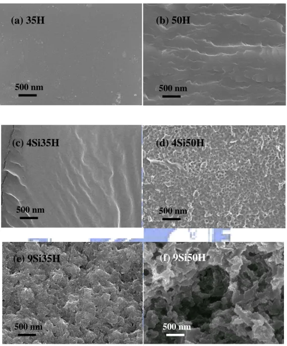
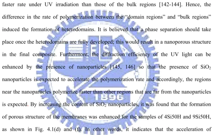
Fig. 4.1 SEM micrographs of cross-section image of various SiO2/pHEMA composites...37
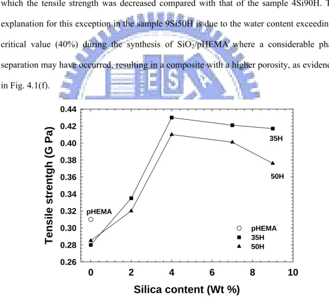
Fig. 4.2 Mechanical properties of pHEMA and SiO2/pHEMA composites ...39
Fig. 4.3 Amount of water absorption of pHEMA and SiO2/pHEMA composites...40
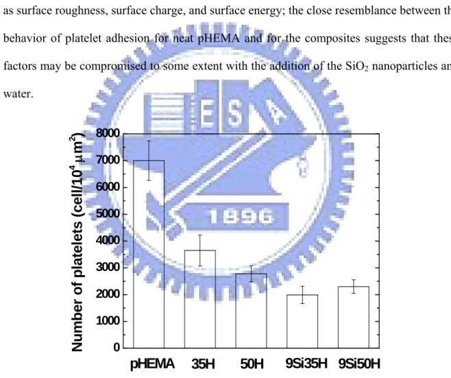
Fig. 4.4 Number of platelets adhering onto surface of pHEMA and SiO2/pHEMA membranes...41
Fig. 4.5 Tapping-mode AFM images of (a) 35H and (b) 9Si35H...42
Fig. 4.6 DSC thermogram of pHEMA and SiO2/pHEMA composites...43
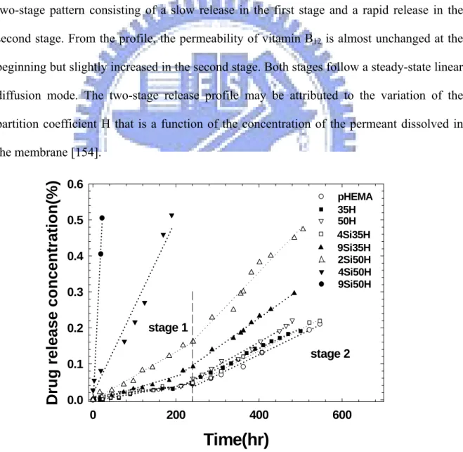
Fig. 4.7 Drug release profiles of pHEMA and SiO2/pHEMA composites ...44
Fig. 4.8 SEM photographs of (a) 35H before drug diffusion and after drug diffusion for (b) 35H, (c) 9Si35H, and (d) 50H ...46
Fig. 4.9 Permeability [DH] of B12 at stage 2 of SiO2/pHEMA composite ...47
Fig. 4.10 TEM micrographs of SiO2/pHEMA composite ...48
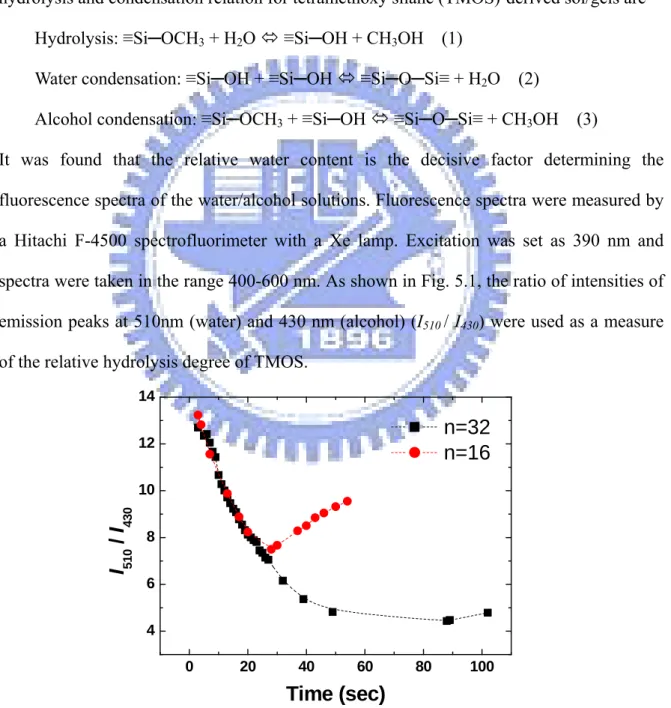
Fig 5.1 Time dependencies of the relative water content in the sol-gel solution of a nominal composition TMOS : H2O = 1:n, where n=16 and 32...51
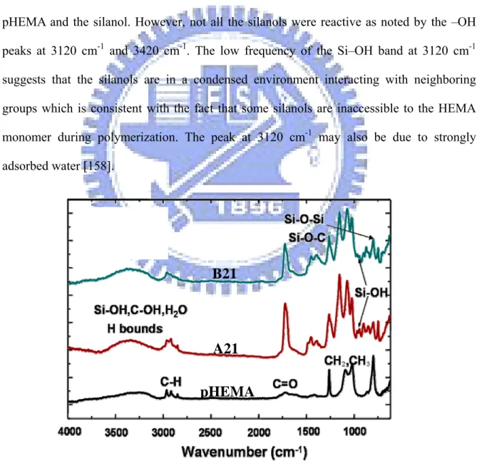
Fig. 5.2 FTIR, Mid-IR, absorption spectra of pHEMA and silanol/pHEMA hybrid ...52
Fig. 5.3 FTIR, Near-IR, absorption spectra of pHEMA and silanol/pHEMA hybrid ...53
Fig. 5.4 SEM micrograph of cross-section image of silanol/pHEMA hybrid ...54
Fig. 5.5 TG thermograms of pure pHEMA and silanol/pHEMA hybrids ...55
Fig. 5.6 Drug release profile of pHEMA and silanol/pHEMA hybrid ...57
Fig. 6.1 (a) FT-IR ATR spectra of the synthesized pHEMA and (b) Cu(II)-pHEMA hybrid The composition of Cu(II)-pHEMA hybrid was the same with Hybrid-4-Cu05 ...61
Fig. 6.2 TGA thermograms of the synthesized pHEMA, Cu(II)-pHEMA hybrid, and Cu(0)-pHEMA hybrid. The composition of Cu(II)-pHEMA and Cu(0)-pHEMA hybrid is the same as indicated in Hybrid-4-Cu05 ...62
Fig. 6.3 The XRD patterns of pHEMA and Cu(0)-pHEMA hybrids...64
Fig. 6.4 TEM photograph of Cu(0)-pHEMA nanocomposite (a) Hybrid-1-Cu05 and (b) Hybrid-4-Cu05. Both marked area are further magnified in (c) and (d), respectively. 65 Fig. 6.5 A schematic drawing of the proposed synthesis scheme with the bonding configuration of Cu ions with surrounding environment of Cu(0)-pHEMA hybrid. The Cu(II) ions are assumed to be coupled with the unpaired O of COOR groups along the pHEMA chain or coupled by the unpaired O of H2O forming a nanophasic Cu domain in place...66
Fig. 6.6Cross-sectional microstructure of the Cu(0)-pHEMA hybrids (a) Hybrid-1-Cu05, and (b) Hybrid-4-Cu05, where microporous morphology is clearly shown as starting water concentration was increased. ...67 Fig. 6.7 Cu(Ⅱ) release from the Cu(0)-pHEMA hybrids with different H2O/HEMA molar ratio. The release patterns showed a relatively slow and sustained kinetics with the
values of k and n determined according to Eq. (2) for Mt/M∞< 0.6 (n = 5). ...68 Fig. 6.8 (a) Curves of Cu(Ⅱ) release rates of in-situ Cu(0)-pHEMA hybrids and
Cu(0)-pHEMA composites in the PBS. (b) Dependence of n value of different copper content for Cu(Ⅱ) release from Cu(0)-pHEMA composites and in-situ
Cu(0)-pHEMA hybrids...69 Fig. 6.9 Effect of copper ion on HUVEC proliferation at various time periods. Cell number was determined by AlamarBlue assay. ...72 Fig. 7.1 TEM photographs of Cu(0)-pHEMA hybrid showing (a) an orderly packing
configuration of Cu(0) nanoparticles distributed in the pHEMA matrix, (b) High resolution image of Cu(0) nano particle in pHEMA matrix where the nanopaticle has a size of about 3 nm in average, and (c) selected area electron diffraction pattern of Cu(0) nano particle, indicated the Cu(0) a poorly crystalline (or
amorphous) structure. ...76 Fig. 7.2 A schematic drawing for the proposed synthesis scheme of Cu(0)-pHEMA hybrid, where (a) the Cu(II) were localized and chemically reduced by N2H4 in-situ to metallic Cu(0) nanoparticles and (b) the Cu(II) ions were assumed to be coupled with the unpaired O of COOR groups along the pHEMA chain network, forming a regularly arranged nanostructure, as illustrated in Fig. 7.1a. ...78 Fig. 7.4 Effect of the nano copper particles on zeta potential of Cu(0)-pHEMA hybrid with different copper particle content (weight %). Concentration of KCl = 1*10-3 M at pH 7.4 ...81 Fig. 7.5 The blood platelet adhesion test showed that the Cu(0)-pHEMA hybrid
demonstrated an improved anti-platelet adhesion behavior by ~70% and 92% compared to that of the neat pHEMA and PSF, respectively. (The scale bar of SEM image is 10μm.)...83 Fig. 8.1 Cyclic voltammograms of pHEMA and Cu(0)-pHEMA in the phosphate-buffered saline solution, v=50mVs-1...88 Fig. 8.2 Nyquist diagram of selected Cu(0)-pHEMA hybrid showing various values of time constant...89 Fig. 8.3 (a) Amperometric responses of NO-generating from Hybrid-10. (b) The correlation between nitrosoglutathione (GSNO) and reduction current...91 Fig. 8.4 Amperometric responses of NO-generating from Composite-05 micro,
Composite-05 nano, Hybrid-2, and Hybrid-4 with 20mM GSNO. ...91 Fig. 8.5 The Cu 2p3/2 peaks of XPS spectra of Cu(II)-pHEMA hybrid after redox reaction with GSNO. ...92 Fig. 8.6 Scheme of the electrochemical reaction for Cu(0)-pHEMA hybrid. ...93
Table captions
Table 4.1 pHEMA and SiO2/pHEMA composites………34
Table 4.2 The permeability [DH] of B12 of pHEMA and SiO2/pHEMA composites.…45
Table 5.1 The compositions of the Silanol/pHEMA hybrid ………48 Table 5.2 The partition coefficient of the pHEMA, B11, A11, B21, and A21 samples..54 Table 6.1 The parameters n and k calculated from Eq. (2) of Cu(0)-pHEMA
Chapter 1
Introduction
Improved compatibility with blood is one of the key features for a variety of medical devices. The materials used currently to manufacture the medical devices are mostly polymers, which are not inherently compatible to human blood but require a subsequent modification for clinical uses. However, conditions become more stringent for a number of implantable devices, such as vascular grafts/stents, artificial heart valves, etc. since those devices may suffer from a variety of stresses, flowing fields, and site-specific physiological conditions. Some of the devices require not only surface but also bulk to be biologically compatible; they also need sufficient strength to keep long-lasting service performance during the life time of the patients. Therefore, development of new biomaterials with improved blood compatibility has continuously attracted great attention.
The design and preparation of hydrogels have attracted a great deal of interest in biomedical engineering, pharmaceutical applications, and biomaterials science because of their tunable chemical and three-dimensional (3D) physical structure, good mechanical properties, high water content, and biocompatibility. These unique properties offer great potential for the utilization of hydrogels in tissue engineering, biomedical implants, drug delivery, and bionanotechnology [1-5]. Among all, it has been noticed that methacrylate monomers consisting of an alkyl group, an acrylate ester group, and a functional carboxyl group can react with a wide range of monomers and functionalized molecules providing flexible polymer chains. Methacrylate hydrogels are prepared by interconnecting the lineal polymeric chains with cross-linkers establishing a three-dimensional network of strong chemical bonds. For example, the synthesis of Poly(2-hydroxyethyl methacrylate), i.e., pHEMA, is a favorable biomaterial because of its excellent biocompatibility and
chemical and hydrolytic stability and good tolerance for entrapped cells. Because of these unique characteristics, pHEMA is one of the most extensively studied materials in tissue engineering, and has also been widely used as the backbone for synthesizing stimuli-responsive hydrogels. The presence of hydroxyl and carboxyl groups makes this polymer compatible with water, whereas the hydrophobic methyl groups and backbone impart hydrolytic stability to the polymer and support the mechanical strength of the matrix [8].
During the last decade, considerable attention was paid to inorganic–organic hybrid materials because their solid state properties could be tailored in relation to the nature and relative content of their constitutive components. These organic–inorganic composites possess a complicated structure, including phases of the organic and the inorganic polymer and a third phase, which is a product of interaction between the organic and the inorganic components [9]. Recently, the development of hydrogel hybrid with nano-sized materials such as inorganic clays [10-11], carbon nanotubes [12], and polyaniline nanosticks [13] has also been of increasing interest in the field of biomaterials science. Composite materials containing metal (M) and semiconductor (SC) nanoparticles, immobilized in polymer matrices are under intense studies due to fundamental novelty and technological importance. The interconnection of inorganic and organic ingredients by either chemical, or physical interaction upon in-situ polymerization provides not only a featured nanostructure in the resulting hybrid, but also correspondingly offers unique properties that are frequently not able to achieve through individual components. Therefore, it is more interesting and potentially clinically important if a hybrid system can be developed where the constituting component is able to provide desirable functional reaction to deactivate clot-forming proteins in human blood and further reduce or eliminate platelet deposition and aggregation. Moreover, drug controlled delivery could be achieved in this hybrid.
In this thesis, the introduction of inorganic (silica) and metal (Cu) nanoparticles into the pHEMA matrix to form inorganic/organic nanocomposites for drug controlled release and
blood compatibility was achieved. It can be categorized into two parts: Part 1. An in-situ method is developed successfully to mix well-dispersed silica colloidal suspension (Chapter 4) and silica sol-gel solution (chapter 5) with a HEMA monomer following photopolymerization to form a nanocomposite. In part 2, a novel in-situ synthesis method is developed where a hybrid system based on the use of HEMA monomers that were photopolymerized in the presence of Cu2+ precursor was prepared through an in-situ synthesis, following an in-situ chemical reduction of the Cu2+ precursor to form resulting metallic Cu-containing hybrid.
1.1 Silica-base pHEMA nanocomposites (Part 1)
It can be expected that the incorporation of a silica nanophase into the pHEMA matrix can not only enhance the mechanical properties of the resulting composites but also regulate the release kinetics of the drug from within. Until today, the effects of the variation of the structure of the composite on the diffusion characteristics of a molecule, i.e., a drug, have not been systematically investigated in detail. In addition, the effect of silica nanophase on the hemocompatibility of pHEMA is still unclear. Hence, in chapter 4, nanoporous SiO2/pHEMA nanocomposites were synthesized in situ by incorporating silica colloid with a HEMA monomer, following a UV-induced photopolymerization. The evolution of nanostructure and drug release behaviors of the hydrogel composite was thoroughly studied.
In addition, the incorporation of polymeric components to sol gel derived materials may constitute an important tool to provide modified chemical reactivity [14] or more compatible media for encapsulation of biological molecules and medicines [15]. The silanol group could provide an active site for further reaction of the surface modification [16] or be a catalyst for the nucleation of other bioactive materials [17]. Because of the activity of silanol, the variation and the modification of the structure and chemical bonding of the hybrid could be expected. Therefore, in chapter 5, silanol group was produced and
The hydroxyl group of the hybrid was verified by controlling the hydrolysis degree of silanol and the composition of the hybrid.
1.2 Copper-base pHEMA nanocomposites (Part2)
Copper has been well-recognized as a physiologically important element for human health and from a number of clinical practices. Copper deficiency has been known to associate with complications such as Wilson’s disease, thrombotic disorder, etc. In the meantime, copper is also a widely known element that is able to enhanced repairing efficacy with optimal dose for skin and bone growth as a result of proangiogenic action that favors the development of new vessels after surgery [18-21]. . Copper has also been widely known to enhance the antifertility effect of an intrauterine device (IUD); great efforts have been made to improve the copper-containing intrauterine device (Cu-IUD) and to investigate corrosion behavior of copper in vivo and in vitro [22]. All of those studies, from in-vivo to clinical practices, have pointed out that a stringent control of copper concentration level, either for the maintenance of health or acting as therapeutic purposes, is essentially critical. For the development of Cu-containing composites, although not extensively studied so far, has been reported for years. However, limitation of copper ion content and the stability of ion release from the hydrogel system are still heavily concerned.
In chapter 6, structural evolution of the Cu-containing composite has been elucidated in terms copper release and its effect on the proliferation or inhibition of human endothelial cells and smooth muscle cells were also investigated.
In chapter 7, the Cu-containing nanocompoistes were characterized by transmission electron microscopy (TEM), X-ray photoelectron spectroscopy (XPS), and streaming potential measurements. Interaction between blood and hybrids were examined in terms of platelet adhesion test.
measurements demonstrated that the electrochemical properties of Cu/pHEMA composite could be changed by alternating the synthesis condition. In this study, the nano-sized metal or metal oxide particles could disperse uniformly in polymer matrix and to react with ionic group in the aqueous solution by the charge transfer from the interfacial surface of nano copper particles. In this paper, an experimental study of different electro-chemical and physical aspects of electrochemical sensitive Cu/pHEMA composite was presented.
Chapter 2
Literature Review and Theory
2.1 pHEMA nanocomposites hydrogelSince the pioneering work of Wichterle and Lim in 1960 on cross-linked HEMA hydrogels [23], and because of their hydrophilic character and potential to be biocompatible; hydrogels have been of great interest to biomaterial scientists for many years. Poly(2-hydroxyethyl methacrylate), i.e., pHEMA, has been extensively studied because of its relatively high capability of water uptake, non-toxicity, and favorable compatibility to tissues and blood, which renders itself as an attractive biomaterials for many biomedical applications such as drug delivery vehicles, contact lenses, antithrombotic devices, and implants [24-25]. However, there are still some disadvantages of the pHEMA hydrogels in utilization such as its poor mechanical properties after swelling. Hence, numerous studies have been conducted to modify pHEMA with the aim of improving its mechanical properties [26-28], electro-responsive properties [29] and to elicit better physiologic responses [30]. To deal with the challenge of producing high mechanical strength hydrogels, researchers have taken many approaches: (1) using special comonomers or altering their composition [31], changing the type and concentration of cross-linking agent [32], and optimizing polymerization conditions [33]; (2) inducing a double network (DN) structure for various combinations of hydrophilic polymers [34]; (3) introducing interpenetrating polymer networks (IPN) into hydrogels [35]; (4) synthesizing organic/inorganic hybrid nanocomposite hydrogels [36]; (5) adopting the method of material reinforcement [26].
In the past few years, nano-composites have attracted considerable attention from both fundamental research and application points of view [37-38]. Nano-structured composites, mixed at the molecular level or near molecular level, are much different from the conventional composites with incorporation of a variety of additives in the polymer
matrices [39]. In inorganic–organic nano-composites, strong chemical bonds (covalent or ionic) or interactions such as van der Waals forces, hydrogen bonding, or electrostatic forces, often exist between the organic and inorganic components. This usually leads to some novel nanocomposites with improved performance properties, which may have large potential applications in the fields of optics [40], electrical devices [41], mechanics [42], photoconductors [43], and so on. In order to meet practical requirements, certain properties of pHEMA such as mechanical properties, thermal stability, wettability, diffusion mechanism of solutes (such as biomolecules or drugs), and affinity to specific biomolecules can be tailored and regulated by the incorporation of nanomaterials and manipulated through controlling the composition of inorganic or organic ingredients, nanostructure, nature of inorganic and/or organic components, etc. [44-46].
2.1.1 Silica/pHEMA nanocomposites
Since the mid-1980s, when the first sol-gel derived hybrids were obtained by mixing linear polymer chains with silica precursors [47], many works have been conducted trying to probe structure and to explore potential applications of this type of materials, as demonstrated by several review papers [48-50]. This low temperature process, which is particularly well suitable to combine with organic species, offers an interesting way of preparing hybrid materials. The most commonly used silica precursor is tetraethoxysilane (TEOS). The sol–gel reactions involving this silicon alkoxide can be summarized as follows: (i) Hydrolysis ≣Si-O-C2H5 + H2O→≣Si-OH+C2H5OH (1) (ii) Condensation ≣Si-OH+HO-Si≣→≣Si-O-Si≣+H2O (2) and/or
If these sol– gel reactions are complete, full condensed silica is obtained through a chemical process that can be summarized by the following equation:
Si(O-C2H5)4+2 H2O→SiO2+4C2H5-OH (4)
At high pH values, where condensation reactions are favored, discrete, large, and highly condensed silica particles are produced, whereas low pH conditions favor hydrolysis reactions and lead to a finer ramified polymeric silicate structure. A wide variety of types of organic polymers have been employed in the syntheses of hybrids of silica [51]. Particularly the polymer pHEMA is an interesting choice because, in addition to being easily soluble in the water-alcohol mixtures employed in sol-gel method, its pendant hydroxyl groups lead to the formation of hydrogen bonds and eventual condensation with silanol groups [52, 45], thus favoring the production of structurally homogeneous materials within a wide range of compositions[53]. Many studies have been devoted to the preparation of pHEMA-based composite materials with organic HEMA monomers and inorganic precursors such as tetramethoxysilane (TMOS) or tetraethoxysilane (TEOS) through sol-gel processes [54-55]. One of the most important problems in studying the hybrid nano-composite materials is their structural analysis. The correlation between material evolution and composition–structure–property dependence, as well as the processes of nanostructure formation, aggregation and development of self-organized materials has been investigated. The nanostructure surface of silica and hybrids has been studied by several research groups [56-57]. The nano-structure of the hybrid, either with the nano-silica uniformly dispersing in the polymer phases or with phase separation of inorganic and organic phases occurring, depends on the processing conditions such as the type of catalyst, the pH value, the water quantity, the solvent system, and the reaction temperature [58]. In order to investigate the type and the extent of the interactions between inorganic and organic domains, which play a determinant role in controlling the properties of hybrid materials, Fontana et al [59] have characterized the hybrids obtained from the polymerization of pHEMA, modified by silicon
tetraethoxide (TEOS) by Raman and Brillouin spectroscopes to explain tentatively the changes of the properties, included of Brillouin scattering and refractive index, were on the basis of the structural change in the polymeric network, due to the presence of the inorganic domains. Moreover, Wu wt al. [60] have used XRD, FT-IR, BET, SEM and AFM techniques to investigate the influence of the main silica precursors and the type and quantity of organic components (HEMA) on the structure of hybrid materials by sol-gel synthesis. It was found that the type of precursors is of paramount importance for the formation of strong chemical bonds between inorganic and organic components of synthesized materials.
In inorganic/organic nano-coomposites, the inorganic molecules and organic molecules interconnect by chemical covalent bonds, hydrogen bonds or physical interaction. The formation of covalent bonds such as Si-C or specific secondary interactions such as hydrogen bonds between organic and inorganic moieties may be a technique for structural control. Structural control by introducing hydrogen bonds into a system depends on the use of polymers containing specific functional groups susceptible to interacting with Si–OH groups of silica gel. Particularly the polymer pHEMA is an interesting choice because its hydroxyl groups, in addition to forming hydrogen bonds, will eventually form Si–O–C bonds by condensation with silanol groups [61-62], although Si–O–C bonds may be, theoretically, unstable to hydrolysis under sol–gel conditions [52]. On the other hand, the formation of covalent bonds between organic and inorganic moieties may be a technique for structural control. Some studies [63] developed during the past years have emphasized the insertion of covalent bonds linking organic polymers and silica network by introducing coupling agents, which contain both polymerizable and hydrolysable groups. In order to improve the understanding of structural control of hybrid materials synthesized from mixtures of tetramethoxysilane, water and pHEMA, Costa and Vasconcelos [64] introduced
reactions to evaluate the effect of primary interactions between organic and inorganic components. The results reveled that thhe presence of MTMOS prevented crack formation and macroscopic phase separation, though the formation of larger fluctuating composition domains with increasing pHEMA content.
An alternative way to synthesize the inorganic/organic hybrids is by directly mixing inorganic colloidal particles with polymers. The inorganic colloidal particles can be pre-produced through the sol–gel process with a pure inorganic precursor such as alkoxysilanes. Vigier et al. [45] compared two types of pHEMA/silica nano-composites prepared by undergoing free radical polymerization of HEMA either in the presence of HEMA-functionalized SiO2 nano-particles (Type 1) or during the simultaneous in situ growing of the silica phase through the acid-catalyzed sol– gel polymerization of tetraethoxysilane (TEOS) (Type 2). They demonstrated that Type 1 systems exhibit classical particle-matrix morphology, but where particles tend to form aggregates. Type 2 systems possess a finer morphology characterized by a very open massfractal silicate structure, which is believed to be bicontinuous with the organic phase at a molecular level. Similar discovers were also observed by Yang et al. [65] that the structure of the colloidal silica/pHEMA hybrid consisted of nano-silica uniformly dispersed in the pHEMA phase with slight inter-molecular hydrogen bonding. Furthermore, the structure of TEOS/pHEMA hybrid was similar to a semi-interpenetrated network with pHEMA chains tethered into the nano-silica network by inter- and intra-molecular hydrogen bonding. Consequently, the TEOS/pHEMA hybrid gels exhibited a smoother surface, higher transparency, and better thermal stability than the colloidal silica/pHEMA hybrid gels.
2.1.2 Metal/pHEMA nanocomposites
Recently, polymer-protected nanoparticles have been synthesized successfully [66, 67]. Polymer-protected, nano-sized metals are the subject of increasing attention because they have high potential for application in many technologies, such as the controlled release of
drugs, catalysis, and electron or energy storage [68]. There are different ways to produce such materials: treatment of polymers by metal vapours; reduction of metal salts or their reactions in polymer solutions; incorporation of metal salt solutions in micropores of stretched polymer films; various polymerisation processes of metal-containing monomer systems, etc. As an intermediate between molecular and bulk states, inorganic nano-particles often exhibit unique properties (e.g., electrical, optical, magnetic, catalytic), which are, in addition, dependent on their size [69]. But, this also makes them perfect building blocks for preparation of composite nanostructures with adjustable performances. A polymer is a good choice for the matrix material since it enables an easy processing of the obtained nanocomposites into technologically useful forms. Furthermore, depending on the preparation procedures, polymer matrices can be used to control the size and shape of the inorganic nanofiller. Under certain conditions, nanoparticles of metals can be induced to undergo self-organization into three-dimensional super-lattices [70]. Such nanoparticle assemblies open the door to “tunable” materials, in which optical and electronic properties can be controlled by both the initial cluster sizes and the manner in which the clusters are organized to form larger structures.
It is important to note that the HEMA monomer can likewise act as a ligand due to the presence of the pendant –OH group. Moreover, the pHEMA chains may intertwine amongst the nanoparticles and act as “polyligands” resulting in supramolecular structures. In addition, the HEMA monomer which is approximately 5 Å in width, can find its way into the interior of the nano-particles through the porous structure/windows. Harmon et al. [71] indicated that the cross-linking density increased in the pHEMA nanocomposites suggesting that there is an interaction taking place between the nanoparticles and HEMA; they believe that this interaction is physical (H bonding). In this case the pHEMA–nanparticle nanocomposites may form structures similar in concept to polyrotaxanes. Polyrotaxanes are macromolecules
component are capped with bulky end groups to prevent slippage [72, 73]. Threading of the linear component through the macrocycle is a statistical event; however, driving forces such as electrostatic attraction, hydrogen bonding and hydrophobic–hydrophilic interactions have been shown to favor threading as well; it should be noted that no covalent bonding exists between the two components in the rotaxane. If the linear component is not capped the system is called a pseudorotaxane; dethreading occurs in pseudorotaxanes and as a result they are much less thermodynamically stable than true rotaxanes.
Currently, several successful methods have been carried out to obtain functional metal nanoparticle/pHEMAl nanocomposites. For example, atom transfer radical polymerization (ATRP) is a recently developed “living” radical polymerization method, which is particularly versatile for the polymerization of various vinyl monomers, such as styrene, methacrylates, acrylates and acrylonitrile [74]. Wang groups [75] developed a kind of polymer-metal complexes which were obtained after the introduction of Cu2+ cations into the pHEMA brushes by the complexing bond between Cu2+ cations and the hydroxyl groups in pHEMA brushes. However, the homogeneous dispersion of metal nanoparticles in these hydrogels is a key challenge due to the easy agglomeration of nanoparticles and the high viscosity of hydrogels [76]. Recently, several approaches have been reported to obtain composites with well-fined size and morphology of the metal nanoparticles [77, 78]. A more effective approach reported by Wang et al. [79] is the in situ reduction of metal ions in the hydrogel. In this method, the metal ions were anchored in the hydrogel network by the functional groups of the hydrogels before in situ reduction. It can effectively inhibit the aggregation of the metal particles. This method requires the particular modification of the hydrogel with functional groups capable of interacting with the metal precursor ions. However, in most cases, those functional groups, such as thiol, pyridyl and amine, are not environmentally friendly. Therefore, the deprotonized carboxylic acid groups (COO-) of the pH responsive hydrogel were used directly as the anchoring reagent of the silver (Ag) ions
[80] to prepared successfully novel pH-responsive Ag nanoparticle / p(HEMA–PEGMA– MAA) composite hydrogels by in situ reducing the anchored Ag+ ions.
2.2 pHEMA nanocomposites for drug delivery system
It would be most desirable for drug release to match a patient’s physiological needs at the proper time and/or the proper site. This is why there is a great interest in the development of controlled delivery systems [81]. In particular, the application of polymeric systems provides, in a great number of selected cases, a clear optimization in the dosage methods to get the desired therapeutic result in the required target, as well as the optimization of the control drug release in order to obtain the maximum result and the minimum adverse effects. The release of a drug incorporated in a polymeric system takes place by migration of the solute to the medium that surrounds the system by molecular diffusion through the support or by diffusion through micropores of the polymeric matrix. This makes the solute solubility in the polymer an important factor in the control of its migration. Drug diffusion from monolithic systems can be analyzed using Fick's Second Law of Diffusion [82].
Diffusion-controlled polymeric matrix devices have been among the most widely used drug delivery systems, mainly due to their low manufacturing cost. However, in conventional matrix devices, where the drug to be released is dispersed or dissolved uniformly through the polymer, the diffusional distance increases with time (as drug is released), and hence, the release rate decreases. To circumvent this disadvantage of first-order diffusion behavior, various approaches have been developed to achieve constant release rates in polymeric matrix devices, including variations in geometry [83], development of surface eroding polymers [84], and design of devices combining several release mechanisms [85]. Swelling-controlled release systems [86] are capable of delivering
delivery is controlled by the balance between drug (solute) diffusion across a concentration gradient, the polymer relaxation occurring as the cross-linked polymer imbibes water, and the osmotic pressure occurring during the swelling process [87]. Swelling-controlled release systems are based on the principles, where a polymeric carrier can counterbalance normal Fickian diffusion by hindering the release of an imbedded solute or drug, leading to an extended period of drug delivery under zero-order release conditions, Case II transport [88]. Swelling and relaxational behavior of p(HEMA-co-MMA)) polymers were observed by Davidson and Peppas [89] who determined polymer relaxation times by mechanical stress relaxation experiments and used them to calculate the diffusional Deborah number (De), a dimensionless parameter relating solvent uptake to macromolecular relaxation. Franson and Peppas [90] observed the swelling front motion using polarized light to view stressed regions in p(HEMA-co-MMA) and related gels when exposed to water. They noted the importance of gel history on the swelling behavior. After a dry sample was swollen to equilibrium, some macromolecular chains could be disentangled to yield a different structure and different swelling kinetics upon subsequent swelling processes. In addition, Water and solute or drug transport in p(HEMA-co-MMA) was investigated to determine the effects of polymer morphology, composition and solute properties on transport behavior [91]. Anseth et al. [92] developed a novel approach to immobilize nonuniform initial drug concentration profiles in multilaminated matrix devices utilizing photopolymerization techniques. Solution polymerization of HEMA and diethylene glycol dimethacrylate (DEGDMA) in the presence of a model compound, acid orange 8 (AO8), was conducted using UV light and photoinitiators to construct a laminated matrix device. The results indicate that a zero-order release pattern can be approximated by employing a suitable nonuniform initial drug concentration profile. Penetrant uptake behavior into crosslinked polymers has been investigated over the past several decades, with notable contributions made to the understanding of deviations from classical Fickian diffusion [93, 94]. This
general behavior, known as anomalous transport, is bound by pure Fickian diffusion and Case II transport which have been observed in several polymer/penetrant systems [95].
Another powerful approach for controlling drug delivery is to incorporate the drug into biodegradable polymeric matrix, which can achieve a controlled and sustained fashion through the drug diffusion or/and the polymeric carrier degradation. Sustained drug release from a degrading hydrogel is obtained when the initial mesh size of the network is smaller than the size of the drug molecules, since the latter cannot leave the gel before the network has been degraded [96]. Currently, there is a major interest in pulsed drug delivery in which the pharmaceutical device releases the drug at a preprogrammed time [97]. Pulsed drug release can be achieved by creating a rigid, semipermeable membrane around the degradable gel particle. During degradation the gel gradually liquefies, and the swelling pressure Πsw increases. When Πsw exceeds the tensile strength of the membrane, it ruptures [98], followed by a sudden release of the drug. Demeester et al. [99] had demonstrated that the chemical composition of the network (dex-HEMA content and the number of HEMA groups on the dextran chains) strongly affects the degradation rate of dex-HEMA hydrogels. These observations are important to design degrading hydrogel systems with tailored swelling pressure profile for pulsed drug delivery.
Drug delivery technology can be brought to the next level by the fabrication of smart materials into a single assembled device that is responsive to the individual patient’s therapeutic requirements and able to deliver a certain amount of drug in response to a biological state. Such smart therapeutics should possess one or more properties such as proper drug protection, local targeting, precisely controlled release, self-regulated therapeutic action, permeation enhancing, enzyme inhibiting, imaging, and reporting. This is clearly a highly challenging task and it is difficult to add all of these functionalities in a single device. Hence, Lee et al. [100] attemped to develop an intelligent system for drug
hydrogels. In the study, a pH-sensitive hydrogel together with a pHEMA barrier was used as a gate to control drug release. In addition, pHEMA coated with poly(ethylene oxide)/poly(propylene oxide)/poly(ethylene oxide) (PEO–PPO–PEO) surfactant was utilized to enhance mucoadhesion on the device surface. Herein, the pHEMA layer not only affects the folding direction but also serves as a barrier to protect the model drugs. In addition, Roma´n et al. [101] designed one kind of “polymeric drugs” based on copolymers of HEMA, and five methacrylic derivatives which incorporate ibuprofen or ketoprofen in their chemical structure by means of labile ester bonds. The use of polymeric systems with pharmacological activity provides very good local activities reducing the toxicological risks, and in addition could act as release systems of the pharmacological active residue, controlled by chemical reactions, mainly hydrolytic processes under enzymatic catalysis [102].
Recently, much attention has been focused on delivery systems which can deliver drugs on demand in response to an external signal. In these systems, release could be pulsatile, periodic or in direct response to some signal generated by the disease state itself. Intelligent materials have been a focus for therapeutic and diagnostic uses [103]. The concept of intelligent drug-delivery systems for clinical therapy could be considered by combining the homeostasis theory and chronopharmacology of drugs [104]. Therefore, intelligent drug-delivery systems not only act as a rate-controlling system for drug release but also deliver the drug when it is required, i.e. they are time-controlled [105]. The use of thermoresponsive hydrogels has received much attention in recent years. Ansari et al. [106] reported the use of thermotropic liquid crystals (LCs) embedded in pHEMA membranes as a temperature-controlled drug release system. The use of thermoresponsive liquid-crystal-embedded membranes has also been studied before [107]. However, the use of thermotropic liquid crystals as thermoresponsive drug-delivery systems is rare. Temperature-related properties of thermotropic liquid crystals are both sharp and positive,
i.e. they can control drug release in response to minute temperature changes.
2.3 Blood compatibility of pHEMA nanocomposites
Antithrombogenic biomaterials have been of great interest in the development of artificial internal organs. When a foreign material is exposed to blood, we first observe the activation of clotting factors or the adhesion and activation of platelets, and finally the formation of non-soluble fibrin network or thrombus. The most popular methods for improving hemocompatibility are the modification of the materials themselves into antithrombogenic materials. To be biocompatible materials used in medical applications must meet certain criteria and regulatory requirements. What actually happens is that when a material comes in contact with the flowing blood, the earliest event that happens at the solid-liquid interface is believed to be the adsorption of blood proteins which is further followed by a number of consequential processes such as the activation of intrinsic coagulation, the adhesion and aggregation of platelets and the activation of the complement system [108]. Since the first attempt in 1950s to develop blood-compatible materials with a negatively-charged surface for artificial vessels, continuous efforts to design biomaterials with superior blood compatibility have been made by various research groups. Such efforts created various polymer surfaces with good blood compatibility, for instance, the surfaces composed of nonionic hydrophilic polymer [109, 110], biomembrane-like polymer [111], and microphase-separated block copolymer [112-113]. To explain the compatibility of these materials, a number of mechanisms have been proposed, because one material has one mechanism different from the others. Moreover, these mechanisms are qualitative and inadequate to explain various phenomena of blood compatibility of the materials. That is, a universal mechanism that can explain the reason for the blood compatibility has not been proposed yet. However, it is known that cells and proteins generally display a low tendency
when in contact with body fluids [114]. One of the most extensively studied hydrogels in biomedical applications is poly(2-hydroxyethyl methacrylate), [p(HEMA)], a thermoset that is not enzymatically degraded or hydrolysed by acidic or alkaline solutions [115-117]. In addition, numerous studies have been conducted to modify p(HEMA) with the aim of improving its mechanical properties [118-120], its electro-responsive properties and to elicit better physiologic responses [121].
A series of complex interactions occur when blood comes into contact with an artificial surface. The interaction between the biological environment (e.g., hard or soft tissue, blood, body liquids, or saliva) and biomaterials take place on the surface of the materials. The biological response of the living tissues against the biomaterials depends on the surface properties, such as the chemical composition, surface energy, resistance to corrosion, and tendency to denature of the neighboring proteins. Therefore, the surfaces of biomaterials are believed to play a vital role in determining biocompatibility [122]. In addition, physicochemical surface properties of biomaterials, such as the chemical composition, wettability, surface energy, semiconductivity, and surface charge, also play important roles in these interactions [123]. Therefore, the surface modification of synthetic polymeric materials in contact with blood is a new research area [124]. Since 1985, HEMA has been polymerized in presence of 2-mercaptoethylamine to an amino-semitelechelic pHEMA [125]. Both prepolymers were linked together by coupling reaction between -NH2 and -NCO groups, resulting in an ABA type block copolymer. Further, animal experiments showed that this ABA type block copolymer has good blood compatibility and excellent permeability for uric poisonous substances. The surface modification method can be grouped into general categories: physical adsorption, grafting coupling, and grafting copolymerization. Grafting copolymerization of hydrophilic polymer chains to the cellulose surface has been considered to be a promising method for improving blood compatibility [126]. Kwon et al. [127] has found that hemodialysis, acrylic acid, HEMA grafted cellulose
film by radiation grafting technique can effectively improve the surface blood compatibility of cellulose film. In addition, synthesis of poly(HEMA-co-acrylic acid) [128] and poly(HEMA-co-acrylamide) [129] in the presence of gelatin results in semi interpenetrating polymer networks (IPNs) showed remarkable water sorption potential and a fair level of blood compatibility.
Protein adsorption on surfaces of biomaterials and medical implants is an essential aspect of the cascade of biological reactions taking place at the interface between a synthetic material and the biological environment [130]. The anticoagulation activity was evaluated using protein adsorption, platelet adhesion and coagulation time including activated partial thromboplastin time (APTT), prothrombin time (PT), fibrinogen time (FT), and thrombin time (TT). Concerning the protein adsorption process, the fundamental electrostatic interactions between ceramic particles and proteins are only fragmentarily investigated and not well understood [131]. Thus, realizing the potential significance of blood protein–material interaction in biomaterial science, Mishra et al. [132] aimed at studying adsorption of bovine serum albumin (BSA) and fibrinogen (Fgn) onto the surfaces of pHEMA-silica nanocomposites. It was indicated that the nanocomposites surfaces show a fair level of blood compatibility especially at higher and lower content of pHEMA and silica, respectively.
2.4 Effect of copper particles on blood compatibility
It has been noticed that the use of organic-metal compound such as lipophilic Cu complex [133] or metallic Cu particles [134] embedded in the polymeric matrix where a catalytic generation of nitric oxide generated from the composite surface has received promising outcomes in-vitro where both the lipophilic Cu complex or copper particle were employed as a catalyst to activate the redox chemistry by reduction of the nitrite or
S-nitrosothiols into nitric oxide in blood and has successfully mimicking the nitric oxide forming activity of endothelial cell or other cells.
In addition, the proangiogenic activity of a hyaluronan-based 50% hydrogel (Hyal-50%) enriched with copper ions (Cu(II)) has also been reported where the Cu-containing hydrogel was mixed with freeze-dried bone and inserted in subcutaneous pockets. The angiogenesis results showed a significant higher vascular density in Hyal-50%- Cu(II) and Hyal-50%- Cu(II) plus freeze-dried bone group when compared to other groups [18].
Besides, it is interesting that using of n-type oxides (e.g., CuO), which behave as a semiconductor, provides a thromboresistant surface [135]. Briefly, Platelets and blood were found to react strongly with p-type biomaterials while little or no sign of interaction with n-type biomaterials was demonstrated. It might be due to that p-type semiconductors have a higher oxidative potential than n-type semiconductors.
Moreover, from the view of nutrition, copper has been well-recognized as a physiologically important element for human health and from a number of clinical practices. The copper content of adult is about 1.4 ~ 2.1mg of copper per kilo of dry body and the copper concentration in the group of apparently healthy human was 0.85 ± 0.19 (μg Cu/ml)[]. The deficiency of copper has been known to associate with complications such as Wilson’s disease, thrombotic disorder, etc. In the meantime, copper is also a widely known element that is able to enhanced repairing efficacy with optimal dose for skin and bone growth as a result of proangiogenic action that favors the development of new vessels after surgery [19-21]. It has to pay attention that copper ions stimulate proliferation of human umbilical artery and vein endothelial cells but not human dermal fibroblasts or arterial smooth muscle cells [22]. Incubation of human umbilical vein endothelial cells for 48 h with 500 μM CuSO4 in a serum-free medium in the absence of exogenous growth factors
results in a twofold increase in cell number, similar to the cell number increase induced by 20 ng/ml of basic fibroblast growth factor under the same conditions.
From these evidence, therefore, either being a catalyst for producing nitric oxide in the blood vessel or a activation factor for blood vessel repairing and the stimulation of endothelial cells, copper particles or copper ion must play an important role in coagulating mechanism.
Chapter 3
Experimental
3.1 Flowchart of Experiment ProcessHEMA
Silica-base pHEMA nanocomposites Copper-base pHEMA nanocomposites
SiO2/HEMA Silanol/HEMA Cu(0)/HEMA
Inorganic/Organic ratio
Solvent/Monomer ratio
ESCA
SEM, TEM
DSC, TGA
Cr
y
stallization
CytotoxityDru
g
Controlled Release
Electrochemical
FT-IR, ATR
Controlled release and
Blood compatibility
3.2 Materials and Equipment Materials
Name Source 2-hydroxyethyl
methacrylate (HEMA)
Tokyo Kasei Kogyo Co., Ltd., Japan
benzoin methyl ether (BME)
Tokyo Kasei Kogyo Co., Ltd.
ethylene glycol dimethacrylate (EGDMA)
Showa Chemical Co., Ltd., Japan
Nanosilica suspension (ChangChun group, Taiwan)
particle size ranging from 10 to 12 nm and 20% solid content
Vitamin B12 Sigma-Aldrich, Inc. USA Phosphate-buffered
saline (PBS)
Sigma-Aldrich, Inc. USA glutaraldehyde Fluka, Sigma-Aldrich, Inc.
USA
Alcohol Sigma-Aldrich, Inc. USA. 99.5%
Darocur 1173 Ciba tetramethoxy silane
(TMOS)
Sigma-Aldrich, Inc. USA Pyranine Fluka, Sigma-Aldrich, Inc.
USA Polyvinylpyrrolidone
(PVP)
Sigma-Aldrich, Inc. USA, Average mol wt 10,000, analytical grade
CuSO4 Sigma-Aldrich, Inc. USA analytic grade Hydrazine Sigma-Aldrich, Inc. USA analytic grade
Heparin Sigma-Aldrich, Inc. USA Grade I-A, cell culture tested, ≥140 USP units/mg, powder Endothelial cell growth
supplement (ECGS)
Sigma-Aldrich, Inc. USA from bovine pituitary, cell culture tested
TES Sigma-Aldrich, Inc. USA. Biotechnology
performance certified, cell culture tested
insulin Sigma-Aldrich, Inc. USA Chemically defined, recombinant from
Saccharomyces cerevisiae,
solution, sterile-filtered, cell culture tested
apo-transferring human Sigma-Aldrich, Inc. USA Powder, γ-irradiated, cell culture tested
sodium selenite Sigma-Aldrich, Inc. USA Lyophilized powder, γ-irradiated, cell culture tested
fetal bovine serum Biological Industries, Israel Lot: 215029 medium 199 GIBCO®, Invitrogen™ cell
culture
Medium 199 (1X), liquid F-12K medium GIBCO®, Invitrogen™ cell
culture
F-12K Nutrient Mixture, Kaighn’s Modification (1X), liquid
L-glutamine GIBCO®, Invitrogen™ cell culture
L-glutamine-200 mM (100X), liquid
HEPES GIBCO®, Invitrogen™ cell culture,
HEPES Buffer solution (1M)
Sodium
diethyldithiocarbamate trihydrate
Sigma-Aldrich, Inc. USA
AlamarBlue™ assay AbD Serotec Ltd, Oxford, UK
Nitrosoglutathione (GSNO)
Equipments Name Type Field-emission scanning electron microscopy (FE-SEM,). JAM-6500F, JAM-6700F Transmission electron microscope (TEM)
Philips Tecnai 20 (Holland, The Netherlands)
operated at 200 keV Ultramicrotome Leica EM UC6
Tensile mechanical test MTS Tytron 250 and TestStar IIs systems Differential scanning calorimeter (DSC) Perkin Elmer Atomic force microscopy (AFM) NanoScope Multimode atomic force microscope (Digital Instruments, Inc.) UV-VIS spectrometer SP-8001, Metertech Inc. digital thickness gauges ASTM D926
Fourier transformed infrared ATR spectra (FTIR-ATR)
Perkin Elmer Spectrum, USA ZnSe prism 16 scans from 4000 to 650 cm- 1 region and 4000 to 8500 cm-1 thermogravimetric analyzer (TGA). DuPont 2050 fluorescence spectrophotometer (F-4500, Hitachi, Ltd. Japan) analytical semi-micro precision balance
PRECISA XS225A Series, Switzerland
precision: 0.0001g
X-ray diffraction Cu Kα X-radiation of
wavelength 1.54 Å ELASA
spectrophotometer
GDV DV990 BV4 potentiostat CH instruments, CHI,
3.3 Characteristics Analysis
Field-emission scanning electron microscopy (FE-SEM)
The morphology and structures of the samples were examined by field-emission scanning electron microscopy (FE-SEM, JAM-6500F).
Transmission electron microscope (TEM)
Microstructure observations were performed using a Philips Tecnai 20 (Holland, The Netherlands) transmission electron microscope operated at 200 keV.
The ultrathin sections of the composites were prepared using a Leica EM UC6 Ultramicrotome with EMFCS cryoattachment at room temperature. The cross-sections measuring approximately 100 nm in thickness were obtained by using a diamond knife. The ultrathin films of the composites were directly placed on the copper grids.
Tensile mechanical test
The tensile mechanical properties of the hybrid composites were measured in wet state by first immersing the composites in water for 2 hr and then evaluating through a complete system of MTS Tytron 250 and TestStar IIs systems in a crosshead speed of 10 mm·min–1 at 25°C. The initial cross section (10 mm2) was used to calculate the tensile strengths and the tensile modulus. Five parallel measurements were carried out for each sample.
Differential scanning calorimeter (DSC)
The phase transitions of water in the polymer were measured with a differential scanning calorimeter (DSC, Perkin Elmer), equipped with a low-temperature cooling apparatus. The hydrated sample (5–10 mg) was placed in an aluminum pan. The sample was first cooled to –60°C and then heated to 20°C at the rate of 5.0°C/min. The heating process was
monitored.
Atomic force microscopy (AFM)
The topology of the pHEMA and hybrids surfaces was studied by atomic force microscopy using a NanoScope Multimode atomic force microscope (Digital Instruments, Inc.). The images were recorded with standard tips in contact mode at a scan rate of 1.0 Hz. The root-mean-square (RMS) surface roughness of the membranes was also determined.
Fourier transformed infrared ATR spectra
Fourier transformed infrared ATR spectra (FTIR-ATR, Perkin Elmer Spectrum, USA) of the hybrids were obtained with a multiple internal reflectance apparatus where ZnSe prism is used as an internal reflection element. The spectra were obtained with an average of 32 scans from 4000 to 650 cm- 1 region and 4000 to 8500 cm-1.
X-ray diffraction (XRD)
The crystal structure of Cu precipitates in the pHEMA matrix was characterized using X-ray diffraction utilizing Cu Kα X-radiation of wavelength 1.54 Å, at scanning rate of 0.1 s-1 in the 2θ range from 20 to 70.
Thermogravimetric analyzer (TGA)
The dynamic weight loss test of hybrids were conducted on a DuPont 2050 thermogravimetric analyzer. All tests were conducted in a N2 purge (25 ml/min) using sample weights of 8~10 mg over a temperature range 40oC to 600 oC at a scan rate of 10 oC/min. The morphology and structures of the Silanol/pHEMA composites were examined
X-ray photoelectron spectroscopy (XPS)
The chemical states of Cu(0)-pHEMA hybrid was determined by X-ray photoelectron spectroscopy (Thermo, VG350, UK) equipped with Mg Kα at 300W power at the anode. A survey scan of varying 0.1 eV for Cu 2p3/2 was taken.
3.4 Methods
In Vitro Release Kinetics
The procedure for drug release tests has been demonstrated in an earlier study [136]. In brief, the test was performed in a vessel containing 2 compartment chambers of equal volume; 1 chamber (considered as the donor side) contained 90 ml of 2000 ppm vitamin B12 in PBS (pH 7.4) and the other chamber was loaded with only drug-free PBS (pH 7.4). Each chamber was mechanically stirred during the test. The concentration of vitamin B12 in the medium was measured by a UV-VIS spectrometer (SP-8001; Metertech Inc.) at 361 nm. The permeability was calculated according to Eqn. (3-1) [136]:
V At DH C C C δ ] [ 2 ) ln( 2 1 10 = − (3-1)
where H is the partition coefficient, D is the effective diffusion coefficient, A is the effective area of the membrane, δ is the thickness of the membrane which was measured by digital thickness gauges (ASTM D926) with 5 parallel measurements for each sample, V is the solution volume (90 ml), C10 is the initial concentration (at t = 0) of the drug in the donor solution. C1 and C2 are the concentrations in the donor and receptor chambers, respectively, at a given time period (t = t) of diffusion. By plotting ln[C10/(C1 – C2)] versus time, the permeability [DH] can be obtained from the slope of the line. The apparent partition coefficient (H) of vitamin B12 for pHEMA, p(water/pHEMA), and SiO2/pHEMA composite membranes can be determined as follows [137]. The wet weight (W) of the membranes was recorded after immersing drug-free membranes (1 g) in the release medium (drug-free PBS,
pH 7.4) for 2 days. The wet membranes were then immersed in 10 ml of 2000 ppm vitamin B12-containing medium. The partition coefficient (H) is determined from the initial (C0) and equilibrium (Ce) concentrations of vitamin B12-containing mediums by Eqn. (3-2):
H = 10(C0 – Ce)/WCe. (3-2)
Platelet Adhesion Assessment
Fresh blood was drawn from a healthy adult volunteer. Care was taken to insure that it was free of aspirin and other drugs, such as anticoagulant, that could bias the results. Fresh blood and platelet adhesion tests were performed in vitro, to investigate the quantity of the adherent platelets on the Cu(0)-pHEMA hybrid. Cu(0)-pHEMA hybrid, i.e., 20C, with 1cm × 1cm in dimensions were immersed in 0.5ml platelet rich plasma (PRP) solution in the 24-well plate and then incubated at 37°C for 2 hr. After terminating the adhesion test by adding 0.5ml PBS into each well, samples were subsequently rinsed with a PBS buffer solution three times to remove the weakly adherent platelets. The firmly adhered blood components or platelets were fixed using 2.5% glutaraldehyde solution at 4 oC for 1 h. Samples with adherent blood components or platelets were then dehydrated in graded ethanol solutions (30%, 50%, 70%, 95%, and 100% for 15 min, respectively, at room temperature) and dried at 37°C for 24 h. Then, the samples were characterized by field-emission scanning electron microscopy (FE-SEM, JAM-6700F) operated at 5 kV. Four SEM images were taken at a magnification of 500X for each sample and five parallel measurements were carried out for each hybrid. The numbers of platelets on the membrane surface were counted for each image and the counting area could be estimated by the scale bar. So the numbers and the volume fraction of platelet on the membrane surface were quantized. Volume fraction of thrombosis coverage on sample surface was counted on 20 fields chosen at random to obtain good statistic analysis.
Copper ion release test
A characteristic coloring is one of the most distinguishing features for cupric ion chelates and visual absorption spectra can be used to quantify the cupric ion content [138, 139]. Sodium diethyldithiocarbamate (copper reagent) was used to form a yellow chelate with the cupric ion. Spectrophotometric measurements were conducted using five concentrations of CuSO4 solution and adding copper reagent chelating agent. Absorbance (Abs) measurements were made at 1 nm intervals from 350 nm to 600 nm. The absorbance was measured using a UV-VIS spectrometer (001; Metertech I SP-8nc.) The calibration curve showed peak height (Abs) as a function of concentration of cupric ions. An absorption maximum can be observed at 450 nm and determinations were performed at this specific wavelength (450 nm). Absorbance measurements were calibrated using cupric sulphate solution and the copper reagent chelating agent. The release of cupric ion was determined by measuring the cupric ion content in the simulated uterine solutions, expressed as mg/L after 24, 48, 72, 96, 120, 144, 168, 192, 216, and 240 hours. Absorbance measurements were performed as follows: chelate solution was added to 10 mL of Cu(0)/pHEMA hybrid sample incubated solutions (pH=7.4 ) and the cupric ion content in the buffer solutions was then calculated from the measured absorbance and the calibration curve.
Cell culture
Endothelial cells (ECs, HUVEC, human umbilical vein endothelial cell, BCRC number: H-UV001) were purchased from Food Industry Research and Development Institute (FIRDI), Taiwan. ECs were grown in 90% medium 199 with 25 U/ml heparin, and 30 μg/ml endothelial cell growth supplement (ECGS) adjusted to contain 1.5 g/l sodium bicarbonate and 10% fetal bovine serum. Smooth muscle cell (SMCs, T/G HA-VSMC smooth muscle cell, human normal aorta smooth muscle cell, BCRC number: 60293) were also purchased from FIRDI and were grown in culture medium containing 90% F-12K
medium with modifications-Ham’s F12K medium with 2mM L-glutamine adjusted to contain 1.5 g/l sodium bicarbonate supplemented with 10mM HEPES, 10mM TES, 0.05 mg/ml ascorbic acid, 0.01 mg/ml insulin, 0.01 mg/ml transferrin, 10 ng/ml sodium selenite and 0.03 mg/ml ECGS and 10% fetal bovine serum. The cells were incubated in 75 cm2 culture flask (for ECs, the flask was coated with 1% gelatin (Sigma-Aldrich, Inc. USA ) before used) in 5% CO2 at 37 oC and culture medium renewal was carried out about every 2~3 days. After 5~10 days, the cells were removed from the 75 cm2 culture flask with trypsin (Gibco, Invitrogen, Taipei, Taiwan) into the 96-well culture plates with a cell concentration of 5× 10³ cells/ml for 6 h and then incubated with pHEMA and Cu(0)-pHEMA hybrids for durations of 1 and 2 days.
Cell proliferation assay
The viability and proliferation of ECs or SMCs culture with pHEMA and Cu(0)-pHEMA hybrids at specific durations were determined by AlamarBlue™ assay (AbD Serotec Ltd, Oxford, UK). AlamarBlue™ assay is designed to quantitatively measure the proliferation of various human and animal cell lines. AlamarBlue™ assay was diluted in an amount equal to 10% of PBS volume. At the specified durations (1 and 2 days), pHEMA or Cu(0)-pHEMA hybrids and culture medium were removed from the 96-well culture plate, and 100μl of diluted AlamarBlue™ assay was added to each well and the plates were incubated at 37 oC under 5% CO2 for 4 h. The absorbance, or called optical density (OD), of AlamarBlue™
assay (color from blue to purple) was measured at 590 nm with an ELASA
spectrophotometer. The proliferation of endothelial cells (HUVEC) and smooth muscle cells (T/G HA-VSMC) cultured in medium without the endothelial cell growth supplement (ECGS) was also tested in contrast. All experiments were repeated five times. Data were taken to be significant, when a p-value of 0.05 or less was obtained (showing a 95%