行政院國家科學委員會專題研究計畫 期末報告
散射實驗在蛋白質結構及動態之研究
計 畫 類 別 : 個別型 計 畫 編 號 : NSC 101-2112-M-009-019- 執 行 期 間 : 101 年 08 月 01 日至 102 年 10 月 31 日 執 行 單 位 : 國立交通大學電子物理學系(所) 計 畫 主 持 人 : 梁耕三 共 同 主 持 人 : 胡宇光 報 告 附 件 : 移地研究心得報告 公 開 資 訊 : 本計畫可公開查詢中 華 民 國 103 年 01 月 22 日
中 文 摘 要 : 過去五年來,新一族同步輻射加速器開始加入運轉,其低束 散度(low emittance)的特質,使利用高亮度及同調性(coherence)的 X 光實驗開始受到極大 的注意,未來幾年,美、日、德等國之自由電子雷射(free electron laser)更將達到 硬 X 光的波段,也開始開啟百年來無法解決的 X 光相位 (phase)實驗。此完全同 調的 X 光光源使 X 光結構研究不再侷限於有序(order)的晶 體條件下。本計畫主 旨在利用本研究群過去五年發展的 X 光散射及影像實驗技 術,進行蛋白質及細 胞之研究。本期計畫之實驗尤其將集中在於一個特定蛋白 中文關鍵詞: 蛋白質、散射、時間解析、光譜
英 文 摘 要 : The availabilities of x-ray sources from low-emittance synchrotrons and new x-ray
free electron lasers allow researchers to explore new class of x-ray scattering
measurements by the utilization of high brilliance and high degree of coherence of the
X-ray beam. In couple with the new developments in x-ray scattering methodologies,
such as over sampling methods of phase problem and iterative phase retrieve methods
for phase imaging, have opened up exciting opportunities in x-ray science to solve the
long standing structural problems of disordered or partially disordered systems in
condensed matter and biology. In the past several years, our work has been focused on
the implementation of x-ray techniques for the studies of nano materials and
biological systems, including small-angle X-ray scattering (SAXS), transmission
X-ray microscopy (TXM), and coherent diffractive imaging (CDI). In the next three
years, my research will be highly aimed to advance our structural understanding of
one complex protein system with very important biomedical functions: the Nitric
Oxide Synthase (NOS), a class of enzymes which produce singling molecule NO.
Other proteins and cell systems may also be explored using these newly available
X-ray probes.
行政院國家科學委員會補助專題研究計畫
□期中進度報告
■期末報告
散射實驗在蛋白質結構及動態之研究
計畫類別:■個別型計畫 □整合型計畫
計畫編號:NSC‐101‐2112‐M‐009 ‐019 ‐
執行期間: 2012 年 8 月 1 日至 2013 年 10 月 31 日
執行機構及系所:國立交通大學電子物理學系
計畫主持人:梁耕三
共同主持人:胡宇光
計畫參與人員:陳佩芬、陳軍佑、羅志偉、D.Y.Noh、翁祖謙、陳威仁、C.Song
本計畫除繳交成果報告外,另含下列出國報告,共 _1_ 份:
■移地研究心得報告
□出席國際學術會議心得報告
□國際合作研究計畫國外研究報告
處理方式:除列管計畫及下列情形者外,得立即公開查詢
□涉及專利或其他智慧財產權,□一年□二年後可公開查詢
中 華 民 國 年 月 日
Introduction
The availabilities of x-ray sources from low-emittance synchrotrons and new x-ray free electron lasers allow researchers to explore new class of x-ray scattering measurements by the utilization of high brilliance and high degree of coherence of the X-ray beam. In couple with the new developments in x-ray scattering methodologies, such as over sampling methods of phase problem and iterative phase retrieve methods for phase imaging, have opened up exciting opportunities in x-ray science to solve the long standing structural problems of disordered or partially disordered systems in condensed matter and biology. In the past several years, our work has been focused on the implementation of x-ray techniques for the studies of nano materials and biological systems, including small-angle X-ray scattering (SAXS), transmission X-ray microscopy (TXM), and coherent diffractive imaging (CDI). In the next three years, my research will be highly aimed to advance our structural understanding of one complex protein system with very important biomedical functions: the Nitric Oxide Synthase (NOS), a class of enzymes which produce singling molecule NO. Other proteins and cell systems may also be explored using these newly available X-ray probes.
Literature review and motivation
Coherent diffraction image (CDI)
For CDI bio imaging, the world record is held by Song’s group at SPring-8 using RIKEN undulator beamline. The 3D image of yeast cell has shown a resolution of ~20 nm and reveled different organelles inside the cell. Based on the experiences gained from our own CDI work, we will construct a new zone-plate based CDI camera to be installed at BL12XU. The main science focus will be on cell imaging because of the resolution. However, our efforts will advance along the progress of XFEL to position our selves for effective access to the free electron laser experiments in the future. As the demand of visualizing biological specimens without any treatment and sectioning increased, various kinds of imaging systems such as a Coherent diffraction image of X-rays (CDI), a confocal microscope, and so forth have been issued in recent decades. Among these methods, CDI is a methodology of extending crystallography to determine the structures of nanocrystals and noncrystalline samples and resolution is about 20 ~ 30nm nanometer. The coherent diffraction patterns are recorded and then converted to real space images by using the oversampling phasing
method [1]. Without of multiple copies of the object from crystal, which can amplify the diffraction intensity by Bragg diffraction, the diffraction patterns are usually weak and continuous. The important potential applications of single-particle diffraction lie in biological materials. However, the Poisson noise would greatly reduce the SNR because of the small scattering cross section of some light molecules in biomolecules. Inspired by the heavy-atom method in X-ray crystallography, we want to know if it helps enhance the SNR of the light atoms when some heavy atoms are added. Magnetospirillum magnetotacticum [2] (MS-1) forms single-domain crystals of Fe3O4
in their body and the crystals are arranged in long chains. By coherent diffraction imaging of MS-1, we hope show that the crystals of Fe3O4 in MS-1 can enhance the
SNR and improve reconstructure image [3]. In addition to using heavy atom in MS-1 body to enhance the SNR, we also tried to put MS-1 on designed golden pattern and hope this improve reconstructure image.
Nitric Oxide Synthases (NOS)
Nitric oxide (NO) has important functions in mammals to control a plethora ofcellular activities including vascular homeostasis, neurotransmission, and host defence. The NO can be biosynthesized by three isoforms of nitric oxide synthases (NOS). Because of the biological importance of the NOS, NOS and NOS-like proteins have been widely studied in various groups. Nevertheless, the origins of the different functionalities of these proteins are still not-fully understood even though they are conventionally studied in their stationary spectra or their quasi-stationary spectra in millisecond time-scales. Previously, the dynamics of the NOS proteins were partially studied by picosecond time-resolved absorption at a few probe wavelengths [4,5]. The information obtained there is not enough to understand the mechanism of the NOS protein dynamics. The chemical activity studies as mentioned above can be observed directly by the photolysis pump-probe technique which is based on the excitation of the Fe in the heme domain from Fe+2 to Fe+3 and watch for time decay. Such studies have been done only in the second scale time domain. [6] We will carry out the photolysis experiments in the mini-sec to femto-sec domain to directly reveal the short time reaction kinetics.
Research method
CDI
The Coherent Diffraction Image (CDI) experiment was setup at Spring-8 SP12XU endstation. The energy of incident beam was 5.465 keV. The coherent X-ray beam generated by undulator passed though the double crystal monochromator, collimating mirror, beamline
slit, JJ-slit, pinhole (OSA), sample, beamstop, and the CCD (with 1340 X 1300 pixels and a pixel size of 20um X 20um )from upstream to downstream. The CCD detector was placed downstream ca. 2.46 m from the sample.
After aiming the sample on the X-ray region, the scattering data from high q and low q area were measured. Both of high q and low q, the scattering data from sample on substrate and only substrate were measured. The patterns from sample in separately q range were obtained after subtracting the pattern from substrate. Then, the high q and low q patterns were combined and recover the missing part from beamstop by the central symmetry. The final diffraction image and the image reconstructure reconstructured from by Hybrid Input-Output Method (HIO) [1].
Sample preparation for CDI
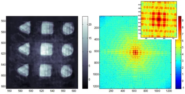
We examined MS-1, a microaerophilic spirillum of size 0.5 x 5μm. MS-1 was fixed by 4 % paraformaldehyde solution. The all samples loaded to a 100 nm thick empty silicon nitride and silicon nitride substrate which contained the designed golden pattern which performed by for e-beam writer (Fig.1). We dropped the MS-1 on the designed pattern by try and error. Before experiment, we checked the sample by optical microscope and found that MS-1 was on the designed pattern. Finally, located relative position between MS-1 and silicon nitride edge.
Fig1.SEM image of designed pattern. The MS-1 is dropped on the center of cross region which contained the designed pattern (3 triangles, 3 squqres and three circules).
Ultrafast transient absorption spectroscopy
The femtosecond transient absorption spectroscopy, shown in figure, operated at 5 KHz and provided excitation pulses (200 nJ at 400 nm) and white light continuum probe pulses of about 100 fs duration. Both beams were focused to a spot of ~ 100 μm and spatially overlapped in the sample cell, which was continuously moved perpendicular to the
beans to ensure sample renewal between the shots. Foe each laser shot, a reference probe intensity was measured simultaneously with the probe intensity passing through the sample in order to calculate the absorbance change. The transient absorption spectra were registered with a polychromator coupled to a fast CCD, and up to 2.5K spectra per second at each delayed time point were averaged.
Fig2. Ultrafast transient absorption spectroscopy and probed spectrum.
Result and discussion
CDI
Fig3. Is the .Recovered diffraction pattern from MS-1 and the reconstructure by HIO. The size and shape of reconstruction result are similar with MS-1 in [3]. However, radiation damage may suffered during x-ray exposure.The blurry reconstruction may also result from the loss of support from sample.
Fig3.Recovered diffraction pattern from MS-1 and the reconstructure by HIO .
Fig4. and 5 show a reconstructed image and the measured diffraction intensity from designed pattern and reconstructed images with different color scale. The preliminary reconstruction does not a quite good, but we could see rough outlines of MS-1. Although the reconstructed image isn’t good to figure out MS-1, the line profiles of
diffraction pattern from reference particles and them with MS-1 is comparable. Further data process and image reconstruction may give better electron density map of MS-1
Fig.4 Diffraction pattern from designed pattern (left) and reconstructed image from that (right).
Fig. 5 Reconstructed image of MS-1 loaded on designed pattern
Ultrafast pump-probe in eNOS
Nitric oxide (NO) is produced from L-arginine by three isoform of nitric oxide synthase (eNOS, iNOS and nNOS). NO production is important function in mammals to control plethora cellular activities. Because of biological importance of the NOS, several publications show the quasi-stationary dynamics of NOS protein in millisecond time-scales and pico-second transient absorption spectroscopy at a few probe wavelengths. However, the origin of different functionalities of these proteins is still not-fully understood. In this work, we studied dynamics of heme-containing
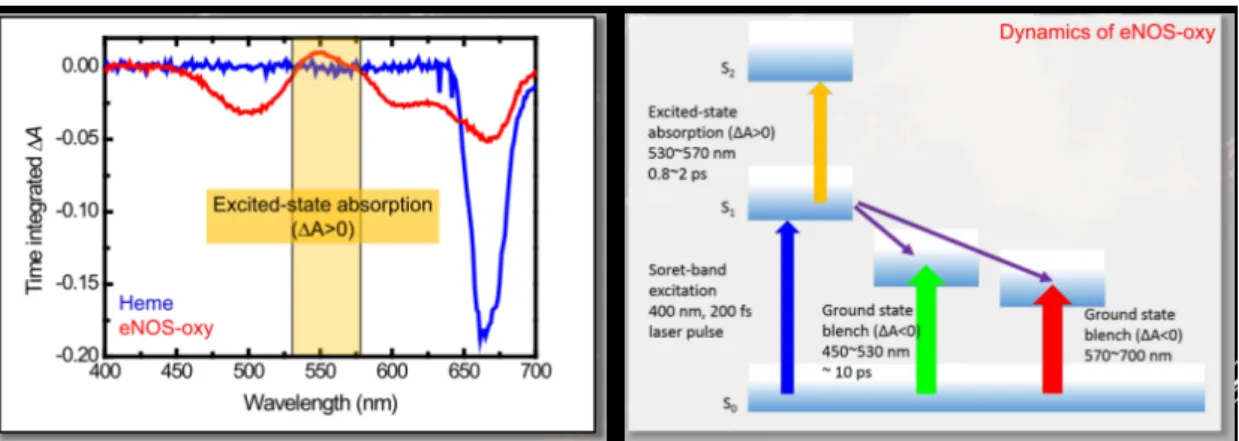
domain of eNOS (oxygenase domain) by ultrafast transient-absorption spectroscopy with 200 fs time resolution (Fig 6). The pump wavelength was centered on 400 nm overlapping the Soret band of heme, which is NO binding site of eNOS, and the time-resolved transient absorption spectrum was probed at visible broadband region extending from 450 nm to 700 nm.
Fig. 6 Experimental diagram for 400 nm pump and supercontinuum white light porbe. The transient absorption trace have shown ultrafast decaying component with lifetime of a few picosecond in both of the probe region (see Fig. 7), which could be assigned to the photo-dissociation and/or recombination dynamics around the heme domain of eNOS. Also, the excited state absorption behavior has observed in wavelength region from 525 nm to 575 nm which has not been shown in previously study (see Fig. 8).
Fig. 7 Two dimensional view of transient absorption change spectra of (a) eNOS oxygenase domain and (b) single heme domain excited at 400 nm by femtosecond pulse. The transient absorption spectra were probed visible broadband around 450 nm to 700 nm in few picosecond time-scale.
Fig. 8 Time integrated transient absorption spectrum and dynamics of eNOS-oxy domain.
Conclusions
CDI
By coherent diffraction imaging of MS-1, we show that the crystals of Fe3O4 in MS-1 can
enhance the SNR and improve reconstructure image. In addition to using heavy atom in MS-1 body to enhance the SNR, we also tried to put MS-1 on designed golden pattern and hope this improve reconstructure image.
Ultrafast pump-probe in eNOS
The time-resolved signal was probed at around 400nm and visible broadband region extending from 450 nm to 750 nm. The observed signal have shown ultrafast decaying components with lifetimes of a few picoseconds in both of the probe regions, which could be assigned to the photo-dissociation and/or recombination dynamics around the heme domain.
Reference
1. C. C. Chen, J. Miao, C. W. Wang, T. K. Lee, Phys. Rev. B 76, 064113 (2007). 2. Blakemore, Richard, Science 190 (4212): 377–379 (1975).
3. Maratea, D., Blakemore, R. P. International Journal of Systematic Bacteriology 31 (4): 452–455 4. W. Belliston-Bittner et al., J. Am. Chem. Soc., 127, 15907-15915 (2005)
5. I. Mikula et al, Biochem. J. 418, 673-682 (2009)
6
國科會補助專題研究計畫移地研究心得報告
日期:2014 年 1 月 18 日一、 移地研究過程
台北時間 7 月 6 日抵達 SSRL,7 月 7 日與 SSRL 研究員翁祖謙博士見面,參觀
SSRL 園區並於當天下午討論 Enos 時間解析光譜的初步結果。7 月 8 日~7 月 16
持續討論 eNOS 時間解析實驗以及未來要在 SSRL 進行 X 光吸收光譜與時間解析
X 光吸收光譜的實驗設計,其中包含維持每次量樣品新鮮度的 continuous flow
cell。7/16 日搭機返台,7/17 日入境台灣。
二、 研究成果
a. Cross-correlation method 在分析 Enos 暫態吸收光譜上的應用
利用此方法我們可以確定 eNOS 時間解析暫態光譜哥波長的響應是否有時間
差,經由對各波長做 cross-correlation 後我們發現:所有的反應的時間差軍
在系統解析度(100fs)以內,故可以視為同時發生。
b. Continuous flow cell 設計
與翁博士討論連續流動樣品盒的設計,此樣品和不僅將使用在可見光波段,未
計畫編號
NSC1012112M009 019
-計畫名稱
散射實驗在蛋白質結構及動態之研究
出國人員
姓名
梁耕三、洪誌彰
服務機構
及職稱
國立交通大學電子物理系
出國時間
2013 年 7 月 5
日至
2013 年 7 月
17 日
出國地點
美國舊金山史丹佛同步輻射中心
(SSRL)
附件四7
來也會使用在 x-ray 波段,進行時間解晰可見光波段與 x-ray 波段 eNOS 超快
動力學探討之用。
Continuous flow cell 以及化學動力學實驗架設
三、 建議
無
四、 其他
國科會補助計畫衍生研發成果推廣資料表
日期:2014/01/21國科會補助計畫
計畫名稱: 散射實驗在蛋白質結構及動態之研究 計畫主持人: 梁耕三 計畫編號: 101-2112-M-009-019- 學門領域: 軟物質及生物物理-實驗無研發成果推廣資料
101 年度專題研究計畫研究成果彙整表
計畫主持人:梁耕三 計畫編號:101-2112-M-009-019-計畫名稱:散射實驗在蛋白質結構及動態之研究 量化 成果項目 實際已達成 數(被接受 或已發表) 預期總達成 數(含實際已 達成數) 本計畫實 際貢獻百 分比 單位 備 註 ( 質 化 說 明:如 數 個 計 畫 共 同 成 果、成 果 列 為 該 期 刊 之 封 面 故 事 ... 等) 期刊論文 0 0 100% 研究報告/技術報告 0 0 100% 研討會論文 0 0 100% 篇 論文著作 專書 0 0 100% 申請中件數 0 0 100% 專利 已獲得件數 0 0 100% 件 件數 0 0 100% 件 技術移轉 權利金 0 0 100% 千元 碩士生 0 0 100% 博士生 0 0 100% 博士後研究員 0 0 100% 國內 參與計畫人力 (本國籍) 專任助理 0 0 100% 人次 期刊論文 0 0 100% 研究報告/技術報告 0 0 100% 研討會論文 2 2 100% 篇 論文著作 專書 0 0 100% 章/本 申請中件數 0 0 100% 專利 已獲得件數 0 0 100% 件 件數 0 0 100% 件 技術移轉 權利金 0 0 100% 千元 碩士生 0 0 100% 博士生 2 2 100% 博士後研究員 0 0 100% 國外 參與計畫人力 (外國籍) 專任助理 0 0 100% 人次其他成果
(
無法以量化表達之成 果如辦理學術活動、獲 得獎項、重要國際合 作、研究成果國際影響 力及其他協助產業技 術發展之具體效益事 項等,請以文字敘述填 列。) 無 成果項目 量化 名稱或內容性質簡述 測驗工具(含質性與量性) 0 課程/模組 0 電腦及網路系統或工具 0 教材 0 舉辦之活動/競賽 0 研討會/工作坊 0 電子報、網站 0 科 教 處 計 畫 加 填 項 目 計畫成果推廣之參與(閱聽)人數 0國科會補助專題研究計畫成果報告自評表
請就研究內容與原計畫相符程度、達成預期目標情況、研究成果之學術或應用價
值(簡要敘述成果所代表之意義、價值、影響或進一步發展之可能性)
、是否適
合在學術期刊發表或申請專利、主要發現或其他有關價值等,作一綜合評估。
1. 請就研究內容與原計畫相符程度、達成預期目標情況作一綜合評估
□達成目標
■未達成目標(請說明,以 100 字為限)
□實驗失敗
□因故實驗中斷
■其他原因
說明:
計畫的初步實驗已達成,包含 coherent diffraction image 實驗技術與數據分析皆建立 好了,以及時間解析暫態吸收光譜架設與在 eNOS 上的應用皆有初步結果,因此自平達成目標 狀況為七成。3 成未達到的原因有:1. SPRING8 的 BEAM TIME 不多,2. 時間解析光譜實驗的 數據需要較多的時間來分析與了解。
2. 研究成果在學術期刊發表或申請專利等情形:
論文:□已發表 □未發表之文稿 □撰寫中 ■無
專利:□已獲得 □申請中 ■無
技轉:□已技轉 □洽談中 ■無
其他:(以 100 字為限)
無3. 請依學術成就、技術創新、社會影響等方面,評估研究成果之學術或應用價
值(簡要敘述成果所代表之意義、價值、影響或進一步發展之可能性)(以
500 字為限)
1. 建立 X-RAY COHERENT DIFFRACTION IMAGE (CDI)在蛋白質樣品上的應用
2. 皆由超快時間解析吸收光譜,我們對於人體必需的一氧化氮合成脢之動力學機制有近 一步的了解,對往後的研究實有助益